
Recommended
More Related Content
What's hot
What's hot (20)
Similar to Anterior abdominal wall.pptx
Similar to Anterior abdominal wall.pptx (20)
More from HARSHIKARIZANI
More from HARSHIKARIZANI (11)
Recently uploaded
Recently uploaded (20)
Anterior abdominal wall.pptx
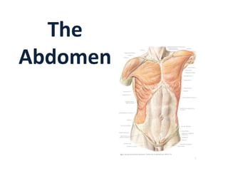
- 1. The Abdomen
- 2. Abdomen • The abdomen is the part of the trunk between the thorax and the pelvis. • It is a flexible, dynamic container, housing most of the organs of the alimentary system and part of the urogenital system. The abdomen consists of: • abdominal walls • abdominal cavity • abdominal viscera
- 3. Anterior Abdominal Wall Boundaries • Superior: • xiphoid process • costal cartilages of the 7th-10th ribs • Inferior: • iliac crest • inguinal fold • pubic symphysis • Lateral: • posterior axillary line
- 4. Surface landmarks and regions of the anterior abdominal wall Topographical divisions of the abdomen are used to describe the location of abdominal organs and the pain associated with abdominal problems. The two schemes most often used are: 1.A four-quadrant pattern 2.A nine-region organizational description.
- 5. Four-quadrant pattern • Transverse Transumbilical plane, passing through the umbilicus (and the intervertebral [IV] disc between the L3 and L4 vertebrae) • Vertical median plane passing longitudinally through the body, dividing it into right and left halves to form four quadrants-the right upper, left upper, right lower, and left lower quadrants
- 6. Using abdominal quadrants to locate major viscera • Liver and gallbladder are in the right upper quadrant. • Stomach and spleen are in the left upper quadrant • Cecum and appendix are in the right lower quadrant • Descending colon and sigmoid colon are in the left lower quadrant.
- 7. Nine-region organizational pattern • The nine regions are delineated by four planes two sagittal (vertical) and two transverse (horizontal) planes. 1. Midclavicular planes that pass from the midpoint of the clavicles (approximately 9 cm from the midline) to the midinguinal points. 2. Subcostal plane is immediately inferior to the costal margins, which places it at the lower border of the costal cartilage of rib X and passes posteriorly through the body of vertebra LIII. 3. Intertubercular plane connects the tubercles of the iliac crests, which are palpable structures 5 cm posterior to the anterior superior iliac spines, and passes through the upper part of the body of vertebra LV.
- 8. The right and left midclavicular lines subdivide it into: Epigastrium: • Epigastric region • Right hypochondric region • Left hypochondric region Mesogastrium: • Umbilical region • Regio lateralis dex. • Regio lateralis sin. Hypogastrium: • Pubic region • Right inguinal region • Left inguinal region
- 9. LAYERS of ABDOMINAL WALL 1- Skin 2- Subcutaneous tissue
- 10. 3. Superficial fascia • Below the umbilicus, it forms two layers: a superficial fatty layer and a deeper membranous layer.
- 11. Superficial fatty layer of superficial fascia (Camper's fascia) • Contains fat and varies in thickness. • It is continuous over the inguinal ligament with the superficial fascia of the thigh and with a similar layer in the perineum. • Continues over the penis and, after losing its fat and fusing with the deeper layer of superficial fascia. • Continues into the scrotum where it forms a specialized fascial layer containing smooth muscle fibers (the dartos fascia)
- 12. Deeper membranous layer of superficial fascia (Scarpa's fascia) • Is thin and membranous, and contains little or no fat. • Inferiorly, it fuses with the deep fascia of the thigh (the fascia lata). • It continues into the anterior part of the perineum where it is referred to as the superficial perineal fascia (Colles fascia).
- 13. 4.Muscles There are five (bilaterally paired) in the anterolateral abdominal wall: three flat muscles - • external oblique, • internal oblique, and •transversus abdominis two vertical muscles – • rectus abdominis and • pyramidalis
- 14. 1- External Oblique m.
- 15. 2- Internal Oblique m.
- 17. 4- Rectus Muscle
- 18. 5- Pyramidalis is a small, insignificant triangular muscle that is absent in approximately 20% of people.
- 20. 6. Peritoneum
- 22. Functions and actions of anterolateral abdominal muscles •Move the trunk and help to maintain posture (resisting lumbar lordosis). • The rectus abdominis is a powerful flexor •Support the abdominal viscera and protect them from most injuries. •Compress the abdominal contents to maintain or increase the intraabdominal pressure •Produce the force required for defecation (discharge of feces), micturition (urination), vomiting, and parturition (childbirth).
- 23. Rectus sheath • The rectus abdominis and pyramidalis muscles are enclosed in an aponeurotic tendinous sheath (the rectus sheath) formed by a unique layering of the aponeuroses of the external and internal oblique, and transversus abdominis muscles
- 24. Organization of the rectus sheath A. Transverse section through the upper ¾ of the rectus sheath B. Transverse section through the lower ¼ of the rectus sheath
- 25. Upper ¾ of the rectus sheath The anterior wall of the rectus sheath consists of • the aponeurosis of the external oblique • & half of the aponeurosis of the internal oblique
- 26. Upper ¾ of the rectus sheath Cont’d The posterior wall of the rectus sheath consists of • The other half of the aponeurosis of the internal oblique • & aponeurosis of the transversus abdominis
- 27. Lower 1/4 of the rectus sheath The anterior wall of the sheath consists of the aponeuroses of the external oblique, internal oblique, and transversus abdominis m. There is no posterior wall at the lower ¼ of the rectus sheath
- 28. Arterial supply and venous drainage Superficially: • The superior part of the wall is supplied by branches from the musculophrenic artery, a terminal branch of the internal thoracic artery. • The inferior part of the wall is supplied by the medially placed superficial epigastric artery and the laterally placed superficial circumflex iliac artery, both branches of the femoral artery.
- 29. Arterial supply and venous drainage At a deeper level: • The superior part of the wall is supplied by the superior epigastric artery, a terminal branch of the internal thoracic artery. • The lateral part of the wall is supplied by branches of the tenth and eleventh intercostal arteries and the subcostal artery.
- 30. Arterial supply and venous drainage At a deeper level: • The inferior part of the wall is supplied by the medially placed inferior epigastric artery and the laterally placed deep circumflex iliac artery, both branches of the external iliac artery. The superior and inferior epigastric arteries both enter the rectus sheath. They are posterior to the rectus abdominis muscle throughout their course, and anastomose with each other
- 31. Veins: In the upper abdomen: • Thoracoepigastric v. In the lower abdomen: • Superficial epigastric v. • Superficial circumflex iliac v. • External pudendal v. Around the umbilicus: • Parumbilical veins Deep veins: • Intercostal vv. • Superior epigastric v. • Inferior epigastric
- 32. Lymphatic drainage From the upper abdominal half to: • Axillary lymph nodes From the lower abdominal half to : •Superficial inguinal lymph nodes
- 33. Innervation • Intercostal nn. Th7 – Th11 • Subcostal n.Th12 •Branches of lumbal plexus Th12 – L4: - Iliohypogastric n. - Ilioinguinal n. - Genitofemoral n
- 34. INGUINAL CANAL • Surgically an important canal because it is the site of inguinal hernias • obliquely located;tubelike • 3-4cm. in length. • Has two openings : • Superficial inguinal ring external oblique apon. -medial • Deep ingunal ring: transversalis fascia - Lateral
- 35. INGUINAL CANAL • Anterior wall • Post. Wall • Superior wall • inferior wall 4-6 cm
- 36. INGUINAL CANAL Anterior wall The anterior wall of the inguinal canal is formed along its entire length by the aponeurosis of the external oblique muscle Posterior wall The posterior wall of the inguinal canal is formed along its entire length by the transversalis fascia Superior wall The roof (superior wall) of the inguinal canal is formed by the arching fibers of the transversus abdominis and internal oblique muscles Inferior wall The floor (inferior wall) of the inguinal canal is formed by the medial one-half of the inguinal ligament
- 37. Contents • The contents of the inguinal canal are: • images the spermatic cord in men, • images the round ligament of the uterus, and • images genital branch of the genitofemoral nerve in women. • These structures enter the inguinal canal through the deep inguinal ring and exit it through the superficial inguinal ring.
- 38. Boundaries of abdomen • Superiorly: The inferior thoracic aperture forms the superior opening to the abdomen, and is closed by the diaphragm. • Inferiorly: the deep abdominal wall is continuous with the pelvic wall at the pelvic inlet.
- 39. Boundaries of abdomen • Anteriorly: anteriorly, a segmented muscle (the rectus abdominis) on each side spans the distance between the inferior thoracic wall and the pelvis • Laterally: lateral parts of the abdominal wall are predominantly formed by three layers of muscles • Posteriorly: vertebral column, the quadratus lumborum, psoas major, and iliacus muscles