SlideShare a Scribd company logo
1 of 58
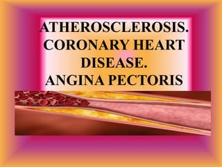
ATHEROSCLEROSIS.
CORONARY HEART
DISEASE.
ANGINA PECTORIS
ATHEROSCLEROSIS
 is a pathological
process leading
to changes in
the arterial
walls due to
accumulation of
lipids,
formation of fibrous tissue and plaques,
constriction of vascular lumen of a vessel.
 Atherosclerosi
s is clinically
manifested in
general and /
or local
circulatory
disorders.
Most commonly, atherosclerotic process
develops in the aorta, femoral, popliteal, tibial,
coronary, internal and external carotid arteries
and in the cerebral arteries.
Risk factors of development
of atherosclerosis
 А. Modified (can be eliminated).
 1. Dislipidemia (hypercholesterinemia, atherogenous
hyperlipoprotein emia).
 2. Smoking.
 3. Arterial hypertension.
 4. Obesity.
 5. Hypodynamia.
 6. Diabetes.
 7. Frequent psychoemotonal stresses.
 8. Hyperhomocycteinemia.
 B. Nonmodified (ineradicable).
 1. The burdened heredity.
 2. The male, age is older than 60 years.
Stages of atherosclerosis
(А. L. Myasnicov, 1965)
 1. Initial (preclinical) period —
formation of atherosclerotic plaques
is observed, lipidemia takes place,
but there are no clinical
manifestations.
 2. The period of clinical
manifestations:
Etiopathogenesis.
Atherosclerot
ic changes
occur in the
endarterium
. This
process has
three
stages:
the fatty streak, the fibrous plaque and
the complex disorders.
1. The fatty streak.
 Spots of
yellowish
color of 1-
2mm in blood
vessels are
detectable
from the
moment of
birth.
These spots are deposits of lipids. They merge and
grow over time. Smooth muscle cells and
macrophages appear in the endartelium; macrophages
accumulate lipids and convert into foam cells.
 In this way a
fatty streak
emerges
consisting of
smooth
muscle cells
and lipid-
containing
macrophages.
fatty streak
2. The fibrous plaque
 is located in the
endarterium and
grows interiorly to
diminish the
vessel’s lumen
over time. A
fibrous plaque has
a thick capsule.
It is composed of endothelial cells, smooth muscle
cells, T-lymphocytes, foam cells (macrophages),
fibrous tissue, and has a soft core containing esters
and cholesterol crystals. Cholesterol comes from
blood.
3. Complex disorders
 are manifested as
reduced thickness
of a capsule of a
fibrous plaque and
its broken
intactness:
development of
cracks, ulcers,
fractures.
 Broken
intactness of
the fibrous
plaque leads
to adhesion
of platelets to
it, their
aggregation
and
thrombosis.
Blood circulation in the affected vessels partially or
completely ceases. Myocardial infarction, ischemic
stroke, etc. develop
Clinic.
 Atherosclerotic process is silent and its
clinical signs appear over time. They depend
on localization of the process and an extent
of obstruction of the vascular bed.
 External signs of atherosclerotic process:
xanthoma (tuberosity in the joints, in the
Achilles tendons caused by cholesterol
deposits), xanthelasma (cholesterol deposits
in the skin of the eyelids, often tuberous)
xanthoma xanthelasma
 senile arc on the cornea (a
strip of yellowish color along
the edge of the cornea).
senile arc
Clinical manifestations of atherosclerosis depend
on localization of the pathological process.
 Atherosclerosis of the aorta. Palpation:
amplified pulsation of the aortic arch in
the jugular fossa.
Percussion: expanded
vascular bundle.
Auscultation: diastolic
shock in the 2nd
intercostal space right
of the sternum,
functional systolic
murmur over the aorta.
 Elevated systolic
blood pressure. Pulse
becomes high over
the radial artery. On
inspection and
palpation of the
atherosclerotic
lesions of arteries
(radial, brachial,
temporal, etc.)
 their density, tortuosity and unevenly thickened
walls are determined. In some cases, there may
be asymmetric blood pressure and pulse in the
arms.
 If distinction in
systolic pressure
on both hands
exceeds 10-15
mm Hg, it may
often suggest
disordered
patency in one
of the branches
of the aortic
arch, caused by
presence of an
atherosclerotic
plaque
 at the place of origin of the subclavian artery
or the brachiocephalic trunk.
CT angiogram showing occlusion of
proximal left subclavian artery
Atherosclerosis of the abdominal
aorta.
 In atherosclerotic
constriction of unpaired
visceral branches of the
abdominal aorta a
specific clinical picture
of abdominal coronary
disease develops in most
cases.
In these patients, moderate tenderness on abdominal
palpation, especially in its superior sections is
present. Functional systolic murmur in the epigastric
region is heard in most patients.
Atherosclerosis of the coronary
arteries (coronary heart disease).
 The physical
examination
is described
in the
relevant
section.
Atherosclerosis of cerebral vessels.
 Dyscirculatory
encephalopathy
develops. In its
first stage,
dysarthria,
abnormal axial
reflexes,
decreased step
length, slowed
walk are revealed
on inspection.
 In the second
stage,
disorders
associated
with
dysfunction
of the frontal
lobes are
revealed:
loss of memory, impaired attention, thinking,
ability to plan and control actions.
 Slightly disordered function of the pelvic organs in the
form of frequent nocturnal urination develop.
Occupational and social adaptation of the patient is
affected; but the patient is still able to take care of
him/herself. The same symptoms are typical for the third
stage, though become are more marked. Dementia,
marked emotional and personality-related disorders, gait
disorders, marked cerebellar disorders, urinary
incontinence develop.
Atherosclerosis of peripheral
arteries.
 On inspection of an affected limb, signs of
impaired trophism are revealed: pale and
cold skin, hypotrophy and atrophy of
muscles,
trophic changes (dry
and flaky skin, nail
dystrophy with
frequent fungal
infection, lack of
body hair).
 Cyanosis or
bluish-red
color of the
skin of the
fingers and
feet is
specific,
especially, in
an upright or
sitting
position.
 Cyanosis is a sign of arterial blood flow
disorders
 Slowly
healing
ulcers
and
necrosis
in the
toes and
feet are
possible.
 On palpation, attenuated pulsation or its
absence determined on the limb arteries.
Atherosclerosis of the renal
arteries.
 Increased blood pressure is specific, the disease does not
very well respond to therapy. Over the area of
constricted renal artery, a systolic murmur is auscultated.
In the later stages of the disease, the signs of chronic
renal failure are revealed.
Laboratory diagnostic studies in
arteriosclerosis
 involve determination of lipid disorders
(dyslipidemias detection) and total
cholesterol in blood plasma.
Variants of hypercholesterolemia
Level Total
cholesterol,
mmol/l
LDL,
mmol/l
Optimal below 5.0 below 3.0
Moderately
elevated
≥ 5.0-5.9 ≥ 3.0-3.9
High ≥ 6.0 ≥ 4.0
CORONARY HEART DISEASE
 (CHD) is a
pathological
condition
that develops
when there is
discrepancy
between the demand of the heart in blood
supply and actual blood supply.
Nonatherosclerotic causes
of coronary failure
 1. Arteritis (nodous periarteritis, Takaiasu disease,
systemic lupus erythematosus, lues, etc.).
 2. Traumas of coronary arteries.
 3. Spastic stricture of coronary arteries.
 4. Embolism of coronary arteries (contagious
endocarditis, intracardiac thrombuses implanted
valves, etc.).
 5. Congenital anomalies of coronary arteries.
 6. Other causes — aortal heart diseases,
thyrotoxicosis, hypercoagulation, complications of
catheterization of heart.
Pathogeny of ischemic heart
disease
The need of myocardium for Oxygen depends
of:
 — Frequencies of cardiac reductions.
 — Myocardial contraction.
 — Strains of the left ventricle during systole.
Supply of the myocardium with Oxygen
depends on the size of coronary blood-flow
which is defined by:
 — The size of resistance of coronary arteries
(diameter, elastance).
 — The size of perfused pressure in the phase of
diastole (the difference between diastolic
pressure in the aorta and diastolic pressure in
the left ventricle).
The pathogenetic factors promoting to the
development of ischemia of the myocardium
 1. Organic narrowing of the lumen of the coronary artery by
atherosclerotic process (plaques, units of thrombocytes,
thrombuses).
 2. The spastic stricture of coronary arteries on the background
of atherosclerosis which alters the reactivity of arteries and
makes their hypersensitive to the influence of neurogenic
stimulants and environmental factors.
 3. Drop of ability of coronary arteries to extend adequately
under the influence of the metabolites arising in conditions of
rising of need of myocardium in Oxygen (adenosine, lactic
acid, Inosinum, hypoxanthine).
 4. The dysfunction of endothelium caused by atherosclerosis,
characterized by predominance of coagulator and
vasoconstrictive factors.
Clinical classification of
coronary heart disease.
• 1. Sudden cardiac death (primary cardiac arrest).
• 2. Angina.
• 2.1. Stable angina (four functional classes).
• 2.2. Unstable angina:
• 2.2.1. Primary angina.
• 2.2.2. Progressive angina.
• 2.2.3. Early post-infarction or post-operative angina.
• 2.3. Spontaneous (vasospastic, variant, Prinzmetal's) angina.
• 3. Painless myocardial ischemia.
• 4. Microvascular angina ("Syndrome X").
• 5. Myocardial infarction.
• 5.1. Myocardial infarction with Q-wave (macrofocal, transmural).
• 5.2. Myocardial infarction without Q-wave (microfocal).
• 6. Postinfarction cardiosclerosis.
• 7. Heart failure (with indicated form and stage).
• 8. Disordered cardiac rhythm and conductivity (with indicated form).
 Primary circulatory arrest is a sudden
death associated with electrical instability
of the myocardium. It is most often
associated with development of
ventricular fibrillation.
 Stable angina (angina of effort) is
characterized by transient episodes of pain
caused by exertion or other factors leading
to increased
demand of the
myocardium in
oxygen (psycho-
emotional stress,
high blood
pressure).
 The pain disappears at
rest or after sublingual
intake of nitroglycerine.
Exertional angina is divided into four functional
classes (FC) based on the extent of physical
exertion (FE), which causes pain.
 FC 1: normal, everyday physical exertion
(walking, stair climbing) does not cause
attacks. They appear only against the patient
extreme or intense exertion.
 FC 2: Slightly restricted usual physical
activity. Attacks occur in normal patient
exertion (walking on a flat ground
covering a distance of more than 200m or
climbing stairs of more than one floor).
 FC 3: markedly
restricted physical
activity. Attacks
occur on performing
insignificant
physical exertion
(walking at a normal
pace on flat ground
at a distance of 100-
200 m or climbing
the stairs of less than
one floor).
 FC 4: inability to tolerate slightest
physical exertion without discomfort. Pain
may also arise at rest.
Unstable angina is subdivided into
three forms:
 1. Primary angina: duration of
the disease is less than 1 month.
It may be a
precursor or a first
manifestation of
myocardial
infarction; it may
transit into a stable
angina or disappear.
 Progressive angina: increased frequency,
severity and duration of attacks of
retrosternal pain in response to normal
exertion.
Onset of angina
attacks in response to
less than normal
effort. Pain becomes
more frequent, more
intense and
prolonged. Patients
have to increase
intake of
nitroglycerin, with its
effect reduced.
 Spontaneous Prinzmetal’s angina is
characterized by retrosternal pain
attacks occurring irrespective of
physical exertion.
The pain is usually
more intense and
prolonged. It can
be hardly relieved
by nitroglycerin
intake.
Clinical picture.
 Angina of effort is characterized by a
squeezing, pressing, non-intensive
retrosternal pain.
To describe its
localization the patient
often puts the fist or
the palm upon the
sternum ("clenched
fist" sign).
 A pain radiating into the left arm,
shoulder, blade and neck is specific. The
more severe angina, the broader an area
of ​​pain irradiation.
 Pain arises on physical or psycho-
emotional stress, cold, after heavy meal,
lasts up to 15-20 minutes, disappears at rest
and after nitroglycerin intake.
Diagnostics.
 The main supplementary technique for diagnostics
of coronary artery is the ECG study. In-between the
attacks, the ECG changes may be absent in
patients. During an angina attack, depending on
localization of myocardial ischemia, negative or
high peaked ("coronary") T-waves are registered in
ECG.

high peaked and negative ("coronary") T-waves
 Simultaneously, especially in long-standing
ischemia, myocardial ischemic lesion develops. It
is manifested in shifted ST-segment upwards or
downwards the isoline . The emerging changes on
the ECG are reversible; they disappear after the
blood flow in the myocardium is restored.
Before attack After attack
Loading electrocardiography-Tests
1. Tests provoking ischemia of myocardium by
rising oxygen consumption:
 — veloergometria;
 — tredmil-test;
 — transesophageal atrial pacing;
2. Tests provoking ischemia of myocardium by
drop of Oxygen delivery:
 — test with dipiridamolum;
 — test with adenosine.
 Veloergometry. It
allows to determine
cardiac exertion
tolerance and to
identify latent
coronary insufficiency.
This test is carried out
in patients with no
visible changes on the
ECG. Exertion
enhances cardiac
performance and,
therefore, increases
myocardial demand in
oxygen.
 In patients with affected coronary arteries, exertion
induces changes on the ECG - depressed or elevated ST-
segment by more than 1 mm from the isoelectric line, or
appearance of a negative T-wave
Other tool methods of ischemic
heart disease diagnostics
 1. Holter’s monitoring — prolonged registration of
electrocardiography (ECG) in conditions of free
(habitual) for a tested activity with the subsequent
analysis of the received findings on the decoder.
 2. Coronary angiography — «the gold standard» of
coronary artery lesion and atherosclerosis
diagnostics. It is carried out with the help of the
angiograph by introduction into of coronary artery
ostium of a contrast agent through a catheter with
the subsequent roentgenography.
Laboratory tests.
 Activity of cardiac specific
enzymes (aspartic
aminotransferase, lactate
dehydrogenase, creatine
phosphokinase) in angina is
not different from that in
normal condition. Only in a
severe course of the disease
against long-standing
myocardial ischemia,
necrosis of some
cardiomyocytes may occur.
In these cases, a slightly
increased activity of these
enzymes is noted.
 The most specific and sensitive laboratory test for
diagnosing cardiomyocyte necrosis a test for
determining the level of troponins in blood serum.
 Echocardiography in patients with angina
reveals signs of local myocardial contractility.
Coronary angiography
 It allows to specify
the location, extent
and nature of
lesion in the
coronary vessels.
These findings are
necessary to make
a decision on the
expediency of
surgical
intervention.
 Coronary angiography of a
critical sub-occlusion of the
common trunk of the left
coronary artery and the
circumflex artery.
Samples of diagnosis formulations:
1. Coronary heart disease. Angina of
effort. FC III. Chronic heart failure,
stage I.
 2. Coronary heart disease.
Progressive angina. Chronic heart
failure, stage I.
Treatment.
 The main objectives:
to reduce the risk of
myocardial infarction
and of a sudden
death; to reduce of
the aftereffects of an
acute ischemia of the
left ventricular
myocardium
 (disturbances in the cardiac rhythm and
conduction, and others).
The following medications and non-
medication measures are used for this
purpose:
 • antianginal medications: β-blockers, nitrates, slow
calcium channel blockers;
 • antithrombin preparations: heparins (unfractionated
and low-molecular), direct thrombin inhibitors;
 • antiplatelet agents, aspirin, adenosine diphosphate
receptor antagonists (thienopyridines), blockers of
glycoprotein IIb / IIIa platelet receptors;
Thank you for
attention!

More Related Content

Similar to атеросклероз лекция.pptx

stroke ( ischemic stroke )
stroke ( ischemic stroke )stroke ( ischemic stroke )
stroke ( ischemic stroke )D.A.B.M
 
Arteriosclerosis and venous disease
Arteriosclerosis and venous diseaseArteriosclerosis and venous disease
Arteriosclerosis and venous diseasepankaj patel
 
CARDIOMYOPATHY.pptx
CARDIOMYOPATHY.pptxCARDIOMYOPATHY.pptx
CARDIOMYOPATHY.pptxZellanienhd
 
ilovepdf_merged (1).pdf
ilovepdf_merged (1).pdfilovepdf_merged (1).pdf
ilovepdf_merged (1).pdfssuser702574
 
Cerebrovascular disease pathology stroke
Cerebrovascular disease pathology strokeCerebrovascular disease pathology stroke
Cerebrovascular disease pathology strokeAppy Akshay Agarwal
 
Pathology of blood vessels
Pathology of blood vesselsPathology of blood vessels
Pathology of blood vesselsmartin osodo
 
strokeppt-170720174010.pdf
strokeppt-170720174010.pdfstrokeppt-170720174010.pdf
strokeppt-170720174010.pdfRiyaSharma295
 
atherosclerosisppt-140124150622-phpapp01.pdf
atherosclerosisppt-140124150622-phpapp01.pdfatherosclerosisppt-140124150622-phpapp01.pdf
atherosclerosisppt-140124150622-phpapp01.pdfSoumyadipPradhan2
 
Ischemic Heart Disease.pptx
Ischemic Heart Disease.pptxIschemic Heart Disease.pptx
Ischemic Heart Disease.pptxAmalRashid10
 
upper limb ischemia ppt.pptx
upper limb ischemia ppt.pptxupper limb ischemia ppt.pptx
upper limb ischemia ppt.pptxPRAGATISHUKLA40
 
MYOCARDIAL INFARACTION.pptx
MYOCARDIAL INFARACTION.pptxMYOCARDIAL INFARACTION.pptx
MYOCARDIAL INFARACTION.pptxDINESH SINGH
 
Cardiovascular system pathology
Cardiovascular system pathologyCardiovascular system pathology
Cardiovascular system pathologyRemix education
 
Cardiovascular Pathophysiology- Coronary Atherosclerosis and Arteriosclerosis
Cardiovascular Pathophysiology- Coronary Atherosclerosis and ArteriosclerosisCardiovascular Pathophysiology- Coronary Atherosclerosis and Arteriosclerosis
Cardiovascular Pathophysiology- Coronary Atherosclerosis and ArteriosclerosisVISHALJADHAV100
 

Similar to атеросклероз лекция.pptx (20)

stroke ( ischemic stroke )
stroke ( ischemic stroke )stroke ( ischemic stroke )
stroke ( ischemic stroke )
 
Arteriosclerosis and venous disease
Arteriosclerosis and venous diseaseArteriosclerosis and venous disease
Arteriosclerosis and venous disease
 
CARDIOMYOPATHY.pptx
CARDIOMYOPATHY.pptxCARDIOMYOPATHY.pptx
CARDIOMYOPATHY.pptx
 
Atherosclerosis
AtherosclerosisAtherosclerosis
Atherosclerosis
 
ilovepdf_merged (1).pdf
ilovepdf_merged (1).pdfilovepdf_merged (1).pdf
ilovepdf_merged (1).pdf
 
Atherosclerosis ppt
Atherosclerosis pptAtherosclerosis ppt
Atherosclerosis ppt
 
Cerebrovascular disease pathology stroke
Cerebrovascular disease pathology strokeCerebrovascular disease pathology stroke
Cerebrovascular disease pathology stroke
 
Pathology of blood vessels
Pathology of blood vesselsPathology of blood vessels
Pathology of blood vessels
 
strokeppt-170720174010.pdf
strokeppt-170720174010.pdfstrokeppt-170720174010.pdf
strokeppt-170720174010.pdf
 
Stroke ppt
Stroke pptStroke ppt
Stroke ppt
 
Atherosclerosis
AtherosclerosisAtherosclerosis
Atherosclerosis
 
atherosclerosisppt-140124150622-phpapp01.pdf
atherosclerosisppt-140124150622-phpapp01.pdfatherosclerosisppt-140124150622-phpapp01.pdf
atherosclerosisppt-140124150622-phpapp01.pdf
 
Ischemic Heart Disease.pptx
Ischemic Heart Disease.pptxIschemic Heart Disease.pptx
Ischemic Heart Disease.pptx
 
upper limb ischemia ppt.pptx
upper limb ischemia ppt.pptxupper limb ischemia ppt.pptx
upper limb ischemia ppt.pptx
 
Atherosclerosis by neha
Atherosclerosis by nehaAtherosclerosis by neha
Atherosclerosis by neha
 
MYOCARDIAL INFARACTION.pptx
MYOCARDIAL INFARACTION.pptxMYOCARDIAL INFARACTION.pptx
MYOCARDIAL INFARACTION.pptx
 
Cardiovascular system pathology
Cardiovascular system pathologyCardiovascular system pathology
Cardiovascular system pathology
 
Cardiovascular Pathophysiology- Coronary Atherosclerosis and Arteriosclerosis
Cardiovascular Pathophysiology- Coronary Atherosclerosis and ArteriosclerosisCardiovascular Pathophysiology- Coronary Atherosclerosis and Arteriosclerosis
Cardiovascular Pathophysiology- Coronary Atherosclerosis and Arteriosclerosis
 
Atherosclerosis
AtherosclerosisAtherosclerosis
Atherosclerosis
 
@ Stroke seminar
@ Stroke seminar@ Stroke seminar
@ Stroke seminar
 

More from HARSHIKARIZANI

Anterior abdominal wall.pptx
Anterior abdominal wall.pptxAnterior abdominal wall.pptx
Anterior abdominal wall.pptxHARSHIKARIZANI
 
3.inflammation 1-1.pptx
3.inflammation 1-1.pptx3.inflammation 1-1.pptx
3.inflammation 1-1.pptxHARSHIKARIZANI
 
Instrumental_and_Laboratory_Techniques_of_Examination_in_Pathology of CVS.ppt
Instrumental_and_Laboratory_Techniques_of_Examination_in_Pathology of CVS.pptInstrumental_and_Laboratory_Techniques_of_Examination_in_Pathology of CVS.ppt
Instrumental_and_Laboratory_Techniques_of_Examination_in_Pathology of CVS.pptHARSHIKARIZANI
 
magnetic_resonance_imaging1.ppt
magnetic_resonance_imaging1.pptmagnetic_resonance_imaging1.ppt
magnetic_resonance_imaging1.pptHARSHIKARIZANI
 
Cell injury_e894658c-e167-4c2d-a3bb-ff7a45959c3b.ppt
Cell injury_e894658c-e167-4c2d-a3bb-ff7a45959c3b.pptCell injury_e894658c-e167-4c2d-a3bb-ff7a45959c3b.ppt
Cell injury_e894658c-e167-4c2d-a3bb-ff7a45959c3b.pptHARSHIKARIZANI
 
congo fever ppt shweta.pptx
congo fever ppt shweta.pptxcongo fever ppt shweta.pptx
congo fever ppt shweta.pptxHARSHIKARIZANI
 
physio. adaptation copy.pptx
physio. adaptation copy.pptxphysio. adaptation copy.pptx
physio. adaptation copy.pptxHARSHIKARIZANI
 
Microbiology bacillus cereus.pptx
Microbiology bacillus cereus.pptxMicrobiology bacillus cereus.pptx
Microbiology bacillus cereus.pptxHARSHIKARIZANI
 

More from HARSHIKARIZANI (11)

4. fever.pptx
4. fever.pptx4. fever.pptx
4. fever.pptx
 
Anterior abdominal wall.pptx
Anterior abdominal wall.pptxAnterior abdominal wall.pptx
Anterior abdominal wall.pptx
 
3.inflammation 1-1.pptx
3.inflammation 1-1.pptx3.inflammation 1-1.pptx
3.inflammation 1-1.pptx
 
Instrumental_and_Laboratory_Techniques_of_Examination_in_Pathology of CVS.ppt
Instrumental_and_Laboratory_Techniques_of_Examination_in_Pathology of CVS.pptInstrumental_and_Laboratory_Techniques_of_Examination_in_Pathology of CVS.ppt
Instrumental_and_Laboratory_Techniques_of_Examination_in_Pathology of CVS.ppt
 
magnetic_resonance_imaging1.ppt
magnetic_resonance_imaging1.pptmagnetic_resonance_imaging1.ppt
magnetic_resonance_imaging1.ppt
 
X-ray.ppt
X-ray.pptX-ray.ppt
X-ray.ppt
 
Cell injury_e894658c-e167-4c2d-a3bb-ff7a45959c3b.ppt
Cell injury_e894658c-e167-4c2d-a3bb-ff7a45959c3b.pptCell injury_e894658c-e167-4c2d-a3bb-ff7a45959c3b.ppt
Cell injury_e894658c-e167-4c2d-a3bb-ff7a45959c3b.ppt
 
congo fever ppt shweta.pptx
congo fever ppt shweta.pptxcongo fever ppt shweta.pptx
congo fever ppt shweta.pptx
 
Hantavirus.pdf
Hantavirus.pdfHantavirus.pdf
Hantavirus.pdf
 
physio. adaptation copy.pptx
physio. adaptation copy.pptxphysio. adaptation copy.pptx
physio. adaptation copy.pptx
 
Microbiology bacillus cereus.pptx
Microbiology bacillus cereus.pptxMicrobiology bacillus cereus.pptx
Microbiology bacillus cereus.pptx
 

Recently uploaded

Unit 3 Emotional Intelligence and Spiritual Intelligence.pdf
Unit 3 Emotional Intelligence and Spiritual Intelligence.pdfUnit 3 Emotional Intelligence and Spiritual Intelligence.pdf
Unit 3 Emotional Intelligence and Spiritual Intelligence.pdfDr Vijay Vishwakarma
 
What is 3 Way Matching Process in Odoo 17.pptx
What is 3 Way Matching Process in Odoo 17.pptxWhat is 3 Way Matching Process in Odoo 17.pptx
What is 3 Way Matching Process in Odoo 17.pptxCeline George
 
TỔNG ÔN TẬP THI VÀO LỚP 10 MÔN TIẾNG ANH NĂM HỌC 2023 - 2024 CÓ ĐÁP ÁN (NGỮ Â...
TỔNG ÔN TẬP THI VÀO LỚP 10 MÔN TIẾNG ANH NĂM HỌC 2023 - 2024 CÓ ĐÁP ÁN (NGỮ Â...TỔNG ÔN TẬP THI VÀO LỚP 10 MÔN TIẾNG ANH NĂM HỌC 2023 - 2024 CÓ ĐÁP ÁN (NGỮ Â...
TỔNG ÔN TẬP THI VÀO LỚP 10 MÔN TIẾNG ANH NĂM HỌC 2023 - 2024 CÓ ĐÁP ÁN (NGỮ Â...Nguyen Thanh Tu Collection
 
How to Manage Call for Tendor in Odoo 17
How to Manage Call for Tendor in Odoo 17How to Manage Call for Tendor in Odoo 17
How to Manage Call for Tendor in Odoo 17Celine George
 
Python Notes for mca i year students osmania university.docx
Python Notes for mca i year students osmania university.docxPython Notes for mca i year students osmania university.docx
Python Notes for mca i year students osmania university.docxRamakrishna Reddy Bijjam
 
QUATER-1-PE-HEALTH-LC2- this is just a sample of unpacked lesson
QUATER-1-PE-HEALTH-LC2- this is just a sample of unpacked lessonQUATER-1-PE-HEALTH-LC2- this is just a sample of unpacked lesson
QUATER-1-PE-HEALTH-LC2- this is just a sample of unpacked lessonhttgc7rh9c
 
How to Manage Global Discount in Odoo 17 POS
How to Manage Global Discount in Odoo 17 POSHow to Manage Global Discount in Odoo 17 POS
How to Manage Global Discount in Odoo 17 POSCeline George
 
FICTIONAL SALESMAN/SALESMAN SNSW 2024.pdf
FICTIONAL SALESMAN/SALESMAN SNSW 2024.pdfFICTIONAL SALESMAN/SALESMAN SNSW 2024.pdf
FICTIONAL SALESMAN/SALESMAN SNSW 2024.pdfPondicherry University
 
REMIFENTANIL: An Ultra short acting opioid.pptx
REMIFENTANIL: An Ultra short acting opioid.pptxREMIFENTANIL: An Ultra short acting opioid.pptx
REMIFENTANIL: An Ultra short acting opioid.pptxDr. Ravikiran H M Gowda
 
Accessible Digital Futures project (20/03/2024)
Accessible Digital Futures project (20/03/2024)Accessible Digital Futures project (20/03/2024)
Accessible Digital Futures project (20/03/2024)Jisc
 
Spellings Wk 4 and Wk 5 for Grade 4 at CAPS
Spellings Wk 4 and Wk 5 for Grade 4 at CAPSSpellings Wk 4 and Wk 5 for Grade 4 at CAPS
Spellings Wk 4 and Wk 5 for Grade 4 at CAPSAnaAcapella
 
Model Attribute _rec_name in the Odoo 17
Model Attribute _rec_name in the Odoo 17Model Attribute _rec_name in the Odoo 17
Model Attribute _rec_name in the Odoo 17Celine George
 
Exploring_the_Narrative_Style_of_Amitav_Ghoshs_Gun_Island.pptx
Exploring_the_Narrative_Style_of_Amitav_Ghoshs_Gun_Island.pptxExploring_the_Narrative_Style_of_Amitav_Ghoshs_Gun_Island.pptx
Exploring_the_Narrative_Style_of_Amitav_Ghoshs_Gun_Island.pptxPooja Bhuva
 
Wellbeing inclusion and digital dystopias.pptx
Wellbeing inclusion and digital dystopias.pptxWellbeing inclusion and digital dystopias.pptx
Wellbeing inclusion and digital dystopias.pptxJisc
 
Details on CBSE Compartment Exam.pptx1111
Details on CBSE Compartment Exam.pptx1111Details on CBSE Compartment Exam.pptx1111
Details on CBSE Compartment Exam.pptx1111GangaMaiya1
 
Economic Importance Of Fungi In Food Additives
Economic Importance Of Fungi In Food AdditivesEconomic Importance Of Fungi In Food Additives
Economic Importance Of Fungi In Food AdditivesSHIVANANDaRV
 
dusjagr & nano talk on open tools for agriculture research and learning
dusjagr & nano talk on open tools for agriculture research and learningdusjagr & nano talk on open tools for agriculture research and learning
dusjagr & nano talk on open tools for agriculture research and learningMarc Dusseiller Dusjagr
 
NO1 Top Black Magic Specialist In Lahore Black magic In Pakistan Kala Ilam Ex...
NO1 Top Black Magic Specialist In Lahore Black magic In Pakistan Kala Ilam Ex...NO1 Top Black Magic Specialist In Lahore Black magic In Pakistan Kala Ilam Ex...
NO1 Top Black Magic Specialist In Lahore Black magic In Pakistan Kala Ilam Ex...Amil baba
 
Interdisciplinary_Insights_Data_Collection_Methods.pptx
Interdisciplinary_Insights_Data_Collection_Methods.pptxInterdisciplinary_Insights_Data_Collection_Methods.pptx
Interdisciplinary_Insights_Data_Collection_Methods.pptxPooja Bhuva
 
Understanding Accommodations and Modifications
Understanding  Accommodations and ModificationsUnderstanding  Accommodations and Modifications
Understanding Accommodations and ModificationsMJDuyan
 

Recently uploaded (20)

Unit 3 Emotional Intelligence and Spiritual Intelligence.pdf
Unit 3 Emotional Intelligence and Spiritual Intelligence.pdfUnit 3 Emotional Intelligence and Spiritual Intelligence.pdf
Unit 3 Emotional Intelligence and Spiritual Intelligence.pdf
 
What is 3 Way Matching Process in Odoo 17.pptx
What is 3 Way Matching Process in Odoo 17.pptxWhat is 3 Way Matching Process in Odoo 17.pptx
What is 3 Way Matching Process in Odoo 17.pptx
 
TỔNG ÔN TẬP THI VÀO LỚP 10 MÔN TIẾNG ANH NĂM HỌC 2023 - 2024 CÓ ĐÁP ÁN (NGỮ Â...
TỔNG ÔN TẬP THI VÀO LỚP 10 MÔN TIẾNG ANH NĂM HỌC 2023 - 2024 CÓ ĐÁP ÁN (NGỮ Â...TỔNG ÔN TẬP THI VÀO LỚP 10 MÔN TIẾNG ANH NĂM HỌC 2023 - 2024 CÓ ĐÁP ÁN (NGỮ Â...
TỔNG ÔN TẬP THI VÀO LỚP 10 MÔN TIẾNG ANH NĂM HỌC 2023 - 2024 CÓ ĐÁP ÁN (NGỮ Â...
 
How to Manage Call for Tendor in Odoo 17
How to Manage Call for Tendor in Odoo 17How to Manage Call for Tendor in Odoo 17
How to Manage Call for Tendor in Odoo 17
 
Python Notes for mca i year students osmania university.docx
Python Notes for mca i year students osmania university.docxPython Notes for mca i year students osmania university.docx
Python Notes for mca i year students osmania university.docx
 
QUATER-1-PE-HEALTH-LC2- this is just a sample of unpacked lesson
QUATER-1-PE-HEALTH-LC2- this is just a sample of unpacked lessonQUATER-1-PE-HEALTH-LC2- this is just a sample of unpacked lesson
QUATER-1-PE-HEALTH-LC2- this is just a sample of unpacked lesson
 
How to Manage Global Discount in Odoo 17 POS
How to Manage Global Discount in Odoo 17 POSHow to Manage Global Discount in Odoo 17 POS
How to Manage Global Discount in Odoo 17 POS
 
FICTIONAL SALESMAN/SALESMAN SNSW 2024.pdf
FICTIONAL SALESMAN/SALESMAN SNSW 2024.pdfFICTIONAL SALESMAN/SALESMAN SNSW 2024.pdf
FICTIONAL SALESMAN/SALESMAN SNSW 2024.pdf
 
REMIFENTANIL: An Ultra short acting opioid.pptx
REMIFENTANIL: An Ultra short acting opioid.pptxREMIFENTANIL: An Ultra short acting opioid.pptx
REMIFENTANIL: An Ultra short acting opioid.pptx
 
Accessible Digital Futures project (20/03/2024)
Accessible Digital Futures project (20/03/2024)Accessible Digital Futures project (20/03/2024)
Accessible Digital Futures project (20/03/2024)
 
Spellings Wk 4 and Wk 5 for Grade 4 at CAPS
Spellings Wk 4 and Wk 5 for Grade 4 at CAPSSpellings Wk 4 and Wk 5 for Grade 4 at CAPS
Spellings Wk 4 and Wk 5 for Grade 4 at CAPS
 
Model Attribute _rec_name in the Odoo 17
Model Attribute _rec_name in the Odoo 17Model Attribute _rec_name in the Odoo 17
Model Attribute _rec_name in the Odoo 17
 
Exploring_the_Narrative_Style_of_Amitav_Ghoshs_Gun_Island.pptx
Exploring_the_Narrative_Style_of_Amitav_Ghoshs_Gun_Island.pptxExploring_the_Narrative_Style_of_Amitav_Ghoshs_Gun_Island.pptx
Exploring_the_Narrative_Style_of_Amitav_Ghoshs_Gun_Island.pptx
 
Wellbeing inclusion and digital dystopias.pptx
Wellbeing inclusion and digital dystopias.pptxWellbeing inclusion and digital dystopias.pptx
Wellbeing inclusion and digital dystopias.pptx
 
Details on CBSE Compartment Exam.pptx1111
Details on CBSE Compartment Exam.pptx1111Details on CBSE Compartment Exam.pptx1111
Details on CBSE Compartment Exam.pptx1111
 
Economic Importance Of Fungi In Food Additives
Economic Importance Of Fungi In Food AdditivesEconomic Importance Of Fungi In Food Additives
Economic Importance Of Fungi In Food Additives
 
dusjagr & nano talk on open tools for agriculture research and learning
dusjagr & nano talk on open tools for agriculture research and learningdusjagr & nano talk on open tools for agriculture research and learning
dusjagr & nano talk on open tools for agriculture research and learning
 
NO1 Top Black Magic Specialist In Lahore Black magic In Pakistan Kala Ilam Ex...
NO1 Top Black Magic Specialist In Lahore Black magic In Pakistan Kala Ilam Ex...NO1 Top Black Magic Specialist In Lahore Black magic In Pakistan Kala Ilam Ex...
NO1 Top Black Magic Specialist In Lahore Black magic In Pakistan Kala Ilam Ex...
 
Interdisciplinary_Insights_Data_Collection_Methods.pptx
Interdisciplinary_Insights_Data_Collection_Methods.pptxInterdisciplinary_Insights_Data_Collection_Methods.pptx
Interdisciplinary_Insights_Data_Collection_Methods.pptx
 
Understanding Accommodations and Modifications
Understanding  Accommodations and ModificationsUnderstanding  Accommodations and Modifications
Understanding Accommodations and Modifications
 

атеросклероз лекция.pptx

  • 2. ATHEROSCLEROSIS  is a pathological process leading to changes in the arterial walls due to accumulation of lipids, formation of fibrous tissue and plaques, constriction of vascular lumen of a vessel.
  • 3.  Atherosclerosi s is clinically manifested in general and / or local circulatory disorders. Most commonly, atherosclerotic process develops in the aorta, femoral, popliteal, tibial, coronary, internal and external carotid arteries and in the cerebral arteries.
  • 4. Risk factors of development of atherosclerosis  А. Modified (can be eliminated).  1. Dislipidemia (hypercholesterinemia, atherogenous hyperlipoprotein emia).  2. Smoking.  3. Arterial hypertension.  4. Obesity.  5. Hypodynamia.  6. Diabetes.  7. Frequent psychoemotonal stresses.  8. Hyperhomocycteinemia.  B. Nonmodified (ineradicable).  1. The burdened heredity.  2. The male, age is older than 60 years.
  • 5. Stages of atherosclerosis (А. L. Myasnicov, 1965)  1. Initial (preclinical) period — formation of atherosclerotic plaques is observed, lipidemia takes place, but there are no clinical manifestations.  2. The period of clinical manifestations:
  • 6. Etiopathogenesis. Atherosclerot ic changes occur in the endarterium . This process has three stages: the fatty streak, the fibrous plaque and the complex disorders.
  • 7. 1. The fatty streak.  Spots of yellowish color of 1- 2mm in blood vessels are detectable from the moment of birth. These spots are deposits of lipids. They merge and grow over time. Smooth muscle cells and macrophages appear in the endartelium; macrophages accumulate lipids and convert into foam cells.
  • 8.  In this way a fatty streak emerges consisting of smooth muscle cells and lipid- containing macrophages. fatty streak
  • 9. 2. The fibrous plaque  is located in the endarterium and grows interiorly to diminish the vessel’s lumen over time. A fibrous plaque has a thick capsule. It is composed of endothelial cells, smooth muscle cells, T-lymphocytes, foam cells (macrophages), fibrous tissue, and has a soft core containing esters and cholesterol crystals. Cholesterol comes from blood.
  • 10. 3. Complex disorders  are manifested as reduced thickness of a capsule of a fibrous plaque and its broken intactness: development of cracks, ulcers, fractures.
  • 11.  Broken intactness of the fibrous plaque leads to adhesion of platelets to it, their aggregation and thrombosis. Blood circulation in the affected vessels partially or completely ceases. Myocardial infarction, ischemic stroke, etc. develop
  • 12. Clinic.  Atherosclerotic process is silent and its clinical signs appear over time. They depend on localization of the process and an extent of obstruction of the vascular bed.
  • 13.  External signs of atherosclerotic process: xanthoma (tuberosity in the joints, in the Achilles tendons caused by cholesterol deposits), xanthelasma (cholesterol deposits in the skin of the eyelids, often tuberous) xanthoma xanthelasma
  • 14.  senile arc on the cornea (a strip of yellowish color along the edge of the cornea). senile arc
  • 15. Clinical manifestations of atherosclerosis depend on localization of the pathological process.  Atherosclerosis of the aorta. Palpation: amplified pulsation of the aortic arch in the jugular fossa. Percussion: expanded vascular bundle. Auscultation: diastolic shock in the 2nd intercostal space right of the sternum, functional systolic murmur over the aorta.
  • 16.  Elevated systolic blood pressure. Pulse becomes high over the radial artery. On inspection and palpation of the atherosclerotic lesions of arteries (radial, brachial, temporal, etc.)  their density, tortuosity and unevenly thickened walls are determined. In some cases, there may be asymmetric blood pressure and pulse in the arms.
  • 17.  If distinction in systolic pressure on both hands exceeds 10-15 mm Hg, it may often suggest disordered patency in one of the branches of the aortic arch, caused by presence of an atherosclerotic plaque  at the place of origin of the subclavian artery or the brachiocephalic trunk. CT angiogram showing occlusion of proximal left subclavian artery
  • 18. Atherosclerosis of the abdominal aorta.  In atherosclerotic constriction of unpaired visceral branches of the abdominal aorta a specific clinical picture of abdominal coronary disease develops in most cases. In these patients, moderate tenderness on abdominal palpation, especially in its superior sections is present. Functional systolic murmur in the epigastric region is heard in most patients.
  • 19. Atherosclerosis of the coronary arteries (coronary heart disease).  The physical examination is described in the relevant section.
  • 20. Atherosclerosis of cerebral vessels.  Dyscirculatory encephalopathy develops. In its first stage, dysarthria, abnormal axial reflexes, decreased step length, slowed walk are revealed on inspection.
  • 21.  In the second stage, disorders associated with dysfunction of the frontal lobes are revealed: loss of memory, impaired attention, thinking, ability to plan and control actions.
  • 22.  Slightly disordered function of the pelvic organs in the form of frequent nocturnal urination develop. Occupational and social adaptation of the patient is affected; but the patient is still able to take care of him/herself. The same symptoms are typical for the third stage, though become are more marked. Dementia, marked emotional and personality-related disorders, gait disorders, marked cerebellar disorders, urinary incontinence develop.
  • 23. Atherosclerosis of peripheral arteries.  On inspection of an affected limb, signs of impaired trophism are revealed: pale and cold skin, hypotrophy and atrophy of muscles, trophic changes (dry and flaky skin, nail dystrophy with frequent fungal infection, lack of body hair).
  • 24.  Cyanosis or bluish-red color of the skin of the fingers and feet is specific, especially, in an upright or sitting position.  Cyanosis is a sign of arterial blood flow disorders
  • 25.  Slowly healing ulcers and necrosis in the toes and feet are possible.  On palpation, attenuated pulsation or its absence determined on the limb arteries.
  • 26. Atherosclerosis of the renal arteries.  Increased blood pressure is specific, the disease does not very well respond to therapy. Over the area of constricted renal artery, a systolic murmur is auscultated. In the later stages of the disease, the signs of chronic renal failure are revealed.
  • 27. Laboratory diagnostic studies in arteriosclerosis  involve determination of lipid disorders (dyslipidemias detection) and total cholesterol in blood plasma.
  • 28. Variants of hypercholesterolemia Level Total cholesterol, mmol/l LDL, mmol/l Optimal below 5.0 below 3.0 Moderately elevated ≥ 5.0-5.9 ≥ 3.0-3.9 High ≥ 6.0 ≥ 4.0
  • 29. CORONARY HEART DISEASE  (CHD) is a pathological condition that develops when there is discrepancy between the demand of the heart in blood supply and actual blood supply.
  • 30. Nonatherosclerotic causes of coronary failure  1. Arteritis (nodous periarteritis, Takaiasu disease, systemic lupus erythematosus, lues, etc.).  2. Traumas of coronary arteries.  3. Spastic stricture of coronary arteries.  4. Embolism of coronary arteries (contagious endocarditis, intracardiac thrombuses implanted valves, etc.).  5. Congenital anomalies of coronary arteries.  6. Other causes — aortal heart diseases, thyrotoxicosis, hypercoagulation, complications of catheterization of heart.
  • 31. Pathogeny of ischemic heart disease The need of myocardium for Oxygen depends of:  — Frequencies of cardiac reductions.  — Myocardial contraction.  — Strains of the left ventricle during systole. Supply of the myocardium with Oxygen depends on the size of coronary blood-flow which is defined by:  — The size of resistance of coronary arteries (diameter, elastance).  — The size of perfused pressure in the phase of diastole (the difference between diastolic pressure in the aorta and diastolic pressure in the left ventricle).
  • 32. The pathogenetic factors promoting to the development of ischemia of the myocardium  1. Organic narrowing of the lumen of the coronary artery by atherosclerotic process (plaques, units of thrombocytes, thrombuses).  2. The spastic stricture of coronary arteries on the background of atherosclerosis which alters the reactivity of arteries and makes their hypersensitive to the influence of neurogenic stimulants and environmental factors.  3. Drop of ability of coronary arteries to extend adequately under the influence of the metabolites arising in conditions of rising of need of myocardium in Oxygen (adenosine, lactic acid, Inosinum, hypoxanthine).  4. The dysfunction of endothelium caused by atherosclerosis, characterized by predominance of coagulator and vasoconstrictive factors.
  • 33. Clinical classification of coronary heart disease. • 1. Sudden cardiac death (primary cardiac arrest). • 2. Angina. • 2.1. Stable angina (four functional classes). • 2.2. Unstable angina: • 2.2.1. Primary angina. • 2.2.2. Progressive angina. • 2.2.3. Early post-infarction or post-operative angina. • 2.3. Spontaneous (vasospastic, variant, Prinzmetal's) angina. • 3. Painless myocardial ischemia. • 4. Microvascular angina ("Syndrome X"). • 5. Myocardial infarction. • 5.1. Myocardial infarction with Q-wave (macrofocal, transmural). • 5.2. Myocardial infarction without Q-wave (microfocal). • 6. Postinfarction cardiosclerosis. • 7. Heart failure (with indicated form and stage). • 8. Disordered cardiac rhythm and conductivity (with indicated form).
  • 34.  Primary circulatory arrest is a sudden death associated with electrical instability of the myocardium. It is most often associated with development of ventricular fibrillation.
  • 35.  Stable angina (angina of effort) is characterized by transient episodes of pain caused by exertion or other factors leading to increased demand of the myocardium in oxygen (psycho- emotional stress, high blood pressure).
  • 36.  The pain disappears at rest or after sublingual intake of nitroglycerine.
  • 37. Exertional angina is divided into four functional classes (FC) based on the extent of physical exertion (FE), which causes pain.  FC 1: normal, everyday physical exertion (walking, stair climbing) does not cause attacks. They appear only against the patient extreme or intense exertion.
  • 38.  FC 2: Slightly restricted usual physical activity. Attacks occur in normal patient exertion (walking on a flat ground covering a distance of more than 200m or climbing stairs of more than one floor).
  • 39.  FC 3: markedly restricted physical activity. Attacks occur on performing insignificant physical exertion (walking at a normal pace on flat ground at a distance of 100- 200 m or climbing the stairs of less than one floor).
  • 40.  FC 4: inability to tolerate slightest physical exertion without discomfort. Pain may also arise at rest.
  • 41. Unstable angina is subdivided into three forms:  1. Primary angina: duration of the disease is less than 1 month. It may be a precursor or a first manifestation of myocardial infarction; it may transit into a stable angina or disappear.
  • 42.  Progressive angina: increased frequency, severity and duration of attacks of retrosternal pain in response to normal exertion. Onset of angina attacks in response to less than normal effort. Pain becomes more frequent, more intense and prolonged. Patients have to increase intake of nitroglycerin, with its effect reduced.
  • 43.  Spontaneous Prinzmetal’s angina is characterized by retrosternal pain attacks occurring irrespective of physical exertion. The pain is usually more intense and prolonged. It can be hardly relieved by nitroglycerin intake.
  • 44. Clinical picture.  Angina of effort is characterized by a squeezing, pressing, non-intensive retrosternal pain. To describe its localization the patient often puts the fist or the palm upon the sternum ("clenched fist" sign).
  • 45.  A pain radiating into the left arm, shoulder, blade and neck is specific. The more severe angina, the broader an area of ​​pain irradiation.
  • 46.  Pain arises on physical or psycho- emotional stress, cold, after heavy meal, lasts up to 15-20 minutes, disappears at rest and after nitroglycerin intake.
  • 47. Diagnostics.  The main supplementary technique for diagnostics of coronary artery is the ECG study. In-between the attacks, the ECG changes may be absent in patients. During an angina attack, depending on localization of myocardial ischemia, negative or high peaked ("coronary") T-waves are registered in ECG.  high peaked and negative ("coronary") T-waves
  • 48.  Simultaneously, especially in long-standing ischemia, myocardial ischemic lesion develops. It is manifested in shifted ST-segment upwards or downwards the isoline . The emerging changes on the ECG are reversible; they disappear after the blood flow in the myocardium is restored. Before attack After attack
  • 49. Loading electrocardiography-Tests 1. Tests provoking ischemia of myocardium by rising oxygen consumption:  — veloergometria;  — tredmil-test;  — transesophageal atrial pacing; 2. Tests provoking ischemia of myocardium by drop of Oxygen delivery:  — test with dipiridamolum;  — test with adenosine.
  • 50.  Veloergometry. It allows to determine cardiac exertion tolerance and to identify latent coronary insufficiency. This test is carried out in patients with no visible changes on the ECG. Exertion enhances cardiac performance and, therefore, increases myocardial demand in oxygen.  In patients with affected coronary arteries, exertion induces changes on the ECG - depressed or elevated ST- segment by more than 1 mm from the isoelectric line, or appearance of a negative T-wave
  • 51. Other tool methods of ischemic heart disease diagnostics  1. Holter’s monitoring — prolonged registration of electrocardiography (ECG) in conditions of free (habitual) for a tested activity with the subsequent analysis of the received findings on the decoder.  2. Coronary angiography — «the gold standard» of coronary artery lesion and atherosclerosis diagnostics. It is carried out with the help of the angiograph by introduction into of coronary artery ostium of a contrast agent through a catheter with the subsequent roentgenography.
  • 52. Laboratory tests.  Activity of cardiac specific enzymes (aspartic aminotransferase, lactate dehydrogenase, creatine phosphokinase) in angina is not different from that in normal condition. Only in a severe course of the disease against long-standing myocardial ischemia, necrosis of some cardiomyocytes may occur. In these cases, a slightly increased activity of these enzymes is noted.  The most specific and sensitive laboratory test for diagnosing cardiomyocyte necrosis a test for determining the level of troponins in blood serum.
  • 53.  Echocardiography in patients with angina reveals signs of local myocardial contractility.
  • 54. Coronary angiography  It allows to specify the location, extent and nature of lesion in the coronary vessels. These findings are necessary to make a decision on the expediency of surgical intervention.  Coronary angiography of a critical sub-occlusion of the common trunk of the left coronary artery and the circumflex artery.
  • 55. Samples of diagnosis formulations: 1. Coronary heart disease. Angina of effort. FC III. Chronic heart failure, stage I.  2. Coronary heart disease. Progressive angina. Chronic heart failure, stage I.
  • 56. Treatment.  The main objectives: to reduce the risk of myocardial infarction and of a sudden death; to reduce of the aftereffects of an acute ischemia of the left ventricular myocardium  (disturbances in the cardiac rhythm and conduction, and others).
  • 57. The following medications and non- medication measures are used for this purpose:  • antianginal medications: β-blockers, nitrates, slow calcium channel blockers;  • antithrombin preparations: heparins (unfractionated and low-molecular), direct thrombin inhibitors;  • antiplatelet agents, aspirin, adenosine diphosphate receptor antagonists (thienopyridines), blockers of glycoprotein IIb / IIIa platelet receptors;