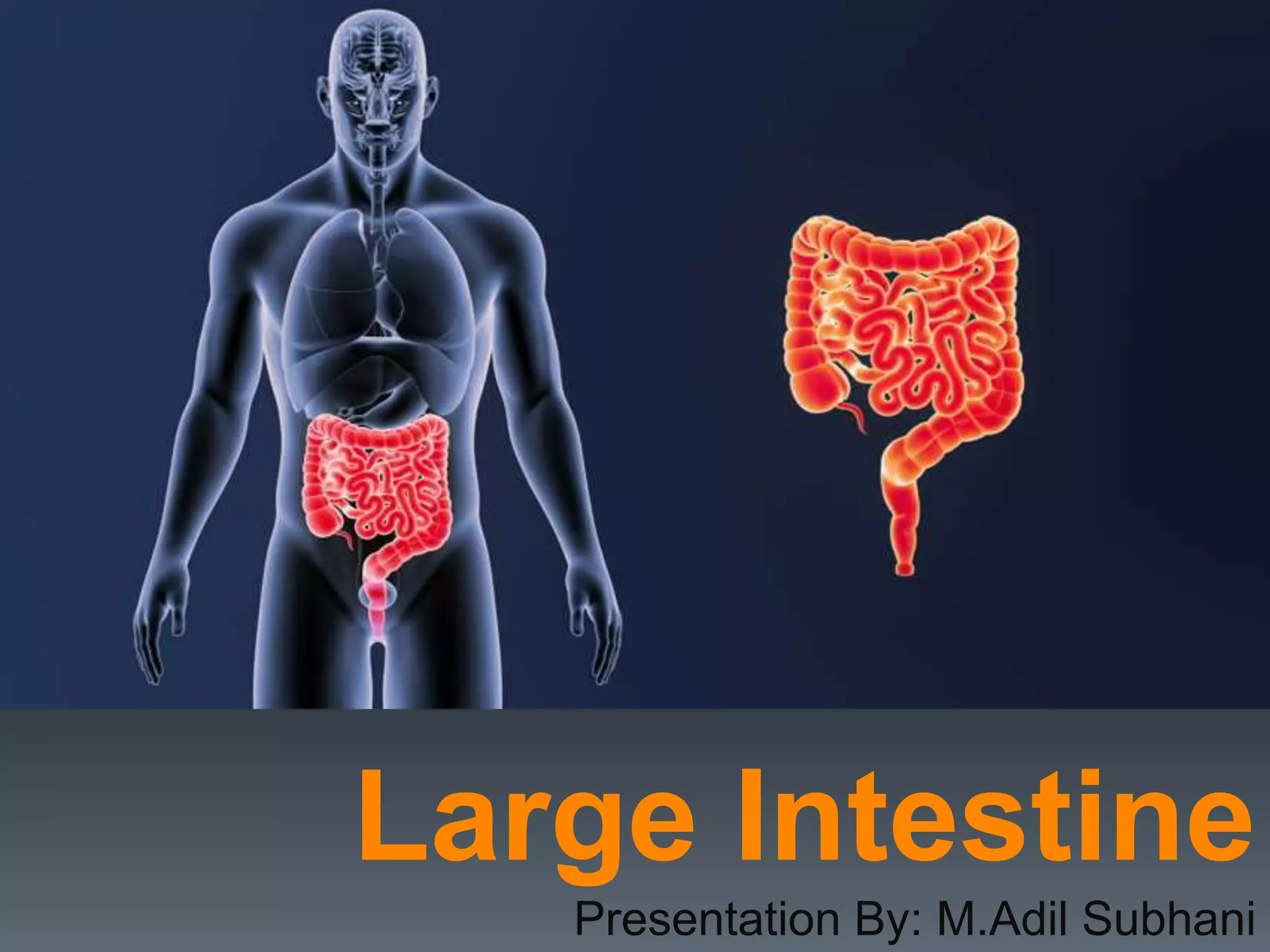
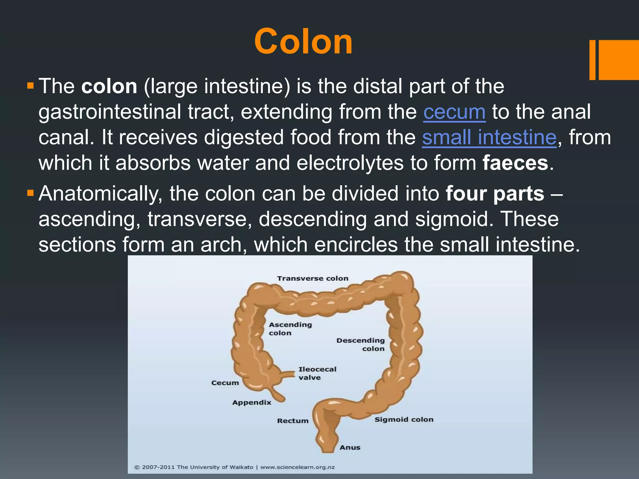
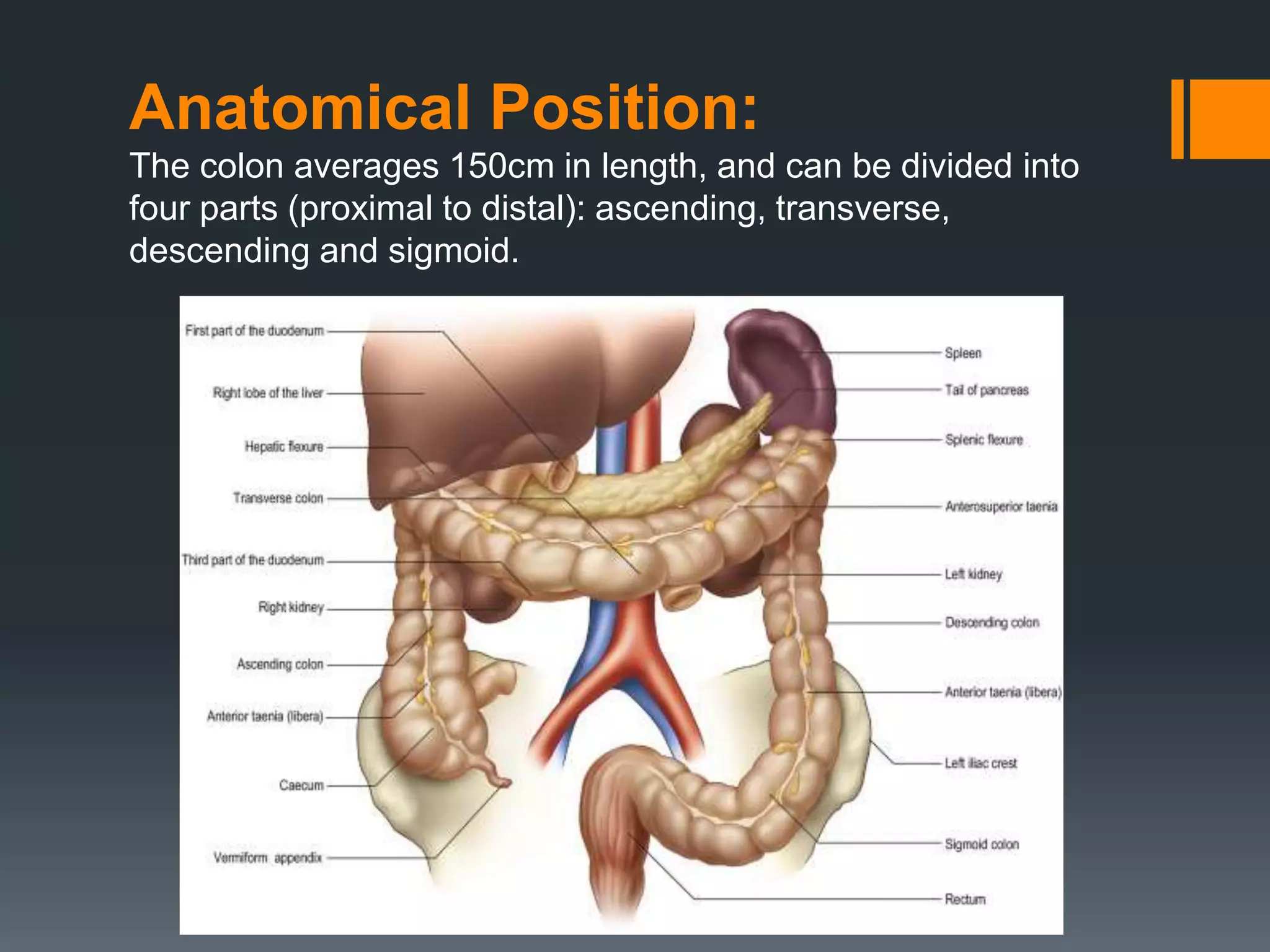
2. Definition:
“The wide lower section of the intestine that extends from
the end of the small intestine to the anus. The large
intestine acts mainly to absorb water from digested
materials and solidify feces. In most vertebrate animals, it
includes the cecum, colon, and rectum.”
Synonym:
The colon
3. Colon
The colon (large intestine) is the distal part of the
gastrointestinal tract, extending from the cecum to the anal
canal. It receives digested food from the small intestine, from
which it absorbs water and electrolytes to form faeces.
Anatomically, the colon can be divided into four parts –
ascending, transverse, descending and sigmoid. These
sections form an arch, which encircles the small intestine.
4. Anatomical Position:
The colon averages 150cm in length, and can be divided into
four parts (proximal to distal): ascending, transverse,
descending and sigmoid.
5. Ascending Colon
The colon begins as the ascending colon, a retroperitoneal
structure which ascends superiorly from the cecum.
When it meets the right lobe of the liver, it turns 90 degrees to
move horizontally. This turn is known as the right colic flexure (or
hepatic flexure), and marks the start of the transverse colon.
Transverse Colon
The transverse colon extends from the right colic flexure to
the spleen, where it turns another 90 degrees to point inferiorly.
This turn is known as the left colic flexure (or splenic flexure). Here,
the colon is attached to the diaphragm by the phrenicocolic
ligament.
The transverse colon is the least fixed part of the colon, and is
variable in position (it can dip into the pelvis in tall, thin individuals).
Unlike the ascending and descending colon, the transverse colon
is intraperitoneal and is enclosed by the transverse mesocolon.
6. Descending Colon:
After the left colic flexure, the colon moves inferiorly towards the pelvis
– and is called the descending colon. It is retroperitoneal in the
majority of individuals, but is located anteriorly to the left kidney,
passing over its lateral border.
When the colon begins to turn medially, it becomes the sigmoid
colon.
Sigmoid Colon:
The 40cm long sigmoid colon is located in the left lower quadrant of
the abdomen, extending from the left iliac fossa to the level of the S3
vertebra. This journey gives the sigmoid colon its characteristic “S”
shape.
The sigmoid colon is attached to the posterior pelvic wall by a
mesentery – the sigmoid mesocolon. The long length of the
mesentery permits this part of the colon to be particularly mobile.
7.
8. Paracolic Gutters:
The paracolic gutters are two spaces between the
ascending/descending colon and the posterolateral abdominal wall.
These structures are clinically important, as they allow material that has
been released from inflamed or infected abdominal organs to
accumulate elsewhere in the abdomen.
9. Anatomical Structure:
The large intestine has a number of characteristic features,
which allows it to be distinguished from the small intestine:
Attached to the surface of the large intestine are omental
appendices – small pouches of peritoneum, filled with fat.
Running longitudinally along the surface of the large bowel
are three strips of muscle, known as the teniae coli. They
are called the mesocolic, free and omental coli.
10. The teniae coli contract to shorten the wall of the bowel,
producing sacculations known as haustra.
The large intestine has a much wider diameter compared to
the small intestine.
These features cease at the rectosigmoid junction, where
the smooth muscle of the teniae coli broaden to form a
complete layer within the rectum.
11. Anterior Posterior
Anatomical Relations
The colon has numerous important anatomical relations in the
abdomen
Ascending colon
Small intestine
Greater omentum
Anterior abdominal
wall
Iliacus and quadratus
lumborum
Right kidney
Iliohypogastric and
ilioinguinal nerves
15. Functions of the Large Intestine
Reabsorb water and compact material into feces
ELECTROLYTE ABSORPTION
Absorb vitamins produced by bacteria
Store fecal matter prior to defecation
NB: Most of the absorption occurs in the proximal half
of the colon (absorbing colon), the distal colon (storage
colon) functions to store faeces until time of excretion
16. Neurovascular Supply
The neurovascular supply to the colon is closely linked to its
embryological origin:
Ascending colon and proximal 2/3 of the transverse colon –
derived from the midgut.
Distal 1/3 of the transverse colon, descending colon and
sigmoid colon – derived from the hindgut.
17. Arterial Supply
As a general rule, midgut-derived structures are supplied by
the superior mesenteric artery, and hindgut-derived
structures by the inferior mesenteric artery.
The ascending colon receives arterial supply from two
branches of the superior mesenteric artery; the ileocolic and
right colic arteries. The ileocolic artery gives rise to colic,
anterior cecal and posterior cecal branches – all of which
supply the ascending colon.
18. The transverse colon is derived from both the midgut and
hindgut, and so it is supplied by branches of the superior
mesenteric artery and inferior mesenteric artery:
Right colic artery (from the superior mesenteric artery)
Middle colic artery (from the superior mesenteric artery)
Left colic artery (from the inferior mesenteric artery)
The descending colon is supplied by a single branch of
the inferior mesenteric artery; the left colic
artery. The sigmoid colon receives arterial supply via the
sigmoid arteries (branches of the inferior mesenteric
artery).
19. Venous Drainage
The venous drainage of the colon is similar to the arterial
supply:
Ascending colon – ileocolic and right colic veins, which
empty into the superior mesenteric vein.
Transverse colon – middle colic vein, which empties
into the superior mesenteric vein.
Descending colon – left colic vein, which drains into
the inferior mesenteric vein.
Sigmoid colon – drained by the sigmoid veins into
the inferior mesenteric vein.
20. The superior mesenteric and inferior mesenteric
veins ultimately empty into the hepatic portal vein.
This allows toxins absorbed from the colon to be
processed by the liver for detoxification.
21. Innervation
The innervation to the colon is dependent on embryological
origin:
Midgut-derived structures (ascending colon and
proximal 2/3 of the transverse colon) receive their
sympathetic, parasympathetic and sensory supply via
nerves from the superior mesenteric plexus.
Hindgut-derived structures (distal 1/3 of the transverse
colon, descending colon and sigmoid colon) receive their
sympathetic, parasympathetic and sensory supply via
nerves from the inferior mesenteric plexus:
Parasympathetic innervation via the pelvic splanchnic
nerves
Sympathetic innervation via the lumbar splanchnic
nerves.
22. Lymphatic Drainage
The lymphatic drainage of the ascending and transverse colon is into
the superior mesenteric nodes. The descending colon and sigmoid
drain into the inferior mesenteric nodes.
Most of the lymph from the superior mesenteric and inferior mesenteric
nodes passes into the intestinal lymph trunks, and on to the cisterna
chyli – where it ultimately empties into the thoracic duct.
23. Colonic Diseases
Also called: Large intestine
diseases
Colorectal cancer
Colonic polyps - extra tissue growing in the colon that can
become cancerous
Ulcerative colitis - ulcers of the colon and rectum
Diverticulitis - inflammation or infection of pouches in the
colon
Irritable bowel syndrome - an uncomfortable condition
causing abdominal cramping and other symptoms