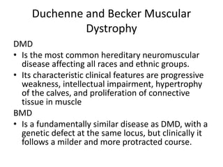
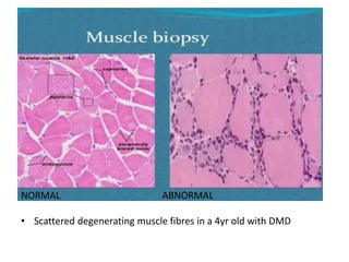
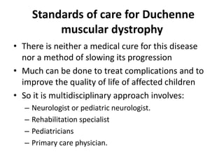
3. Introduction
• Muscular Dystrophy (MD) is a group of inherited diseases in
which the voluntary muscles progressively weaken overtime.
• The term dystrophy means abnormal growth,
• A muscular dystrophy is distinguished from all other
neuromuscular diseases by four obligatory criteria:
– It is a primary myopathy
– it has a genetic basis
– the course is progressive
– and degeneration and death of muscle fibers occur at some stage in
the disease
• Heart and other organs can also be affected.
4. Epidemiology
• It is estimated that between 50,000 -250,000
are affected annually. 1 per 3500 live male
births.
• Can occur at any age
• Most common in young males.
• Type depends on how severe the disease is,
which muscles it affects, rate of progression
7. Duchenne and Becker Muscular
Dystrophy
DMD
• Is the most common hereditary neuromuscular
disease affecting all races and ethnic groups.
• Its characteristic clinical features are progressive
weakness, intellectual impairment, hypertrophy
of the calves, and proliferation of connective
tissue in muscle
BMD
• Is a fundamentally similar disease as DMD, with a
genetic defect at the same locus, but clinically it
follows a milder and more protracted course.
8. Genetic Etiology and Pathogenesis:
• DMD and BMD are inherited as an X-linked
recessive trait. The abnormal gene is at the Xp21
locus and is one of the largest genes.
• Despite the X-linked recessive inheritance, about
30% of cases are new mutations, and the mother
is not a carrier
• The female carrier usually shows no clinical signs,
but occasionally there are girls who show mild
muscle weakness; where the normal X
chromosome becomes inactive and the one with
gene deletion is active
9. • A cytoskeletal protein known as dystrophin is encoded
by the gene at the Xp21.
• Dystrophin links muscle cells to extracellular matrix
stabilising the membrane and protecting the
sarcolemma from the stresses that develop during
muscle contraction.
• Mechanical damage through eccentric contractions
puts high stress on fragile membranes, provoke micro
lesions that eventually lead to loss of calcium
homeostatis and cell death
10. Clinical features in DMD
Early phase
• In infants:
rarely symptomatic at birth or in early infancy,
although some are mildly hypotonic,
other gross motor skills(sitting, standing) maybe
mildly delayed
Poor head control is usually the first sign, walking is
usually at normal age(12mo)
11. Transitional phase
• Toddler:
– By the 2nd year have hip girdle weakness
– Delayed walking, falling, toe walking and trouble
running or walking upstairs
– Developmental delay
– Lordotic posture when standing to compensate for
gluteal weakness
12. Transitional phase:
• In 3-9yrs:
– Early Gowers sign-hands pushing on legs to stand
– Trendelenburg gait/hip waddle
– Pseudohypertrophy of calf muscles
– Contractures mostly of ankles, knees hips and
elbows
13. Loss of ambulation
• In 10-14yrs:
– Children are using wheelchair
– Scoliosis due to sitting and back weakness
– Upper limb weakness and difficult to retain use of
fingers makes daily activities difficult
Though in Beckers children remain ambulatory until
early adult life
14. Late stage
• 15yrs+:
– Cardiomyopathies ; peristent tachycardia, dyspnea and LL
edema
– Respiratory problems; ineffective cough, frequent
pulmonary infections and decreasing respiratory reserve
– Pharyngeal weakness; ep of aspiration, nasal regurgitation
of liquids and nasal voice quality
– CNS; learning disability but can fxn in regular
classroom(20-30% have an IQ of <70)(less common in
Becker), ep of epilepsy
18. Physical examination
• Involves examination of all systems:
– CNS
– CVS
– RS
– Per Abdomen
• Special tests:
– Thomas test:
• Test for hip flexor contracture/shortness
• Pt lies supine and one of the hips is fully flexed if the opposite leg lifts of the
examination table then it is a posiive test
– Obers test:
• Test for iliotibial band tightness
• With the pt lying in the lateral position, support the the knee and flex it to 90
degrees then extend and abduct the hip and then release the knee support
• Failure of the to abduct is a positive test
19.
20. Investigations
Primary investigations:
Serum CK
– usually greatly elevated in DMD
– (15,000-35000IU/L),norm(<160IU/L)
– But can be low in terminal stage
Cardiac assesment:
– ECHO,ECG,chest x-ray done periodically
22. Invx…
MUSCLE BIOPSY
• Diagnostic & its done when PCR is normal but clinical
suspicion is high.
• Immunohistochemical staining of frozen sections of
muscle biopsy tissue is done.
• Shows myopathic changes such as; connective tissue
proliferation, scattered degenerating and regenerating
myofibres
• Common muscles sampled are the vastus lateralis
(quadriceps femoris ) and the gastrocnemius
24. Differential Diagnosis
Neurogenic Non hereditary Non progressive-
non necrotizing
Hereditary –non
progressive
Spinal muscular
atrophy
Dermatomyositis congenital muscle
fiber-type
disproportion
(CMFTD
Metabolic
myopathies
25. Standards of care for Duchenne
muscular dystrophy
• There is neither a medical cure for this disease
nor a method of slowing its progression
• Much can be done to treat complications and to
improve the quality of life of affected children
• So it is multidisciplinary approach involves:
– Neurologist or pediatric neurologist.
– Rehabilitation specialist
– Pediatricians
– Primary care physician.
26. Neurology
The use of corticosteroids in DMD: prednisone,
prednisolone, deflazacort, or other steroids.
– Decrease the rate of apoptosis of myotubes during
ontogenesis and can decelerate the myofiber
necrosis
• Timing- Physical performance of the child
plateaus (4-6yrs),
– Less functional when initiated close to the loss of
ambulation.
27. Neuro…
• Regimen:
– 0.75 mg/kg/day prednisone/prednisolone and 0.9
mg/kg/day deflazacort (less effect of weight gain).
– Alternating day dosing to reduce steroid-associated
side-effects.
– Dose reduction even withdrawal if severe and/or
unacceptable side effects occur in a tapering manner
– Continuation of steroids beyond the loss of
ambulation is common practice in some centres for
the possible protective effect on spinal alignment,
respiratory and cardiac function.
28. GI-Nutrition
• Adequate dietary advice should be offered
from a young age with specific focus on
weight control using(BMI)
• focused especially at:
– Initiation of steroids
– loss of ambulation
– Adequate calcium and vitamin D intake
– Controlled sodium intake
29. GI…
• Overweight- aim for a weight loss of 0.5 kg per
month.
• Undernutrition are most likely after the boy
starts using a wheelchair (approximately 12-13
years of age) and may be multifactorial.
• The first step is to evaluate intake and to optimize if
necessary the existing diet with energy and protein.
• The next step with more severe malnutrition is enteral
nutrition during night time.
30. GI…
Esp. in steroid users: if the diet is not adequate in
supply of calcium and vitamin D, these should be
added separately to reach a recommended intake:
• calcium(4-8 years: 800 mg/d; 9-18 years: 1300 mg/d) &
• vitamin D (400 IU)
Later stages of the disease there can be difficulty
swallowing and where this leads to aspiration
and/or undernutrition:
• feeding by tube or
• percutaneous endoscopic gastrostomy (PEG) is indicated
31. RESPIRATORY CARE
• Respiratory surveillance: Serial measurement of
forced vital capacity(FVC) to detect progression of
respiratory muscle weakness.
• FVC <40% predicted value serial measurement of
overnight oximetry allows the recognition of the
development of nocturnal respiratory failure.
• Cough effectiveness: Peak cough flow (PCF)
• Cough assist machine should be considered when the PCF is
below 270 l/ min in non-ambulant boys and introduced
before PCF is less than 160 l/min
32. Resp…
• Prophylaxis and management of chest infections:
• Through vaccinations and antibiotics should be provided
promptly.
• Chest physiotherapy such as postural drainage and assisted
coughing should be taught when coughing is ineffective
• Management of nocturnal hypoventilation
Elective non-invasive nocturnal ventilation (NIV)
33. CARDIAC CARE
• Surveillance: Cardiac investigation
(echocardiogram and ECG)- 2yrly/10years
then annually and prior to GA at any age.
• Prophylaxis: ACE-inhibitors should be started
at a subclinical deterioration of cardiac
function as detected by echocardiography.
• Early prophylactic treatment with ACE-
inhibitors at a pre-clinical stage is
recommended from 5-10 years of age.
34. Cardiac…
• Treatment: Dependent on type and stage of
cardiomyopathy. DCM is the most common form.
– ACE-inhibition and beta-blocker both or ACEI starts
– Add diuretics and other with onset of heart failure.
– Anticoagulation therapy in severe cardiac dysfunction to
prevent systemic thromboembolic events
• Arrhythmias: Occur in late stage hence
antiarrhytmic treatment should be introduced,
bearing in mind the possible negative inotropic
effect of agent chosen
35. ORTHOPEDIC CARE
• Splinting: In ambulant children night splints
should be provided when there is loss of
dorsiflexion at the ankle and before the foot
can only achieve plantagrade.
• AFOs(ankle-foot orthosis)/brace & Standing
frames can delay contracture development in
non-ambulant children.
37. ORTHO…
• DMD spine deformity A fusion surgery can
be recommended as soon as there is clear
progression and the Cobb angle passes 25 –
30 degrees
38. PSYCHOLOGICAL CARE
• Social (information, advocacy and advice) and
psychological support should be offered at
times of changing needs and crisis.
• Learning difficulties/ autism spectrum
disorders should be identified early and
recommendations given to parents and
educators about managing these difficulties.
39. REHABILITATION
• Annual neurological, respiratory and cardiological
assessments should ideally be co-ordinated.
• Physiotherapist and occupational therapist are to
encourage activity (i.e. self hygiene care) and
promote function.
• Exercise: Resisted exercises may accelerate muscle
damage. Moderate levels of active exercise is
recommended.
• Wheelchairs: to improve mobility and
independence.
41. Prognosis
• Most of these patients have a poor prognosis
• Patients with DMD have the worst prognosis;
death occurs usually at about 18-20yrs of age.
The causes of death are:
– respiratory failure in sleep
– intractable heart failure
– pneumonia
– aspiration
– airway obstruction.