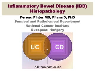
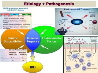
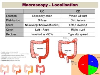
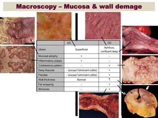
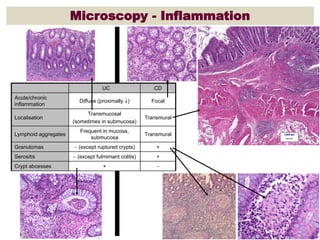
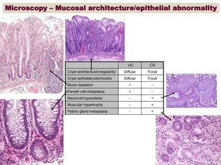
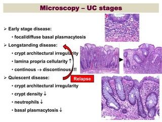
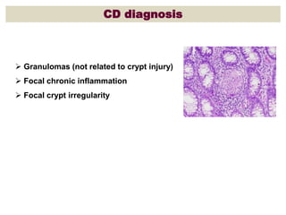
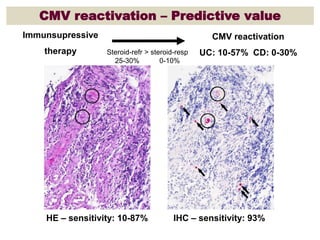
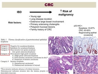
Inflammatory Bowel Disease (IBD)
Histopathology
Ferenc Pinter MD, PharmD, PhD
Surgical and Pathological Department
National Cancer Institute
Budapest, Hungary
Incidence
ECCO-EpiCom
2010 & 2011:
Crohns: 37%
UC: 53%
IBD unclassified: 10%
Eastern Europe:
11,3 / 100 000
Ulcerative Colitis Crohn’s Disease
Age-Specific Incidence of IBD *
Incidence in both CD and UC have 2 peaks
( in 3 rd and 6 th decades ).
10
0
2
4
6
8
0 20 40 60 80
10
0
2
4
6
8
0 20 40 60 80
Age (yrs) Age (yrs)
Etiology + Pathogenesis
Etiology: Smoking
Surgical therapy: UC vs. CD
Requirements for Dx
 ≥5 sites x ≥2 biopsies
• rectum  terminal ileum
• colonoscopy + ileoscopy
 2-3 tissue levels (step sections) x ≥5 serial sections
 Clinical information
• endoscopic findings
• age
• duration
• treatment
• comorbidities
• travel history
Macroscopy - Localisation
CDUC
Whole GI tractEspecially colonLocation
Skip lesionsDiffuseDistribution
Often involvedNo (except backwash ileitis)Ileum
RightLeftLeftRightColon
Typically sparedInvolved in >90%Rectum
Macroscopy – Mucosa & wall demage
CDUC
Aphtous,
confluent deep
SuperficialUlcers
+Mucosal atrophy
+Inflammatory polyps
+Cobblestone pattern
+ (except fulmimant colitis)Deep fissures
+ (except fulmimant colitis)Fistulae
NormalWall thickness
+Fat wrapping
+Strictures
Microscopy - Inflammation
CDUC
FocalDiffuse (proximally )
Acute/chronic
inflammation
Transmural
Transmucosal
(sometimes in submucosa)
Localisation
Transmural
Frequent in mucosa,
submucosa
Lymphoid aggregates
+ (except ruptured crypts)Granulomas
+ (except fulmimant colitis)Serositis
+Crypt abcesses
Microscopy – Mucosal architecture/epithelial abnormality
CDUC
FocalDiffuseCrypt architectural irregularity
FocalDiffuseCrypt epithelial polymorphs
+Mucin depletion
+Paneth cell metaplasia
+Neuronal hyperplasia
+Muscular hypertrophy
+Pyloric gland metaplasia
Microscopy – UC stages
 Early stage disease:
• focal/diffuse basal plasmacytosis
 Longstanding disease:
• crypt architectural irregularity
• lamina propria cellularity 
• continous  discontinous !!!
 Quiescent disease:
• crypt architectural irregularity
• crypt density 
• neutrophils 
• basal plasmacytosis 
Relapse
CD diagnosis
 Granulomas (not related to crypt injury)
 Focal chronic inflammation
 Focal crypt irregularity
CMV reactivation – Predictive value
Immunsupressive
therapy
CMV reactivation
UC: 10-57% CD: 0-30%Steroid-refr > steroid-resp
25-30% 0-10%
HE – sensitivity: 10-87% IHC – sensitivity: 93%
CRC
Risk factors:
IBD  Risk of
malignancy
• Young age
• Long disease duration
• Extensive large bowel involvement
• Primary sclerosing cholangitis
• Polypoid mucosal lesions
• Family history of CRC
p53 IHC+:
Dysplasia: 33-67%
CRC: 83-95%
Regenerating epithel:
occasional
Summary
 UC/CD  different therapy  differentiation is important
 Differentiation: - granuloma formation: +/-
- inflammation: focal/diffuse
- wall structure change: focal/diffuse
 Diagnostics is impacted by IBD stage (early/chronic/quiscent)
 Dysplasia is the most reliable marker of CRC risk.
This should be evaluated in areas with chronic inflammation and
confirmation by an independent expert GI pathologist is
recommended.

Inflammatory Bowel Disease (IBD) Histopathology

  • 1.
    Inflammatory Bowel Disease(IBD) Histopathology Ferenc Pinter MD, PharmD, PhD Surgical and Pathological Department National Cancer Institute Budapest, Hungary
  • 3.
    Incidence ECCO-EpiCom 2010 & 2011: Crohns:37% UC: 53% IBD unclassified: 10% Eastern Europe: 11,3 / 100 000
  • 4.
    Ulcerative Colitis Crohn’sDisease Age-Specific Incidence of IBD * Incidence in both CD and UC have 2 peaks ( in 3 rd and 6 th decades ). 10 0 2 4 6 8 0 20 40 60 80 10 0 2 4 6 8 0 20 40 60 80 Age (yrs) Age (yrs)
  • 5.
  • 6.
  • 7.
  • 8.
    Requirements for Dx ≥5 sites x ≥2 biopsies • rectum  terminal ileum • colonoscopy + ileoscopy  2-3 tissue levels (step sections) x ≥5 serial sections  Clinical information • endoscopic findings • age • duration • treatment • comorbidities • travel history
  • 9.
    Macroscopy - Localisation CDUC WholeGI tractEspecially colonLocation Skip lesionsDiffuseDistribution Often involvedNo (except backwash ileitis)Ileum RightLeftLeftRightColon Typically sparedInvolved in >90%Rectum
  • 10.
    Macroscopy – Mucosa& wall demage CDUC Aphtous, confluent deep SuperficialUlcers +Mucosal atrophy +Inflammatory polyps +Cobblestone pattern + (except fulmimant colitis)Deep fissures + (except fulmimant colitis)Fistulae NormalWall thickness +Fat wrapping +Strictures
  • 11.
    Microscopy - Inflammation CDUC FocalDiffuse(proximally ) Acute/chronic inflammation Transmural Transmucosal (sometimes in submucosa) Localisation Transmural Frequent in mucosa, submucosa Lymphoid aggregates + (except ruptured crypts)Granulomas + (except fulmimant colitis)Serositis +Crypt abcesses
  • 12.
    Microscopy – Mucosalarchitecture/epithelial abnormality CDUC FocalDiffuseCrypt architectural irregularity FocalDiffuseCrypt epithelial polymorphs +Mucin depletion +Paneth cell metaplasia +Neuronal hyperplasia +Muscular hypertrophy +Pyloric gland metaplasia
  • 13.
    Microscopy – UCstages  Early stage disease: • focal/diffuse basal plasmacytosis  Longstanding disease: • crypt architectural irregularity • lamina propria cellularity  • continous  discontinous !!!  Quiescent disease: • crypt architectural irregularity • crypt density  • neutrophils  • basal plasmacytosis  Relapse
  • 14.
    CD diagnosis  Granulomas(not related to crypt injury)  Focal chronic inflammation  Focal crypt irregularity
  • 15.
    CMV reactivation –Predictive value Immunsupressive therapy CMV reactivation UC: 10-57% CD: 0-30%Steroid-refr > steroid-resp 25-30% 0-10% HE – sensitivity: 10-87% IHC – sensitivity: 93%
  • 16.
    CRC Risk factors: IBD Risk of malignancy • Young age • Long disease duration • Extensive large bowel involvement • Primary sclerosing cholangitis • Polypoid mucosal lesions • Family history of CRC p53 IHC+: Dysplasia: 33-67% CRC: 83-95% Regenerating epithel: occasional
  • 17.
    Summary  UC/CD different therapy  differentiation is important  Differentiation: - granuloma formation: +/- - inflammation: focal/diffuse - wall structure change: focal/diffuse  Diagnostics is impacted by IBD stage (early/chronic/quiscent)  Dysplasia is the most reliable marker of CRC risk. This should be evaluated in areas with chronic inflammation and confirmation by an independent expert GI pathologist is recommended.