Neurocysticercosis ppt irin1
•Download as PPTX, PDF•
1 like•513 views

This document presents a case of neurocysticercosis in a 38-year-old male. On examination, he had decreased consciousness, unequal pupils, spastic muscles, and decreased strength. Investigations showed cysts in the brain and spleen. Neurocysticercosis is caused by the pork tapeworm Taenia solium and can infect the brain, eye, muscle or subcutaneous tissue. Symptoms depend on the location of cysts and include seizures, headaches, strokes and hydrocephalus. Diagnosis involves blood tests, imaging and biopsy. Treatment involves anti-parasitic drugs and corticosteroids.

Recommended
More Related Content
What's hot
What's hot (20)
Similar to Neurocysticercosis ppt irin1
Similar to Neurocysticercosis ppt irin1 (20)
Recently uploaded
Recently uploaded (20)
Neurocysticercosis ppt irin1
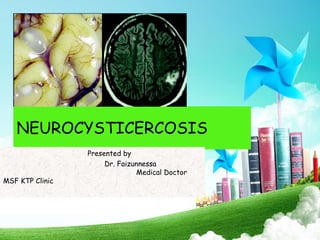
- 1. NEUROCYSTICERCOSIS Presented by Dr. Faizunnessa Medical Doctor MSF KTP Clinic
- 2. OBJECTIVES • Case presentation • Introduction • Pathogenesis • Classification • Clinical presentations • Investigations • Differential diagnosis • Treatment
- 3. CASE PRESENTATION • MD. Hossain Ahmed aged 38yrs a muslim unmarried nondiabetic normotensive unmarried male heiled from KMS EXT has been referred from BKL to our facilities as a case of somatoform disorder & evaluated under mental health where he was suspected as a case of Organic Psychosis. On evaluation of his history we came to know that 10 days back he had only a history of Diarrhoea & URTI for which he took some medication and completely cured but recently for last 4 days he presented with complaints of • a) Giddiness followed by drowsiness • b) Headache • c) slurred speech followed by aphasia • D) Abnormal movement of limbs
- 4. • Patient an young adult male aged 38 yr moderately built & nourished. • His vitals were stable, • He is not anemic not jaundiced, no cyanosis, no clubbing ,no generalized lymphadenopathy. • System examination revealed no significant cardiovascular / respiratory abnormal findings. • Neurological Examination: – HPF: Semiconcious; GCS:8-9/15. – Pupils : Size R 4mm; L 3mm B/L brisk reaction to light & accommodation Fundus Normal. – No other significant cranial nerve palsies observed. – Lead pipe rigidity present, Muscle tone spastic, R+J=E; & Planter extensor B/L with MP 3/5. CASE PRESENTATION----O/E
- 5. • Later on patients attendence with there own interest done few investigations from outside and got readmission in our facility & diagnosed as a case of secondary mets of brain according to MRI & received Dexamethasone + palliative Rx. But as his USG showed Splenic cyst & MRI mets lesions are quiet different from brain mets we suspected it as a case of Neurocysticercosis & started our Rx with a hope that some response may occur. CASE PRESENTATION
- 10. INTRODUCTION • NCC is the infection of the CNS by the larvae of Taenia solium. • Neurocysticercosis (NCC) is the most common parasitic disease of the nervous system. • It is the leading cause of adult onset epilepsy (29% of epilepsy in endemic regions world wide). • It is endemic in Central and South America, sub-Saharan Africa, and in some regions of the Far East, including most area of the Asian subcontinent, Indonesia, and China, reaching an incidence of 3.6% in some regions. • Of note is the near absence of infection in Muslim countries, where the consumption of pork is forbidden by Islam
- 12. Pathogenesis Cestodes/ Tapeworm Human Definitive Host: Taenia Saginata Diphylobrothium Hymenolepis Dipyllidium Cannninum Human Intermediate Host: Echinococcus Either Taenia solium
- 13. Mode of infection • Hetero-inoculation Eggs from the environment • Internal auto-inoculation Regurgitation of the proglottids into the stomach • External auto-inoculation From self??
- 14. Pathogenesis
- 16. Neuropathology • Asymptomatic viable cysts • Dying cysts • Calcified cysts • Racemose cysts • Meningitis • Vasculitis • Hydrocephalus • Intraventricular cysts • Spinal disease • Disseminated disease
- 17. Types of Cyst • Cysticercus cellulosae: Less virulent. Small (<2cm, round, thin walled). In the parenchyma or Subarachnoid space. Often remain silent. • Cysticercus racemose: The racemose (ie, appearing like a cluster of grapes) form refers to the presence of multiple cysts without a scolex. May form giant vesicles up to 10 cm in diameter with predilection for basal cisterns Cysticercotic arachnoiditis Presents as hydrocephalus / meningitis Can occlude vessels stroke Intense inflammation and seizures
- 18. Neurocysticercosis: Stages (Escobar) • Vesicular: No Inflammatory Response • Colloid-vesicular : Larva Begins to Degenerate, Scolex Shrinks, Fluid Turbid, Surrounding Edema: Enhancement • Nodulo-granular: Capsule Thickens, Fluid Reabsorbed; Scolex Mineralized • Shrinks to Calcified Nodule
- 19. Neurocysticercosis • While in the nervous system, the T solium parasite goes through different stages of involution, which include the following:
- 20. CLASSIFICATION • Parenchymal NC • Ventricular NC • Basal Meningites • Mixed forms
- 21. Extra Neural Cysticercosis • Muscle and subcutaneous tissue: Multiple subcutaneous nodules • Neck, Arm, anterior chest wall, • Calf, Thigh. • Ocular Extra ocular (muscle). Intra ocular
- 24. Clinical features • Neurocysticercosis is a pleomorphic disease. • Most symptomatic patients are 15–40 years old, and the disease has no gender or race predilection. • Many are asymptomatic (80%). • Peak is estimated to occur 3-5 years after infection. • The onset of symptoms is usually subacute to chronic, with the exception of seizures, which present in an acute fashion
- 25. • Cysticerci can be found anywhere in the body but are most commonly detected in the brain, cerebrospinal fluid (CSF), skeletal muscle, subcutaneous tissue, or eye. • Physical findings depend on where the cyst is located in the nervous system. • Symptoms are mainly due to mass effect, inflammatory response, or obstruction of foramina and ventricular system of brain. Clinical features
- 26. Parenchymal NC • Epilepsy It is the most common presentation (70%) of neurocysticercosis It is the leading cause of adult-onset epilepsy. SPS, GTC >> CPS Risk of seizures in seropositive individuals 2-3 times higher than seronegative controls. • Headache, nausea, vomiting • Strokes Lacunar infarcts and large cerebral infarcts due to occlusion or vascular damage. Hemorrhage can also occur as a result of rupture of mycotic aneurysms of the basilar artery.
- 27. • Frontal lobe involvement Psychosis, dementia, parkinsonism, intellectual impairement • Cerebellar ataxia • Encephalitis and diffuse brain edema Common in children and young females Risk of developing severe neurological sequelae Parenchymal NC
- 28. Intraventricular - Constitutes 5-10% - 4 th ventricle most common site of obstruction - Lateral ventricular cysts less likely to cause obstruction - Hydrocephalus without localizing signs - Bruns’ syndrome : Unattached cysts may cause sudden positional mechanical obstruction causing nausea, vomiting and vertigo. Meningeal cyst - Meningeal Irritation signs - Raised ICT from inflammation, edema
- 29. Presentations of other forms of neurocysticercosis • Intracranial hypertension • Neuropsychiatric disturbances • Hydrocephalus(10-30%) • Intrasellar neurocysticercosis • Spinal neurocysticercosis --- rare 1% to3%
- 30. INVESTIGATIONS • Peripheral eosinophilia only if cyst is leaking. • Raised IgE level. • Immunologic Testing ELISA (87% sensitive & 95% specific) EITB (95% sensitive & 100% specific) • CSF Analysis: Mononuclear pleocytosis, usually not exceeding 200-300 cells/mm3 Normal glucose levels, Elevated protein levels,(50-200 mg/dL) Eosinophilia High immunoglobulin G (IgG) index, Oligoclonal bands( in some).
- 31. • Stool Examination: Taeniasis may be established by detecting T solium eggs and proglottids in a patient's stool. Taeniasis and neurocysticercosis coexist in 10-15% of patients with neurocysticercosis. Intestinal taeniasis is very common in patients with massive infestation with cysticerci but without cysticercotic encephalitis. Tapeworm carriers may be identified by examining the stool of the relatives of a patient with cysticercosis encephalitis. CT Scan +MRI/MRI SPET INVESTIGATIONS
- 32. Differential Diagnosis • Tuberculoma: Usually Irregular, Greater Than 20 mm in Size. Often Associated with Severe Perifocal Edema and Focal Neurological Deficit • Secondary Mets of Brain with Occult Primary: No evidence of Lung, Liver & bone mets clinically. • Neuropsychiatric Manifestation of Wilson’s Disease: Young patient with EPS, KF ring, Previous history of jaundice, AST • Ecchinococcosis: • Somatoform disorder.
- 33. • Cryptococcosis: presents as chronic or subacute meningitis. Associated with papilloedema, hydrocephalus, focal deficits, seizures and cryptococcomas. Cranial neuropathies, especially of the lower cranial nerves, affecting one or more cranial nerves Immunocompromised HIV-Sero+ve . • Toxoplasmosis Differential Diagnosis
- 34. Treatment • Cysticidal therapy + steroids • Corticosteroids alone • Supportive – Anticonvulsants – Anti edema – Analgesics • Surgery
- 35. Thank you!