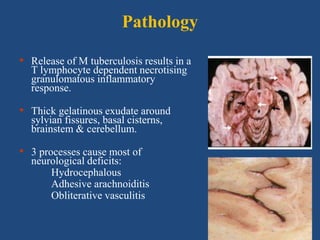
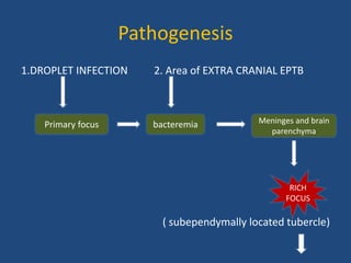
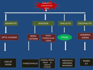
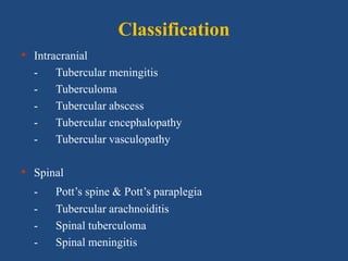
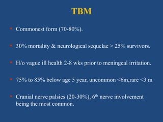
CNS TUBERCULOSIS
By- DR.RATAN LAL MEENA
Moderator- DR. SUBHRA JAIN
Introduction
• One of the most devastating clinical manifestation
of EPTB.
• 5-10% of extra-pulmonary & 1% of all TB cases.
• Male predominance
• The case fatality rate is 100% on untreated case
and delayed in treatment may lead to permanent
neurological damage.
CONTENT
• Etiology
• Pathogenesis
• Classification
• Diagnosis
• Treatment
Etiology
Risk factors;
• Children> adults
• HIV co-infection
• Malnutrition
• Alcoholics
• Malignancies
• Use of
immunosuppressive
agents
Causative agent;
• Most common-
Mycobacterium tuberculosis
• In immunocomromised
patient -
Atypical mycobacteria ;
MAC, mycobacterium
intracellulare
Pathology
• Release of M tuberculosis results in a
T lymphocyte dependent necrotising
granulomatous inflammatory
response.
• Thick gelatinous exudate around
sylvian fissures, basal cisterns,
brainstem & cerebellum.
• 3 processes cause most of
neurological deficits:
Hydrocephalous
Adhesive arachnoiditis
Obliterative vasculitis
Pathogenesis
1.DROPLET INFECTION 2. Area of EXTRA CRANIAL EPTB
( subependymally located tubercle)
Primary focus bacteremia Meninges and brain
parenchyma
RICH
FOCUS
Rupture in
subarachnoid
space
ADHESION VASCULITIS ENCEPHALITIS
STROKE
INTER-
PENDUCULAR
FOSSA
BASAL
CISTERNS
CEREBERAL
EDEMA
HYDROCEPHALUS
CRANIAL NERVE
PALSY &
CAROTID
STENOSIS
PARALYSIS &
ABNORMAL
MOVEMENT
RAISED
ICT
MENINGITIS
OPTIC CHIASMA
LOSS OF
VISION
Classification
• Intracranial
- Tubercular meningitis
- Tuberculoma
- Tubercular abscess
- Tubercular encephalopathy
- Tubercular vasculopathy
• Spinal
- Pott’s spine & Pott’s paraplegia
- Tubercular arachnoiditis
- Spinal tuberculoma
- Spinal meningitis
TBM
• Commonest form (70-80%).
• 30% mortality & neurological sequelae > 25% survivors.
• H/o vague ill health 2-8 wks prior to meningeal irritation.
• 75% to 85% below age 5 year, uncommon <6m,rare <3 m
• Cranial nerve palsies (20-30%), 6th nerve involvement
being the most common.
Clinical features
Sign
• Neck rigidity
• Kerning sign
• Brudzinski sign
• Hemiplagia
• Papilloedema
• Abducens nerve palsy
• Optic atrophy
• Facial nerve palsy
• Oculomotor nerve palsy
• Chhoroidal tubercle
Symptoms
• Fever
• Altered sensorium
• Headache
• Vomiting
• Behavioural changes
• Seizure
• Weight loss
Staging of TBM
British Medical Research Council criteria
Stage I: Prodromal phase with no definite neurologic
symptoms.
Stage II: Signs of meningeal irritation with slight or no
clouding of sensorium & minor or no
neurological deficit.
Stage III: Severe clouding of sensorium, convulsions, focal
neurological deficit & involuntary movements.
Modified MRC criteria
Grade I: Alert and oriented (GCS 15) without focal
neurological deficit.
Grade II: GCS 14-10 with or without focal neurological
deficit or GCS 15 with focal neurological deficit.
Grade III: GCS less than 10 with or without focal
neurological deficit.
Spinal TBM
• May result from rupture of Rich foci in the spinal arachnoid
space
• The acute form presents with fever, headache, and root pains
accompanied by myelopathy
• The chronic form presents with spinal cord compression
Complications
• Hydrocephalus
communicating , non communicating
• Ocular lesion
papiloedema, optic atrophy, vision loss,
• Hypothalamic pituiatary syndrome
SIADH hyponatraemia (49%)
Diabetes incipidus persistent pyrexia
• Cranial nerve palsies
3rd,4th, 5th,6th,7th, horizontal & vertical gaze palsy
Internuclear ophthalmoplegia
• Paralysis & abnormal movement
• Stroke ,seizure
Tuberculoma
• Mass of granulation tissue made up of conglomeration of
microscopic small tubercles.
• Firm, avascular, 2-8cm, spherical granulomatous mass.
• Locations -
infratentorial- <20 year
supratentorial - >20 year
• Solitary more frequent than multiple.
• Symptoms related to size & location.
• Low grade fever, headache, vomiting, seizures,
focal neurological deficit & papilloedema
•CT/MRI
A central nidus of calcification with
surrounding ring like enhancement
known as target sign .
•Histology:- consist of a central core of caseating necrosis with
with a surrounding wall of florid granulomatus reaction
containing lymphocyte, giant cells,
epitheliod cells ,histiocytes.
TUBERCULOMA NEUROCYSTICERCOSIS
At any age Rare before 3 year
Progressive neurological deficit No progressive neurological deficit
Size >20mm,irregular outline Smaller size, regular rounded with less
cerebral edema
Supratentorial or infratentorial Usually supratentorial
Midline shift seen Midline shift not seen
Tubercular brain abscess
• It is characterised by an encapsulated collection of pus containg
viable bacilli without evidence of a classic tubercular
granuloma.
• 4-7.5% of pt with CNS TB.
• More common in elderly & immunocompromised.
• Usually solitary, uni/multi-loculated of variable size.
• Clinical features: partial seizures, focal neurological deficit &
↑ intracranial tension.
•Differentiation b/w tuberculous & pyogenic abcess is made
by MR spectroscopy.
•CT & MRI show large size lesion with marked
surrounding edema.
Tubercular
abscess
tuberculoma
Central core pus Caseating
necrosis
surrounded by
granulomatus
reaction
Peripheral rim Comparative
thicker
thin
Mycobacterium
tuberculosis
Must be
present
May or may
not present
Tubercular encephalopathy
• Seen in infants & children.
• Characterized by convulsions, stupor & coma with signs of
meningeal irritation or focal neurological deficit
• CSF is normal.
• MRI- diffuse brain edema & demyelination which is extensive.
• Responsive to corticosteroids.
Spinal TB
• Infection starts in cancellous bone usually adjacent to an inter-
vertebral disc or anteriorly under the periosteum
• Regional distributions
Thoracic (65%) most common
lumbar (20%)
cervical (10%)
thoraco-lumbar (5%), atlanto-axial region (< 1%)
• type of vertebral lesion:-
Paradiscal most common
Central
Anterior
Appendical
articular
Clinical features-
back pain most commen
Spine stiffness: spasm of paravertebral muscle
Deformity:-knuckle/gibbus/kyphosis
Cold abscess
Paraplagia
Other constitutinal symptoms like fever, weight
loss, cough
Spinal TB
• Radiological changes
Reduced disc space
Destruction of bodies
Loss of trabecular pattern
Skipped lesion
Fusiform paravertebral shadow suggest abcess formation
Tubercular arachnoiditis
• Features of spinal cord or nerve involvement may
predominate, but most often mixed picture.
• Subacute paraparesis, radicular pain & bladder
dysfunction.
• Hallmark of diagnosis is characteristic myelographic
picture, showing poor flow of contrast material with
multiple irregular filling defects, cyst formation &
sometimes spinal block.
Differential diagnosis of TBM
• Fungal meningitis (cryptococcosis, histoplasmosis,
blastomycosis, coccidioidal mycosis)
• Viral meningoencephalitis (herpes simplex, mumps)
• Partially treated bacterial meningitis
• Neurosyphills
• Focal parameningeal infection
• CNS toxoplasmosis
• Neoplastic meningitis (lymphoma, carcinoma)
• Neurosarcoidosis
Diagnostic methods
• CSF examination
• CSF smear examination
• CSF CULTURE examination
• Molecular& immunological test
• Adjunctive test
• Radiology
Diagnostic criteria for TBM
Patients with at least four of the following:
i. History of tuberculosis.
ii. Predominance of lymphocytes in the cerebrospinal fluid.
iii. A duration of illness of more than six days.
iv. A ratio of CSF glucose to plasma glucose of less than 0.5.
v. Altered consciousness
vi. Turbid cerebrospinal fluid.
vii. Focal neurologic signs.
Possible
Patients with one or more of the following:
i. Suspected active pulmonary TB on chest radiograph
ii. AFB found in any specimen other than the CSF.
iii. Clinical evidence of extrapulmonary tuberculosis.
Probable
Acid-fast bacilli seen in the cerebrospinal fluid.Definite
DefinitionClass
TBM
CSF examination :-
• Appearance-slightly opaque,
-cobweb formation on long standing
• leucocytosis (10-1000 x 103 cells/ml
mostly lymphocytes)
• ↑ protein 50-300mg/dl
• Glucose<50mg/dl
• CSF : plasma glucose <50%.
• In immune-suppressed CSF can be acellular or
predominant neutrophils.
Test Appearance Pressure WBC/μL Protein
mg/dL
Glucose
mg/dL
Chlori
de
Normal CSF Clear 90 – 180 mm
hg
0-5 15-45 50-80 115-
130
mEq/L
Acute
bacterial
meningitis
Turbid Increased 1000 -10000 100 – 500 < 40 Decrea
sed
Viral
meningitis
Clear Normal to
moderate
increase
5-300, rarely
>1000
Normal to
mild
increased
Normal Normal
Tuberculous
meningitis
Slightly
opaque
cobweb
formation
Increased/
decreased,
10-1000
mixed
Initially
neutrophillic
& later
lymphocytic
50-300 Decreased Decrea
sed
Fungal
meningitis
Clear Increased 40-400 mixed 50-300 Decreased Decrea
sed
Gram’s and India ink stain-
• a negative Gram’s & india ink stain are pre-requisites for
the diagnosis of TBM
Zeihl –Nelson’s stain-
• Rapid, inexpensive,specific for AFB
• Low sensitivity (12-69%)
• 6 ml CSF should be taken for mycobacterial study
Egg or agar based solid medium:-
• L-J media ; sensitivity(25-80%)
• Takes 2-8 week to isolate the organism.
Automated liquid based:-
• BACTAC ; sensitivity(55%)
• growth detect by production of
Radioactive C-14 in 2 week.
Molecular method
• Sensitivity (56%)
• Diagnostic yield of NAA ↑ when large volm CSF
processed
• NAA more useful than conventional bacteriology
after start of ATT
• NAA assays (CBNAAT) detect R resistance
• PCR for MTB is not affected by presence of other
bacteria
Immunological test-
by using RIA,ELISA, immunoblot method
• Direct test:- detect Ag like;
culture filtrate antigen
antigen-5
• Indirect test :- detect Ab against
glycolipids&Protein isolates from MTB
Ag-5 ,BCG,Lipoarabimannon
Tuberculin skin testing:
• sensitivity ; adult ( 40-65%)
children ( 85-90%)
• Lacks of specificity because cross react with BCG vaccination.
Interferon-gamma release assay:- ( blood test)
• IFN-gamma is cytokine,classic marker of Th-1 type cellular
immune response.
• By using tuberculous specific antigen (ESAT-6,CFP-10).
• Measured by:- ELISA technique (quantiferon test)
ELISPOT technique(T-SPOT)
• No cross react with BCG vaccination so good sensitivity &
specificity.
CSF tuberculostearic acid level:-
•Fatty acid component of Mycobacterial cell wall.
•Identified by gas chromatography or mass spectroscopy.
•Sensitivity -90%, specificity -90%
•Expensive equipment & complex technique has limited its
clinical use.
CSF ADA level
• raised >(5-15 iu/l)
• high ADA also in malaria, lymphoma, pyogenic abcess
• lack specificity, so not recommended as routine
Sensitivity & specificity of various diagnostic
tests for TBM
Menzies et al, Ann Int Med. 2007; 146: 340-354.
Diagnostic test Sensitivity Specificity
ZN staining 10-20% 100%
LJ Culture 15% (25-80) 100%
BACTEC Culture 55% 100%
ELISA 52.3% 91.6%
TB PCR 56% 98%
TST 73% 56%
QTF-GOLD 76% 98%
ELISPOT 87% 92%
Radiology
CHEST X RAY:
•50% TBM pt have CXR
suggesting active/previous PTB.
•10% have miliary disease.
CT /MRI BRAIN
•Confirm the presence and extent of basal arachnoiditis, cerebral
oedema, infarction, ventriculitis and hydrocephalus.
•Abnormalities depend upon stage of disease:
I (normal in 30%), II (Normal in 10%), III (Abnormal in all).
•Hydrocephalus (70-85%), basal meningeal enhancement (40%),
infarction (15-30%), tuberculoma (5-10%).
•Meningeal enhancement, tuberculoma or both have a sensitivity
of 89% and specificity of 100%
•Precontrast hyperdensity in basal cisterns is the most specific
radiological sign.
• Radiological findings also help in prognostication.
 CT brain of every TBM case either before starting
or within 48 hr of Rx (A,II).
 Pts with suspected cerebral tuberculoma/spinal
cord TB investigated by MRI (A,II), whether
surgery indicated & follow response to therapy
(A,II)
British Infection Society guidelines
Recommendation
• Tissue biopsy ↑ diagnostic yield than CSF for
diagnosis of tuberculoma & spinal TB (A,II).
• Careful search should be made for extra-neural
disease that may be biopsied safely(A,II).
• Stereotactic brain biopsy should be considered for
diagnosis of tuberculoma if other investigations fail
(A,II).
Principles of treatment of TBM
Treatment should be started early in suspected TBM.
Drugs must adequately cross blood-CSF barrier to
achieve therapeutic concentrations.
H & Z penetrates CSF freely & potent early
bactericidal activity.
R penetrates CSF less (max concn 30% of plasma)
Treatment
• RNTCP- CAT1/CAT2.
• BTS & ATS recommend 9-12 mth ATT, extend to 18,
who do not tolerate Z.
Adjunctive steroid therapy
INDICATIONS
CLINICAL:-
• Clinical stages 2 and above
• Evidence of raised intracranial pressure
• Focal neurological deficit suggesting arteritis.
RADIOLOGICAL:-
• Cerebral oedema
• Hydrocephalus
• Infarcts
• Opticochiasmatic pachymeningitis.
Adjunctive steroid therapy
Rationale behind use:
• ↓ inflammation within
subarachnoid space,
• ↓ mortality & long term
neurological complications &
permanent sequelae reduced.
 Largest RCT recommends
steroids in pt with TBM for 6-
8 wk.
Main argument:
they ↓ meningeal
inflammation & can affect
CSF penetration of ATT.
Thwaites GE et al. N Engl J Med 2004; 351: 1741-51;
Lancet Neurol 2007; 6: 280-6.
British Infection Society guidelines
Recommendation
• Pts with TBM receive adjunctive corticosteroids
regardless of disease severity at presentation (A,I).
• >14 yr dexamethasone 0.4 mg/kg/d, ≤14 yr: 0.6
mg/kg/d for 4 wks, ↓ course over 4 wks (A,I).
• In tuberculoma without meningitis or with spinal cord
TB, may be helpful in those with uncontrolled
symptoms, worsening on ATT or who have acute
spinal cord compression secondary to vertebral TB
(B,II).
Monitoring therapy
• CSF cell count ↓ by 50% during 1st mth but may not
become normal for a yr.
• CSF glucose normalize in 1-2 mth & protein becomes
normal by 12 mth or longer.
• CSF cultures should be sterile by 1st mth.
Role of surgery in CNS TB
 Aim: ↓ size of space-occupying lesion, thereby diminish
intracranial pressure & eradicate pathogen.
 Indication: hydrocephalus, TB cerebral abscess & vertebral TB
with paraparesis.
Early V-P shunting should be considered in those with non-
communicating hydrocephalus & in those with communicating
hydrocephalus failing medical management.
Urgent surgical decompression should be considered in extra-
dural lesions causing paraparesis.
Prognosis
Pts with no focal deficits & only minor lethargy
recover without sequelae.
Comatose pts- 50% mortality & ↑ incidence of
residual disability.
Late sequelae:
• Cranial nerve deficits,
• Gait disturbance, hemiplegia,
• Blindness, deafness,
• Dementia & pychiatric disturbance
• Various syndromes of hypothalamic or pituitary
dysfunction.
Poor prognostic factors
• Stage of disease.
• Presence of miliary disease
• Severe disease on admission
• Delay in initiation of treatment
• Extremes of age, preexisting debilitating condition
• Very abnormal CSF (very ↓ glucose or ↑ protein)
Empirical treatment: when to
start, when to stop?
British Infection Society guidelines
Recommendation
o Low sensitivity of all currently available rapid
diagnostic tests means empirical therapy need to be
started in many with suspected CNS TB.
o Therapeutic response should not be used to determine
when to stop treatment (B,III).
o Safest approach: complete course of treatment in all
unless alternative diagnosis is made (B,III).
Conclusion
• CNS TB is serious disease with 100% mortality in
untreated cases
• Early diagnosis & treatment prevent further
deterioration.
• CSF studies, CT & MRI & brain biopsy, if
necessary, aid in diagnosis.
Thank you
Cerebral tuberculoma without meningitis
• Clinical features depend on anatomical location, but
often asymptomatic.
• CSF examination- not specific.
Spinal TB
• Can affect any part of spinal cord: nerve roots,
therefore can present with upper or lower motor
neuron involvement or mixed clinical picture.
1.Prodromal phase (early)
Last for 2-3 week, symptoms nonspecific i.e. Low grade fever,
anorexia, irritability vomiting, headache, but conscious.
2.Meningitic phase ( intermediate)
neurologic feature: cranial nerve palsy , motor deficits
sign of raised ICT ,sign of meningeal irritation , impaired
conciousness .
3.Paralytic phase (advanced)
sign of brain stem compression (opisthotonic posture, neck
retraction,decorticate and decerebrate posture,hemiplagia
,paraplagia,
deep coma and death.

Cns tuberculosis (tbm)

  • 1.
    CNS TUBERCULOSIS By- DR.RATANLAL MEENA Moderator- DR. SUBHRA JAIN
  • 2.
    Introduction • One ofthe most devastating clinical manifestation of EPTB. • 5-10% of extra-pulmonary & 1% of all TB cases. • Male predominance • The case fatality rate is 100% on untreated case and delayed in treatment may lead to permanent neurological damage.
  • 3.
    CONTENT • Etiology • Pathogenesis •Classification • Diagnosis • Treatment
  • 4.
    Etiology Risk factors; • Children>adults • HIV co-infection • Malnutrition • Alcoholics • Malignancies • Use of immunosuppressive agents Causative agent; • Most common- Mycobacterium tuberculosis • In immunocomromised patient - Atypical mycobacteria ; MAC, mycobacterium intracellulare
  • 5.
    Pathology • Release ofM tuberculosis results in a T lymphocyte dependent necrotising granulomatous inflammatory response. • Thick gelatinous exudate around sylvian fissures, basal cisterns, brainstem & cerebellum. • 3 processes cause most of neurological deficits: Hydrocephalous Adhesive arachnoiditis Obliterative vasculitis
  • 6.
    Pathogenesis 1.DROPLET INFECTION 2.Area of EXTRA CRANIAL EPTB ( subependymally located tubercle) Primary focus bacteremia Meninges and brain parenchyma RICH FOCUS
  • 7.
    Rupture in subarachnoid space ADHESION VASCULITISENCEPHALITIS STROKE INTER- PENDUCULAR FOSSA BASAL CISTERNS CEREBERAL EDEMA HYDROCEPHALUS CRANIAL NERVE PALSY & CAROTID STENOSIS PARALYSIS & ABNORMAL MOVEMENT RAISED ICT MENINGITIS OPTIC CHIASMA LOSS OF VISION
  • 8.
    Classification • Intracranial - Tubercularmeningitis - Tuberculoma - Tubercular abscess - Tubercular encephalopathy - Tubercular vasculopathy • Spinal - Pott’s spine & Pott’s paraplegia - Tubercular arachnoiditis - Spinal tuberculoma - Spinal meningitis
  • 9.
    TBM • Commonest form(70-80%). • 30% mortality & neurological sequelae > 25% survivors. • H/o vague ill health 2-8 wks prior to meningeal irritation. • 75% to 85% below age 5 year, uncommon <6m,rare <3 m • Cranial nerve palsies (20-30%), 6th nerve involvement being the most common.
  • 10.
    Clinical features Sign • Neckrigidity • Kerning sign • Brudzinski sign • Hemiplagia • Papilloedema • Abducens nerve palsy • Optic atrophy • Facial nerve palsy • Oculomotor nerve palsy • Chhoroidal tubercle Symptoms • Fever • Altered sensorium • Headache • Vomiting • Behavioural changes • Seizure • Weight loss
  • 11.
    Staging of TBM BritishMedical Research Council criteria Stage I: Prodromal phase with no definite neurologic symptoms. Stage II: Signs of meningeal irritation with slight or no clouding of sensorium & minor or no neurological deficit. Stage III: Severe clouding of sensorium, convulsions, focal neurological deficit & involuntary movements.
  • 12.
    Modified MRC criteria GradeI: Alert and oriented (GCS 15) without focal neurological deficit. Grade II: GCS 14-10 with or without focal neurological deficit or GCS 15 with focal neurological deficit. Grade III: GCS less than 10 with or without focal neurological deficit.
  • 13.
    Spinal TBM • Mayresult from rupture of Rich foci in the spinal arachnoid space • The acute form presents with fever, headache, and root pains accompanied by myelopathy • The chronic form presents with spinal cord compression
  • 14.
    Complications • Hydrocephalus communicating ,non communicating • Ocular lesion papiloedema, optic atrophy, vision loss, • Hypothalamic pituiatary syndrome SIADH hyponatraemia (49%) Diabetes incipidus persistent pyrexia • Cranial nerve palsies 3rd,4th, 5th,6th,7th, horizontal & vertical gaze palsy Internuclear ophthalmoplegia • Paralysis & abnormal movement • Stroke ,seizure
  • 15.
    Tuberculoma • Mass ofgranulation tissue made up of conglomeration of microscopic small tubercles. • Firm, avascular, 2-8cm, spherical granulomatous mass. • Locations - infratentorial- <20 year supratentorial - >20 year • Solitary more frequent than multiple. • Symptoms related to size & location. • Low grade fever, headache, vomiting, seizures, focal neurological deficit & papilloedema
  • 16.
    •CT/MRI A central nidusof calcification with surrounding ring like enhancement known as target sign . •Histology:- consist of a central core of caseating necrosis with with a surrounding wall of florid granulomatus reaction containing lymphocyte, giant cells, epitheliod cells ,histiocytes.
  • 17.
    TUBERCULOMA NEUROCYSTICERCOSIS At anyage Rare before 3 year Progressive neurological deficit No progressive neurological deficit Size >20mm,irregular outline Smaller size, regular rounded with less cerebral edema Supratentorial or infratentorial Usually supratentorial Midline shift seen Midline shift not seen
  • 18.
    Tubercular brain abscess •It is characterised by an encapsulated collection of pus containg viable bacilli without evidence of a classic tubercular granuloma. • 4-7.5% of pt with CNS TB. • More common in elderly & immunocompromised. • Usually solitary, uni/multi-loculated of variable size. • Clinical features: partial seizures, focal neurological deficit & ↑ intracranial tension.
  • 19.
    •Differentiation b/w tuberculous& pyogenic abcess is made by MR spectroscopy. •CT & MRI show large size lesion with marked surrounding edema. Tubercular abscess tuberculoma Central core pus Caseating necrosis surrounded by granulomatus reaction Peripheral rim Comparative thicker thin Mycobacterium tuberculosis Must be present May or may not present
  • 20.
    Tubercular encephalopathy • Seenin infants & children. • Characterized by convulsions, stupor & coma with signs of meningeal irritation or focal neurological deficit • CSF is normal. • MRI- diffuse brain edema & demyelination which is extensive. • Responsive to corticosteroids.
  • 21.
    Spinal TB • Infectionstarts in cancellous bone usually adjacent to an inter- vertebral disc or anteriorly under the periosteum • Regional distributions Thoracic (65%) most common lumbar (20%) cervical (10%) thoraco-lumbar (5%), atlanto-axial region (< 1%) • type of vertebral lesion:- Paradiscal most common Central Anterior Appendical articular
  • 22.
    Clinical features- back painmost commen Spine stiffness: spasm of paravertebral muscle Deformity:-knuckle/gibbus/kyphosis Cold abscess Paraplagia Other constitutinal symptoms like fever, weight loss, cough
  • 23.
    Spinal TB • Radiologicalchanges Reduced disc space Destruction of bodies Loss of trabecular pattern Skipped lesion Fusiform paravertebral shadow suggest abcess formation
  • 24.
    Tubercular arachnoiditis • Featuresof spinal cord or nerve involvement may predominate, but most often mixed picture. • Subacute paraparesis, radicular pain & bladder dysfunction. • Hallmark of diagnosis is characteristic myelographic picture, showing poor flow of contrast material with multiple irregular filling defects, cyst formation & sometimes spinal block.
  • 25.
    Differential diagnosis ofTBM • Fungal meningitis (cryptococcosis, histoplasmosis, blastomycosis, coccidioidal mycosis) • Viral meningoencephalitis (herpes simplex, mumps) • Partially treated bacterial meningitis • Neurosyphills • Focal parameningeal infection • CNS toxoplasmosis • Neoplastic meningitis (lymphoma, carcinoma) • Neurosarcoidosis
  • 26.
    Diagnostic methods • CSFexamination • CSF smear examination • CSF CULTURE examination • Molecular& immunological test • Adjunctive test • Radiology
  • 27.
    Diagnostic criteria forTBM Patients with at least four of the following: i. History of tuberculosis. ii. Predominance of lymphocytes in the cerebrospinal fluid. iii. A duration of illness of more than six days. iv. A ratio of CSF glucose to plasma glucose of less than 0.5. v. Altered consciousness vi. Turbid cerebrospinal fluid. vii. Focal neurologic signs. Possible Patients with one or more of the following: i. Suspected active pulmonary TB on chest radiograph ii. AFB found in any specimen other than the CSF. iii. Clinical evidence of extrapulmonary tuberculosis. Probable Acid-fast bacilli seen in the cerebrospinal fluid.Definite DefinitionClass
  • 28.
    TBM CSF examination :- •Appearance-slightly opaque, -cobweb formation on long standing • leucocytosis (10-1000 x 103 cells/ml mostly lymphocytes) • ↑ protein 50-300mg/dl • Glucose<50mg/dl • CSF : plasma glucose <50%. • In immune-suppressed CSF can be acellular or predominant neutrophils.
  • 29.
    Test Appearance PressureWBC/μL Protein mg/dL Glucose mg/dL Chlori de Normal CSF Clear 90 – 180 mm hg 0-5 15-45 50-80 115- 130 mEq/L Acute bacterial meningitis Turbid Increased 1000 -10000 100 – 500 < 40 Decrea sed Viral meningitis Clear Normal to moderate increase 5-300, rarely >1000 Normal to mild increased Normal Normal Tuberculous meningitis Slightly opaque cobweb formation Increased/ decreased, 10-1000 mixed Initially neutrophillic & later lymphocytic 50-300 Decreased Decrea sed Fungal meningitis Clear Increased 40-400 mixed 50-300 Decreased Decrea sed
  • 30.
    Gram’s and Indiaink stain- • a negative Gram’s & india ink stain are pre-requisites for the diagnosis of TBM Zeihl –Nelson’s stain- • Rapid, inexpensive,specific for AFB • Low sensitivity (12-69%) • 6 ml CSF should be taken for mycobacterial study
  • 31.
    Egg or agarbased solid medium:- • L-J media ; sensitivity(25-80%) • Takes 2-8 week to isolate the organism. Automated liquid based:- • BACTAC ; sensitivity(55%) • growth detect by production of Radioactive C-14 in 2 week.
  • 32.
    Molecular method • Sensitivity(56%) • Diagnostic yield of NAA ↑ when large volm CSF processed • NAA more useful than conventional bacteriology after start of ATT • NAA assays (CBNAAT) detect R resistance • PCR for MTB is not affected by presence of other bacteria
  • 33.
    Immunological test- by usingRIA,ELISA, immunoblot method • Direct test:- detect Ag like; culture filtrate antigen antigen-5 • Indirect test :- detect Ab against glycolipids&Protein isolates from MTB Ag-5 ,BCG,Lipoarabimannon
  • 34.
    Tuberculin skin testing: •sensitivity ; adult ( 40-65%) children ( 85-90%) • Lacks of specificity because cross react with BCG vaccination. Interferon-gamma release assay:- ( blood test) • IFN-gamma is cytokine,classic marker of Th-1 type cellular immune response. • By using tuberculous specific antigen (ESAT-6,CFP-10). • Measured by:- ELISA technique (quantiferon test) ELISPOT technique(T-SPOT) • No cross react with BCG vaccination so good sensitivity & specificity.
  • 35.
    CSF tuberculostearic acidlevel:- •Fatty acid component of Mycobacterial cell wall. •Identified by gas chromatography or mass spectroscopy. •Sensitivity -90%, specificity -90% •Expensive equipment & complex technique has limited its clinical use. CSF ADA level • raised >(5-15 iu/l) • high ADA also in malaria, lymphoma, pyogenic abcess • lack specificity, so not recommended as routine
  • 36.
    Sensitivity & specificityof various diagnostic tests for TBM Menzies et al, Ann Int Med. 2007; 146: 340-354. Diagnostic test Sensitivity Specificity ZN staining 10-20% 100% LJ Culture 15% (25-80) 100% BACTEC Culture 55% 100% ELISA 52.3% 91.6% TB PCR 56% 98% TST 73% 56% QTF-GOLD 76% 98% ELISPOT 87% 92%
  • 37.
    Radiology CHEST X RAY: •50%TBM pt have CXR suggesting active/previous PTB. •10% have miliary disease.
  • 38.
    CT /MRI BRAIN •Confirmthe presence and extent of basal arachnoiditis, cerebral oedema, infarction, ventriculitis and hydrocephalus. •Abnormalities depend upon stage of disease: I (normal in 30%), II (Normal in 10%), III (Abnormal in all). •Hydrocephalus (70-85%), basal meningeal enhancement (40%), infarction (15-30%), tuberculoma (5-10%). •Meningeal enhancement, tuberculoma or both have a sensitivity of 89% and specificity of 100% •Precontrast hyperdensity in basal cisterns is the most specific radiological sign.
  • 39.
    • Radiological findingsalso help in prognostication.  CT brain of every TBM case either before starting or within 48 hr of Rx (A,II).  Pts with suspected cerebral tuberculoma/spinal cord TB investigated by MRI (A,II), whether surgery indicated & follow response to therapy (A,II)
  • 41.
    British Infection Societyguidelines Recommendation • Tissue biopsy ↑ diagnostic yield than CSF for diagnosis of tuberculoma & spinal TB (A,II). • Careful search should be made for extra-neural disease that may be biopsied safely(A,II). • Stereotactic brain biopsy should be considered for diagnosis of tuberculoma if other investigations fail (A,II).
  • 42.
    Principles of treatmentof TBM Treatment should be started early in suspected TBM. Drugs must adequately cross blood-CSF barrier to achieve therapeutic concentrations. H & Z penetrates CSF freely & potent early bactericidal activity. R penetrates CSF less (max concn 30% of plasma)
  • 43.
    Treatment • RNTCP- CAT1/CAT2. •BTS & ATS recommend 9-12 mth ATT, extend to 18, who do not tolerate Z.
  • 44.
    Adjunctive steroid therapy INDICATIONS CLINICAL:- •Clinical stages 2 and above • Evidence of raised intracranial pressure • Focal neurological deficit suggesting arteritis. RADIOLOGICAL:- • Cerebral oedema • Hydrocephalus • Infarcts • Opticochiasmatic pachymeningitis.
  • 45.
    Adjunctive steroid therapy Rationalebehind use: • ↓ inflammation within subarachnoid space, • ↓ mortality & long term neurological complications & permanent sequelae reduced.  Largest RCT recommends steroids in pt with TBM for 6- 8 wk. Main argument: they ↓ meningeal inflammation & can affect CSF penetration of ATT. Thwaites GE et al. N Engl J Med 2004; 351: 1741-51; Lancet Neurol 2007; 6: 280-6.
  • 46.
    British Infection Societyguidelines Recommendation • Pts with TBM receive adjunctive corticosteroids regardless of disease severity at presentation (A,I). • >14 yr dexamethasone 0.4 mg/kg/d, ≤14 yr: 0.6 mg/kg/d for 4 wks, ↓ course over 4 wks (A,I). • In tuberculoma without meningitis or with spinal cord TB, may be helpful in those with uncontrolled symptoms, worsening on ATT or who have acute spinal cord compression secondary to vertebral TB (B,II).
  • 47.
    Monitoring therapy • CSFcell count ↓ by 50% during 1st mth but may not become normal for a yr. • CSF glucose normalize in 1-2 mth & protein becomes normal by 12 mth or longer. • CSF cultures should be sterile by 1st mth.
  • 48.
    Role of surgeryin CNS TB  Aim: ↓ size of space-occupying lesion, thereby diminish intracranial pressure & eradicate pathogen.  Indication: hydrocephalus, TB cerebral abscess & vertebral TB with paraparesis. Early V-P shunting should be considered in those with non- communicating hydrocephalus & in those with communicating hydrocephalus failing medical management. Urgent surgical decompression should be considered in extra- dural lesions causing paraparesis.
  • 49.
    Prognosis Pts with nofocal deficits & only minor lethargy recover without sequelae. Comatose pts- 50% mortality & ↑ incidence of residual disability. Late sequelae: • Cranial nerve deficits, • Gait disturbance, hemiplegia, • Blindness, deafness, • Dementia & pychiatric disturbance • Various syndromes of hypothalamic or pituitary dysfunction.
  • 50.
    Poor prognostic factors •Stage of disease. • Presence of miliary disease • Severe disease on admission • Delay in initiation of treatment • Extremes of age, preexisting debilitating condition • Very abnormal CSF (very ↓ glucose or ↑ protein)
  • 51.
    Empirical treatment: whento start, when to stop?
  • 52.
    British Infection Societyguidelines Recommendation o Low sensitivity of all currently available rapid diagnostic tests means empirical therapy need to be started in many with suspected CNS TB. o Therapeutic response should not be used to determine when to stop treatment (B,III). o Safest approach: complete course of treatment in all unless alternative diagnosis is made (B,III).
  • 53.
    Conclusion • CNS TBis serious disease with 100% mortality in untreated cases • Early diagnosis & treatment prevent further deterioration. • CSF studies, CT & MRI & brain biopsy, if necessary, aid in diagnosis.
  • 54.
  • 55.
    Cerebral tuberculoma withoutmeningitis • Clinical features depend on anatomical location, but often asymptomatic. • CSF examination- not specific. Spinal TB • Can affect any part of spinal cord: nerve roots, therefore can present with upper or lower motor neuron involvement or mixed clinical picture.
  • 56.
    1.Prodromal phase (early) Lastfor 2-3 week, symptoms nonspecific i.e. Low grade fever, anorexia, irritability vomiting, headache, but conscious. 2.Meningitic phase ( intermediate) neurologic feature: cranial nerve palsy , motor deficits sign of raised ICT ,sign of meningeal irritation , impaired conciousness . 3.Paralytic phase (advanced) sign of brain stem compression (opisthotonic posture, neck retraction,decorticate and decerebrate posture,hemiplagia ,paraplagia, deep coma and death.

Editor's Notes

  • #6 TBM preferentially involves the meninges and basal cisterns of the brain and spinal cord. Infection starts in a subpial or subependymal cortical focus (ie, Rich focus), resulting in a granuloma that erodes into the subarachnoid space causing basal leptomeningitis. Hydrocephalus may be communicating due to obstruction of the arachnoid granulations, or it may result from obstruction of the cerebral aqueduct or fourth ventricular foramina by tuberculous exudate in the acute phase and by pachymeningitis in the chronic phase of the disease. Infarction is common (>50% of patients) in the acute phase and results from a vasculitis. Small infarcts are common in the basal ganglia and brainstem, where they are responsible for the morbidity associated with the disease. Spinal cord involvement rare.
  • #16 Target sign- a central calcification or nidus surrounded by a ring that enhances after contrast administration
  • #35 Raised ADA in lymphomas, malaria, brucellosis and pyogenic meningitides. Detected by gas chromatography/mass spectrometry
  • #41 brain showing hydrocephalus (star) with VP shunt track in right parietal region (arrow). MR image showing collapse of L1 vertebral body with irregularity of superior end plate of L2 along with bilateral psoas abscesses