Assignment on Secondary messengers and intracellular signaling
•
35 likes•5,968 views

Assignment on Secondary messengers: cyclic AMP, cyclic GMP, calcium ion, inositol 1,4,5- trisphosphate, (IP3), NO, and diacylglycerol. Detailed study of following intracellular signaling pathways: cyclic AMP signaling pathway, mitogen-activated protein kinase (MAPK) signaling, Janus kinase (JAK)/signal transducer and activator of transcription (STAT) signaling pathway.

Recommended
More Related Content
What's hot
What's hot (20)
Similar to Assignment on Secondary messengers and intracellular signaling
Similar to Assignment on Secondary messengers and intracellular signaling (20)
More from Deepak Kumar
More from Deepak Kumar (20)
Recently uploaded
Recently uploaded (20)
Assignment on Secondary messengers and intracellular signaling
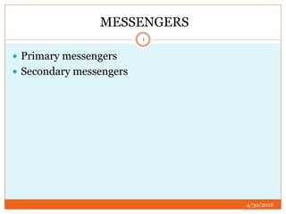
- 1. MESSENGERS 4/30/2018 1 Primary messengers Secondary messengers
- 2. PRIMARY MESSENGERS SECONDARY MESSENGERS 4/30/2018 Extracellular factor like hormones, or neurotransmitter such as epinephrine, growth hormones and serotonin. Peptide hormones and neurotransmitter are hydrophilic. Physically cannot cross the phospholipids bilayer These are intermediate molecules like cyclic AMP or cyclic GMP When hormones bind to the target cell receptor, then the cell release or creates these intermediates which then stimulate cell response Pathway involve- SIGNAL TRANSDUCTION PATHWAY 2 TYPES OF MESSENGERS
- 3. 4/30/20183 HYDROPHOBIC HYDROPHILIC GASES DIACYLGLYCEROL CYCLIC AMP NITRIC OXIDE PHOSPHATIDYL INOSITOL CYCLIC GMP,IP3 HYDROGN SULPHIDE CALCIUM IONS CARBON MONOXIDE TYPES OF SECONDARY MESSENGERS
- 4. HYDROPHOBIC MESSENGERS 4/30/2018 4 Diacylglycerol –stimulate protein kinase c activity by greatly increasing the affinity of the enzyme for calcium ion Known target protein include calmodulin the glucose transporter, HMG-COA reductase
- 5. PHOSPHATIDYLINOSITOL 4/30/2018 5 Phosphatidylinositol negative charged phospholipid and a minor component in eukaryotic cell membranes The inositol can be phosphorylated to form: Phosphatidylinositol-4-phosphate Phosphatidylinositol-4,5-biphosphate Phosphatidylinositol-3,4,5-triphosphate
- 6. CONTINUED…….. 4/30/2018 6 Intracellular enzyme phospholipase Hydrolyzes PIP2 which is found in the inner of the plasma membrane Two product form: Diacylglycerol and PIP3
- 7. G- PROTEIN 4/30/2018 7 Level of middle management in the cellular organization and are able to communicate between receptor and the effector enzymes or ion channels They were called G-PROTEIN because of their interaction with the guanine nucleotides, GTP and GDP The G protein are bound to the cytoplasmic surface of the plasma membrane Heterotrimeric molecules consisting 2α, β,γ
- 8. 4/30/20188 subtype Location of receptor Type of G protein coupled receptor Basic pathways α1 Smooth muscle Gq Increase in PLC, Increase in intracellular calcium ion contraction α2 Presynaptic nerves Gi Decrease in activation of AC Decrease in cyclic AMP β1 β2 β3 Heart Smooth muscle Fat tissue Gs Increase in activation of AC, Increase in cyclic AMP, increase intracellular signaling pathways
- 9. 4/30/20189
- 10. GPCR SIGNALING PATHWAY 4/30/2018 10 Ligand bind to the receptor altering its conformation and increasing its affinity for the G protein to which its binds G subunit release it GDP which is replaced by GTP Alpha subunit dissociate from the G complex and bind to an effector activating the effector
- 11. CONTINUED…… 4/30/2018 11 Activated AC produce cyclic AMP GTPase activity of G protein hydrolyzes the bound GTP deactivating G G reassociates with G reforming the trimeric G protein and the effector ceases its activity
- 12. MAJOR PATHWAYS 4/30/2018 12 ADENYL CYCLASE: CYCLIC AMP PATHWAY PHOSPHOLIPASE C: IP3 DAG PATHWAY CHANNEL REGULATION
- 13. ADENYLYL CYCLASE:CAMP PATHWAY 4/30/2018 13 cAMP is a secondary messenger that is synthesized from ATP by the action of the cAMP- dependent protein kinase Which increase contractility, impulse generation, lipolysis
- 14. 4/30/201814
- 15. IP3- DAG PATHWAY 4/30/2018 15 IP3 located on endoplasmic reticulum Responsible for protein and lipid synthesis DAG- directly activate protein kinase c and control phosphorylation of amino acid of variety of intracellular protein Causes smooth muscle contraction
- 17. CYCLIC GMP 4/30/2018 17 Cyclic guanosine monophosphate is a cyclic nucleotide derived from GTP Common regulator of ion channel, conductance, glycogenolysis, and cellular apoptosis It also relaxes smooth muscles tissue In blood vessels relaxation of vascular smooth muscles lead to vasodilation and increase blood flow
- 18. 4/30/201818
- 19. 4/30/201819
- 20. CALCIUM ION 4/30/2018 20 Great important amongest many other intracellular secondary messengers Calcium ion bind with calmodulin(intracellular) Activate MLCK(myosin light chain kinase)
- 21. Continued……….. 4/30/2018 21 Smooth muscle contraction Calcium ion involved in release of arachidonic acid from membrane phospholipid by activated phospholipase and initiate the synthesis of prostaglandin and leukotrienes Calcium ion synergize with PKC Activation of cellular function like hepatocyte, glycogenolysis, insulin release from pancreas Imp. Role in contraction of smooth muscles
- 22. 4/30/201822 REGULATION OF CALCIUM ION IN PANCREATIC BETA CELL
- 23. GASEOUS SIGNALING 4/30/2018 23 Either synthesized internally in the organism, tissue, or cell or are received by the organism Induce certain physiological or biochemical changes in the organism, tissue or cell Ex carbon monoxide, nitric oxide, hydrogen sulphide
- 24. NITRIC OXIDE 4/30/2018 24 Known as endothelium derived relaxing factor is biosynthesized from L-arginine, oxygen & NADPH by various NO synthase enzymes The endothelium(inner lining) of blood vessel use NO to signal the surrounding smooth muscle Result in vasodilation and increase blood flow
- 25. CONTINUED….. 4/30/2018 25 NO highly reactive its life time is of few sec yet it diffuses freely across membrane For body to generate nitric oxide through nitrate- nitrite-nitric oxide pathway Reduction of nitrate to nitrite occur in mouth by bacteria Monitoring NO status by saliva testing detect the bioconversion of plant derived nitrate into nitric oxide
- 26. CONTINUED…….. 4/30/2018 26 Production of NO is elevated in population living at high altitudes which help these people to avoid hypoxia by aiding in pulmonary vasodilation Nitroglycerin and amyl nitrite serves as vasodilator because they converted to NO in the body Vasodilating antihypertensive drug minoxidil contain an NO moiety act as an NO agonist
- 27. HYDROGEN SULPHIDE 4/30/2018 27 Produced in small amount by some cell of the mammalian body and has a number of biological signaling function The gas is produced from beta- synthase and cysta thionine gamma lyase Act as an relaxant of smooth muscle and as an vasodilator
- 28. CONTINUED….. 4/30/2018 28 It also active in the brain where it increase the response of the NMDA receptor and facilitates long term potentiation
- 29. 29
- 30. 4/30/201830
- 31. 4/30/201831 MECHANISM OF ACTION OF NITRIC OXIDE
- 32. 4/30/201832
- 33. 4/30/201833 CELLULAR EFFECT OF NITRIC OXIDE
- 34. 4/30/201834
- 35. INTRACELLULAR SIGNALING PATHWAY 4/30/2018 35 Cyclic AMP signaling Pathway Mitogen activated protein kinase signaling Janus kinase (JAK) /Signaling transducer and activator of transcripton (STAT) signaling pathway
- 36. Mitogen activated protein kinase signaling 4/30/2018 36 Mitogen activated protein kinases is an enzyme that translocates the signals to the nucleus and activates many transcriptional factor by phosphorylating many different proteins that regulate expression of important cell cycle and differentiation specific protein
- 37. 4/30/201837
- 38. ACTIVATION 4/30/2018 38 Epidermal growth factor (EGF) bind to the (EGFR) epidermal growth factor receptor in the cell membrane g Starting the cascade of signals Activates MAPK(mitogen activated protein kinase) also known (ERK)
- 39. CONTINUED….. 4/30/2018 39 Signal enter the cell nucleus and causes transcription of DNA Then expressed as protein GRB2 growth receptor bound protein 2 is a adapter protein which involve in signal transduction/cell communication
- 40. CONTINUED……. 4/30/2018 40 encoded by this gene bind receptor such as the epidermal growth factor receptor and contain one SH2 domain and two SH3 domain Its two SH3 domain direct complex formation with/other protein and bind to tyrosine phosphorylated sequences
- 41. PATHWAY 4/30/2018 41 Signal pass from activated Ras to a cascade of protein kinases this cascade transmit signals downstream from activated Ras protein to MAP kinase Then MAP kinase translocates the signal to the nucleus and activates trancriptional factor this whole phenomenon called as MAP kinase pathway
- 42. ACTIVATION OF RAS PROTEIN 4/30/2018 42 RAS is a monomeric GTP binding switch protein that alternates between active on state with a bound GTP and inactive off state with a bound GTP It is not directly linked to receptor Its activation is accelerated by a guanosine nucleotide transfer factor
- 43. 4/30/201843 ACTIVATION OF RAS PROTEIN
- 44. HOW (AMPK) GET ACTIVATED 4/30/2018 44 It is activated by increases in the cellular AMP:ATP ratio caused by metabolic stresses Muscle contraction leads to increase in AMP/ATP level to increase in AMPK activity
- 45. FACTORS INVOVED IN ACTIVATION 4/30/2018 45 Heat shock/hypoxia Metabolic changes Glucose deprivation Exercise
- 46. Requirement for signal transduction 4/30/2018 46 Signal –that is to be passed Receptor-to which ligand binds Adapter protein-form link between membrane and bound receptors and protein is to be activated Protein cascade- that would lead to the activation of transcriptional factors Transcriptional factors
- 47. LIGANDS 4/30/2018 47 Ligands for receptor tyrosine kinase (RTKs) including nerve growth factor, platelet derived growth(PDGF), fibroblast growth factor, insulin, epidermal growth factor(EDRF)
- 48. RECEPTORS 4/30/2018 48 Receptor tyrosine kinases- this type of receptor contain intrinsic protein tyrosine kinase activity in their cytosolic domain These have one extracellular domain which serves as ligand and one cytosolic domain to which adapter molecule bind
- 49. ACTIVATION OF RTKs 4/30/2018 49 Most RTKs are monomeric but ligand binding induces dimerization of receptors Formation of ligand receptor complex Alteration and activation of receptor
- 50. CONTINUED….. 4/30/2018 50 This conformational changes facilitates binding of ATP In dimeric receptor the kinases in one subunit can phosphorylate one or more tyrosine residues Phosphorylates other site in the cytosolic domain Activated RTKs interact with adaptor protein
- 51. 4/30/201851
- 52. ADAPTER PROTEIN 4/30/2018 52 Small protein that contain sos, GRB2 domain but have no intrinsic enzymatic or signaling activites These protein couple activated RTKs
- 53. PROTEIN INVOVED 4/30/2018 53 RAS RAF MEK MAP kinase These protein are in inactive state they are need to be activated
- 54. 4/30/201854 FUNCTION OF AMPK PATHWAY
- 55. 4/30/201855
- 56. THERAPEUTIC POTENTIAL 4/30/2018 56 Type 2 diabetic patient are often associated with hypertriglyceridaemia and high cholesterol The potential risk factor for CV problems Activated AMPK could reduce this risk
- 57. JAK-STAT SIGNALING PATHWAY 4/30/2018 57 Janus kinase signal transducers and activators of transcription pathway used to transduce a multitude of signal for development and homeostasis JAK activation stimulates cell proliferation, differentiation, cell migration and apoptosis involve in various processes such as hematopoiesis immune development
- 58. JAK (Janus kinase) 4/30/2018 58 In Mammal the JAK family comprises of 4 member 1) JAK1 2) JAK2 3) JAK3 4) TYK2
- 59. STAT ( signal transduction and activator of transcriptional factor) 4/30/2018 59 STAT are latent transcriptional factor that reside in the cytoplasm until activated STAT conserve tyrosine residue near the c- terminus that is phosphorylated by JAK Phosphorylated STAT enter the nucleus by mechanism that is dependent on nucleoprotein interactor
- 60. CONTINUED…. 4/30/2018 60 Dimerization of STAT bind to the specific regulatory sequence activate or repress transcription of target genes
- 61. MUTATION IN PATHWAY….. 4/30/2018 61 Fail to regulate JAK signaling cause inflammatory disease, erythrocytosis
- 62. COMPONENT 4/30/2018 62 A receptor Janus kinase Signal transducer and activator of transcription
- 63. JAK ACTIVATION 4/30/2018 63 JAK activation occur upon ligand mediated receptor multimerization because two JAK are brought into close allowing phosphorylation
- 65. MECHANISM 4/30/2018 65 JAK have tyrosine kinase activity bind to some cell surfaces cytokine receptor, the binding of the ligand to the receptor triggers activation of JAK With increased kinase activity they phosphorylate tyrosine residues on the receptor STATs Possessing SH2 domain are recruited to the receptor and are themselves tyrosine phosphorylated by JAKs
- 66. CONTINUED….. 4/30/2018 66 Activated STAT dimers accumulate in the cell nucleus and transcription of their target genes STAT may also be tyrosine phosphorylated directly by receptor tyrosine kinases such as the epidermal growth factor receptor as well as by non receptor tyrosine kinases such as c-src The receptor is activated by signal from interferon, interleukin growth factor or other chemical messengers
- 67. REFERENCES 4/30/2018 67 Kobila .K. B ,G protein coupled receptor structure and activation HHS public access Murad.F Nitric oxide: the coming of secondary messenger rambam maimonides medical journal Rawling.S.J,Rosler.M.K Harrison A.D.The JAK- STAT signaling pathway journal of cell science 2004117:128,-1283 1292/jcs.00963 Tripathi.K.D Textbook of medical pharmacology sixth edition jaypee brothers medical publisher page no 22-37
- 68. CONTINUED….. 4/30/2018 68 Yan.k, Gao.N, Zhou X, the cyclic AMP signaling pathway, molecular medicine report spandidos publication
- 69. 4/30/201869