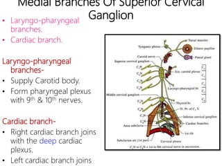
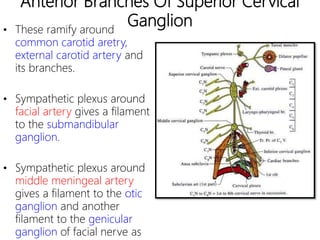
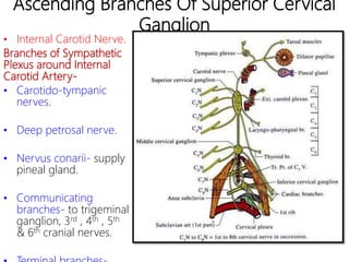
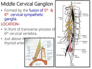
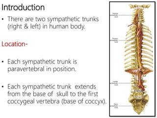
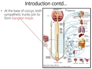
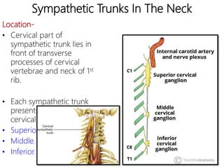
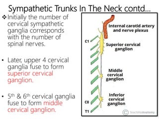
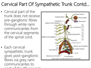
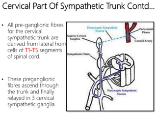
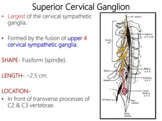
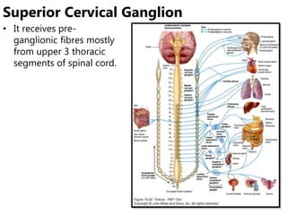
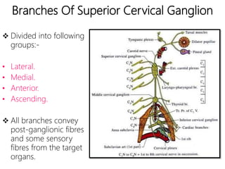
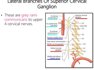
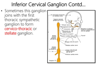
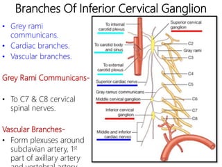
CERVICAL PART OF SYMPATHETIC TRUNK
https://www.slideshare.net/DRCAPRICORN/slideshelf
VESSICO-BULLOUS DISORDER LECTURE : https://youtu.be/lgizglcWJ9I
HOOVER SIGN for leg paresis/ copd=
https://youtu.be/v-rT80AksZw
BEEVOR SIGN = https://youtu.be/QTBqQ31KqUA
ALL PERIPHERAL SIGN'S OF AORTIC REGURGITATION=
https://youtu.be/JZBQGsmK4dY
SUBSCRIBE US ON YOUTUBE : www.youtube.com/c/DrCapricorn
2. Introduction
• There are two sympathetic trunks
(right & left) in human body.
Location-
• Each sympathetic trunk is
paravertebral in position.
• Each sympathetic trunk extends
from the base of skull to the first
coccygeal vertebra (base of coccyx).
3. Introduction contd…
• At the base of coccyx, both
sympathetic trunks join to
form Ganglion Impar.
4. Sympathetic Trunks In The Neck
Location-
• Cervical part of
sympathetic trunk lies in
front of transverse
processes of cervical
vertebrae and neck of 1st
rib.
• Each sympathetic trunk
presents 3 ganglia in the
cervical part:-
• Superior.
• Middle.
• Inferior.
5. Sympathetic Trunks In The Neck
contd…
• Sometimes, inferior cervical and first thoracic
sympathetic ganglion are fused to form a cervico-
thoracic or stellate ganglion.
6. Sympathetic Trunks In The Neck contd…
Initially the number of
cervical sympathetic
ganglia corresponds
with the number of
spinal nerves.
• Later, upper 4 cervical
ganglia fuse to form
superior cervical
ganglion.
• 5th & 6th cervical ganglia
fuse to form middle
cervical ganglion.
7. Cervical Part Of Sympathetic Trunk Contd…
• Cervical part of the
trunk does not receive
pre-ganglionic fibres
through white rami
communicantes from
the cervical segments
of the spinal cord.
• Each cervical
sympathetic trunk
gives post-ganglionic
fibres via grey rami
communicantes to
8.
9. Cervical Part Of Sympathetic Trunk Contd…
• All pre-ganglionic fibres
for the cervical
sympathetic trunk are
derived from lateral horn
cells of T1-T5 segments
of spinal cord.
• These preganglionic
fibres ascend through
the trunk and finally
relayed in 3 cervical
sympathetic ganglia.
10. Superior Cervical Ganglion
• Largest of the cervical sympathetic
ganglia.
• Formed by the fusion of upper 4
cervical sympathetic ganglia.
SHAPE- Fusiform (spindle).
LENGTH- ~2.5 cm.
LOCATION-
• In front of transverse processes of
C2 & C3 vertebrae.
11. • It receives pre-
ganglionic fibres mostly
from upper 3 thoracic
segments of spinal cord.
Superior Cervical Ganglion
12. Branches Of Superior Cervical Ganglion
Divided into following
groups:-
• Lateral.
• Medial.
• Anterior.
• Ascending.
All branches convey
post-ganglionic fibres
and some sensory
fibres from the target
organs.
13. Lateral Branches Of Superior Cervical
Ganglion
• These are grey rami
communicans to upper
4 cervical nerves.
14. Medial Branches Of Superior Cervical
Ganglion• Laryngo-pharyngeal
branches.
• Cardiac branch.
Laryngo-pharyngeal
branches-
• Supply Carotid body.
• Form pharyngeal plexus
with 9th & 10th nerves.
Cardiac branch-
• Right cardiac branch joins
with the deep cardiac
plexus.
• Left cardiac branch joins
15. Anterior Branches Of Superior Cervical
Ganglion• These ramify around
common carotid aretry,
external carotid artery and
its branches.
• Sympathetic plexus around
facial artery gives a filament
to the submandibular
ganglion.
• Sympathetic plexus around
middle meningeal artery
gives a filament to the otic
ganglion and another
filament to the genicular
ganglion of facial nerve as
16. Ascending Branches Of Superior Cervical
Ganglion
• Internal Carotid Nerve.
Branches of Sympathetic
Plexus around Internal
Carotid Artery-
• Carotido-tympanic
nerves.
• Deep petrosal nerve.
• Nervus conarii- supply
pineal gland.
• Communicating
branches- to trigeminal
ganglion, 3rd , 4th , 5th
& 6th cranial nerves.
17. Middle Cervical Ganglion
• Formed by the fusion of 5th &
6th cervical sympathetic
ganglia.
LOCATION-
• In front of transverse process of
6th cervical vertebra.
• Just above the loop of inferior
thyroid artery .
18. Middle Cervical Ganglion Contd…
COMMUNICATIONS-
• Connected with inferior
cervical ganglion by
ansa subclavia.
• Ansa subclavia loops in
front and below the first
part of subclavian
artery.
19. Branches Of Middle Cervical Ganglion
• Lateral.
• Medial.
Lateral branches-
• These are grey rami
comminicans to 5th &
6th spinal nerves.
Medial branches-
• Thyroid branches.
• Cardiac branches.
20.
21. Inferior Cervical Ganglion
• Formed by the fusion of 7th &
8th cervical ganglia.
LOCATION-
• Between transverse process of
C7 vertebra and neck of 1st rib.
22. Inferior Cervical Ganglion Contd…
• Sometimes this ganglion
joins with the first
thoracic sympathetic
ganglion to form
cervico-thoracic or
stellate ganglion.
23. Branches Of Inferior Cervical Ganglion
• Grey rami
communicans.
• Cardiac branches.
• Vascular branches.
Grey Rami Communicans-
• To C7 & C8 cervical
spinal nerves.
Vascular Branches-
• Form plexuses around
subclavian artery, 1st
part of axillary artery
24. Applied Anatomy
HORNER’S SYNDROME-
• A lesion affecting the pre-
ganglionic fibres from T1 & T2
segments of spinal cord.
Clinical Features-
• Constriction of pupil (miosis).
• Drooping of upper eyelid (ptosis).
• Enophthalmos.
• Absence of sweating on affected
half of face and head (anhydrosis).