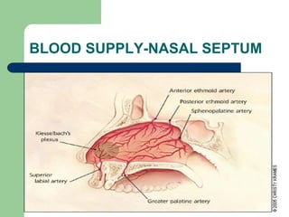
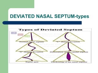
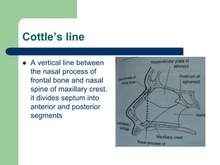
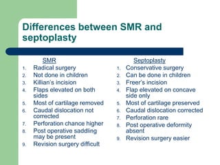
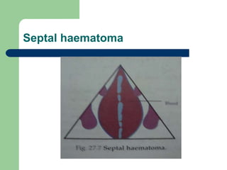
NASAL SEPTUM AND ITS
DISEASES
Nasal septum and its diseases
Anatomy of nasal septum:
Nasal septum consists of three parts:
1. Columellar septum
2. Membranous septum
3. Septum proper: principle constituents of septum proper are
a)perpendicular plate of ethmoid
b)vomer
c)septal(quadrilateral cartilage)
minor contributions from crest of nasal bone,nasal spine of
frontal bone,rostrum of sphenoid,crest of palatine and maxilla
and anterior nasal spine of maxilla.
Nasal septum and its diseases
o Septal cartilage forms a partition between right and left
nasal cavities and provides support to tip and dorsum of
cartilagenous part of nose.
o Septal destruction may occur in septal abscess, injuries,
tuberculosis, excess removal during SMR leads to
depression of lower part of nose and drooping of tip.
o Septal cartilage lies in a groove in the anterior edge of
vomer and rests anteriorly on anterior nasal spine. during
trauma, it may get dislocated from nasal spine or vomer
causing caudal septal deviation and spur respectively.
BLOOD SUPPLY-NASAL SEPTUM
NERVE SUPPLY-NASAL SEPTUM
DEVIATED NASAL SEPTUM
AETIOLOGY:
1) Trauma:
lateral blow-displacement of septal cartilage from vomer.
blow from front-buckling, fracture, duplication of septum with
telescoping of fragments.
2) Developmental: the septum should grow at the same rate as
that of face. if septum grows at faster rate it becomes buckled.
unequal growth between palate and base of skull may also
cause buckling (high arched palate)
3) Congenital: abnormal intrauterine posture cause compressing
forces acting on nose and upper jaw.
4) Hereditary
5) Racial: Caucasians are more affected
6) Secondary: to a tumour, mass or polyp.
DEVIATED NASAL SEPTUM
Types:
1) Deviations: upper or lower, anterior or posterior, C
shaped, S shaped. nasal cavity on the concave
side of the septum will be wider and may show
compensatory hypertrophy of turbinates.
2) Anterior Dislocation: seen on tilting the patients
head backwards.
3) Spurs: shelf like projection at the junction of bone
and cartilage. may predispose for epistaxis and
headache.
4) Thickening: it may be due to organized haematoma
or over-riding of dislocated septal fragments
DEVIATED NASAL SEPTUM-types
Clinical features
 Nasal obstruction: depending on the type it
may be unilateral or bilateral. It is the most
common symptom
 headache
 Recurrent attacks of cold
 Epistaxis
 Anosmia
 External deformity
 Middle ear infection
Clinical features
 Cottle’s test: used in nasal obstruction due to
abnormality of nasal valve. In this test cheek
is drawn laterally while the patient breathes
quietly. If the nasal airway improves on test
side the test is positive and indicates
abnormality of nasal valve
Differential diagnosis
 Polyps
 Septal haematoma
 Hypertrophied turbinates
Treatment- surgery
 Submucous resection of nasal septum (SMR)
 It is generally done in adults
 It consists of elevating mucoperichondrial
and mucoperiosteal flap on either side of the
septum, removing the deflected parts of bony
and cartilagenous septum and then
repositioning the flaps
SMR
 Indications
 Deviated nasal septum causing nasal obstruction and
recurrent headaches
 Deviated nasal septum causing obstruction to
ventilation of paranasal sinuses and middle ear
resulting in recurrent infections
 Recurrent epistaxis from septal spur
 As a part of septorhinoplasty
 Harvesting cartilage graft for tympanoplasty and
rhinoplasty
 As an approach to surgeries of sphenoidal sinus,
vidian nerve and pituitary gland
SMR
 Contraindications
 Acute URTI
 Patient below 17 yrs of age
 Bleeding disorders
 Uncontrolled hypertension and diabetes
mellitus
SMR
 Anesthesia - Local anesthesia/ general
anesthesia
 Positioning: reclining position with head end
of the table raised
SMR - STEPS
 Infiltration: subperichondrial infiltration with 2%
xylocaine with adrenaline
 Incision: killian’s incision- curvilinear incision 2-
3mm behind the anterior end of septal cartilage
 Elevation of flaps: the mucoperichondrial and
mucoperiosteal flap is elevated
 Incision of the cartilage- cartilage is incised just
posterior to the first incision
 Elevation of opposite mucoperichondrial and
mucoperiosteal flap
SMR – STEPS (cont…)
 Removal of cartilage and bone - cartilage
can be removed with Ballinger swivel knife or
luc’s forceps. Bony spur is removed using
gouge and hammer
 Preserve a strip of 1cm wide cartilage along
the dorsal and caudal borders (struts)
 Nasal packing
SMR – STEPS
complications
 Bleeding
 Septal haematoma
 Damage to surrounding structures
 Septal abscess
 Septal Perforation
 Depression of bridge
 Retraction of columella
 Synichae
 Flapping septum
 Infection- sinus and middle ear
 CSF rhinorrhoea
Cottle’s line
 A vertical line between
the nasal process of
frontal bone and nasal
spine of maxillary crest.
it divides septum into
anterior and posterior
segments
Septoplasty
 It is a conservative approach to septal surgery as much
of the septal framework is retained
 Indications:
 Deviated nasal septum causing nasal obstruction and
recurrent headaches
 Deviated nasal septum causing obstruction to ventilation
of paranasal sinuses and middle ear resulting in recurrent
infections
 Recurrent epistaxis from septal spur
 As a part of septorhinoplasty
 As an approach to surgeries of sphenoidal sinus, vidian
nerve and pituitary gland
Septoplasty (cont…)
 Contraindications
 Acute URTI
 Bleeding disorders
 Uncontrolled hypertension and diabetes
mellitus
Septoplasty (cont…)
 Anesthesia: local or general anesthesia
 Position: same as SMR
 Steps :
 Infiltration
 Incision: Freer’s incision– a unilateral
hemitransfixation incision at the caudal
border of the septum
 Exposure: the mucoperichondrial and
mucoperiosteal flap is elevated on only one
side
Septoplasty (cont…)
 Separate septal cartilage from vomer and
ethmoid plate
 Inferior strip of cartilage is removed
 Correct the bony septum by removing deformed
parts
 Minor deviations of cartilage are corrected by
criss cross incision which breaks spring action of
cartilage
 Nasal packing
Post-operative complications
 Bleeding
 Septal haematoma
 Damage to surrounding structures
 Septal abscess
 Septal Perforation
 Depression of bridge
 Retraction of columella
 Synechiae
 Infection- sinus and middle ear
 CSF rhinorrhoea
Differences between SMR and
septoplasty
SMR
1. Radical surgery
2. Not done in children
3. Killian’s incision
4. Flaps elevated on both
sides
5. Most of cartilage removed
6. Caudal dislocation not
corrected
7. Perforation chance higher
8. Post operative saddling
may be present
9. Revision surgery difficult
Septoplasty
1. Conservative surgery
2. Can be done in children
3. Freer’s incision
4. Flap elevated on concave
side only
5. Most of cartilage preserved
6. Caudal dislocation corrected
7. Perforation rare
8. Post operative deformity
absent
9. Revision surgery easier
Septal haematoma
 It is collection of blood under the perichondrium or
periosteum of nasal septum
 Etiology: nasal trauma, post-operative, in bleeding
disorder
 Clinical features:
 Bilateral nasal obstruction
 Frontal headache
 Sense of pressure over nasal bridge
 Examination reveals smooth rounded swelling of the
septum in both the nasal cavity. Palpation may show
the mass to be soft and fluctuant
Septal haematoma
 Treatment: small haematomas can be
aspirated with a wide bore needle, larger
haematomas are incised and drained.
Excision of small piece of mucosa from the
edge of the incision gives better drainage.
Nose is packed on both sides to prevent re-
accumulation. Systemic antibiotics to prevent
septal abscess
Septal haematoma
 Complications
 If not drained may organize into fibrous
tissue leading to a permanently thickened
septum
 If secondary infection supervenes leads to
septal abscess with necrosis of cartilage and
saddling
Septal haematoma
Septal abscess
 Etiology
 Secondary infection of septal haematoma
 Furuncle of the nasal vestibule
Clinical features
 Severe bilateral nasal obstruction with pain and
tenderness over bridge of nose
 Fever with chills
 Frontal headache
 Skin over the nose may be red and swollen
 Smooth bilateral swelling of the nasal septum
 Congested septal mucosa
 Submandibular nodes may be enlarged and tender
Septal abscess
 Treatment
 Abscess should be drained as early as
possible
 Pus and necrosed cartilage removed by
suction
 Incision may required to be re-opened daily
for 2-3 days to drain any pus or remove any
necrosed piece of cartilage
 Systemic antibiotics to be started as soon as
possible and continued for two weeks
Septal abscess
 Complications
 Depression of the cartilagenous dorsum
 Septal perforation
 Meningitis and cavernous sinus thrombosis
(rare)
Nasal septal perforation
 Etiology
 Traumatic - post surgical, habitual nose picking, cauterization of
septum with chemicals or galvano-cautery for epistaxis
 Pathological perforation
a) Septal abscess
b) Nasal myasis
c) Rhinolith or neglected foreign body
d) Chronic granulomatous conditions like TB, lupus, leprosy, syphilis,
wegener’s
 Inhalant irritants- snuff and cocaine irritant, industrial toxins
 Malignancy
 idiopathic
Nasal septal perforation
 Clinical features
 Whistling sound
 Irritation and crusting
 Epistaxis
 Nasal obstruction
Nasal septal perforation
 Treatment
 Treat the root cause
 Inactive small perforation can be surgically
closed by plastic flaps or septal mucosal
flaps
 Larger perforations are difficult to close: their
treatment is aimed to keep the nose crust
free by alkaline nasal douch and application
of lubricants, silastic obturator may also be
used
nasal septum.pptx

nasal septum.pptx

  • 1.
    NASAL SEPTUM ANDITS DISEASES
  • 2.
    Nasal septum andits diseases Anatomy of nasal septum: Nasal septum consists of three parts: 1. Columellar septum 2. Membranous septum 3. Septum proper: principle constituents of septum proper are a)perpendicular plate of ethmoid b)vomer c)septal(quadrilateral cartilage) minor contributions from crest of nasal bone,nasal spine of frontal bone,rostrum of sphenoid,crest of palatine and maxilla and anterior nasal spine of maxilla.
  • 5.
    Nasal septum andits diseases o Septal cartilage forms a partition between right and left nasal cavities and provides support to tip and dorsum of cartilagenous part of nose. o Septal destruction may occur in septal abscess, injuries, tuberculosis, excess removal during SMR leads to depression of lower part of nose and drooping of tip. o Septal cartilage lies in a groove in the anterior edge of vomer and rests anteriorly on anterior nasal spine. during trauma, it may get dislocated from nasal spine or vomer causing caudal septal deviation and spur respectively.
  • 6.
  • 7.
  • 8.
    DEVIATED NASAL SEPTUM AETIOLOGY: 1)Trauma: lateral blow-displacement of septal cartilage from vomer. blow from front-buckling, fracture, duplication of septum with telescoping of fragments. 2) Developmental: the septum should grow at the same rate as that of face. if septum grows at faster rate it becomes buckled. unequal growth between palate and base of skull may also cause buckling (high arched palate) 3) Congenital: abnormal intrauterine posture cause compressing forces acting on nose and upper jaw. 4) Hereditary 5) Racial: Caucasians are more affected 6) Secondary: to a tumour, mass or polyp.
  • 9.
    DEVIATED NASAL SEPTUM Types: 1)Deviations: upper or lower, anterior or posterior, C shaped, S shaped. nasal cavity on the concave side of the septum will be wider and may show compensatory hypertrophy of turbinates. 2) Anterior Dislocation: seen on tilting the patients head backwards. 3) Spurs: shelf like projection at the junction of bone and cartilage. may predispose for epistaxis and headache. 4) Thickening: it may be due to organized haematoma or over-riding of dislocated septal fragments
  • 10.
  • 11.
    Clinical features  Nasalobstruction: depending on the type it may be unilateral or bilateral. It is the most common symptom  headache  Recurrent attacks of cold  Epistaxis  Anosmia  External deformity  Middle ear infection
  • 12.
    Clinical features  Cottle’stest: used in nasal obstruction due to abnormality of nasal valve. In this test cheek is drawn laterally while the patient breathes quietly. If the nasal airway improves on test side the test is positive and indicates abnormality of nasal valve
  • 13.
    Differential diagnosis  Polyps Septal haematoma  Hypertrophied turbinates
  • 14.
    Treatment- surgery  Submucousresection of nasal septum (SMR)  It is generally done in adults  It consists of elevating mucoperichondrial and mucoperiosteal flap on either side of the septum, removing the deflected parts of bony and cartilagenous septum and then repositioning the flaps
  • 15.
    SMR  Indications  Deviatednasal septum causing nasal obstruction and recurrent headaches  Deviated nasal septum causing obstruction to ventilation of paranasal sinuses and middle ear resulting in recurrent infections  Recurrent epistaxis from septal spur  As a part of septorhinoplasty  Harvesting cartilage graft for tympanoplasty and rhinoplasty  As an approach to surgeries of sphenoidal sinus, vidian nerve and pituitary gland
  • 16.
    SMR  Contraindications  AcuteURTI  Patient below 17 yrs of age  Bleeding disorders  Uncontrolled hypertension and diabetes mellitus
  • 17.
    SMR  Anesthesia -Local anesthesia/ general anesthesia  Positioning: reclining position with head end of the table raised
  • 18.
    SMR - STEPS Infiltration: subperichondrial infiltration with 2% xylocaine with adrenaline  Incision: killian’s incision- curvilinear incision 2- 3mm behind the anterior end of septal cartilage  Elevation of flaps: the mucoperichondrial and mucoperiosteal flap is elevated  Incision of the cartilage- cartilage is incised just posterior to the first incision  Elevation of opposite mucoperichondrial and mucoperiosteal flap
  • 19.
    SMR – STEPS(cont…)  Removal of cartilage and bone - cartilage can be removed with Ballinger swivel knife or luc’s forceps. Bony spur is removed using gouge and hammer  Preserve a strip of 1cm wide cartilage along the dorsal and caudal borders (struts)  Nasal packing
  • 20.
  • 21.
    complications  Bleeding  Septalhaematoma  Damage to surrounding structures  Septal abscess  Septal Perforation  Depression of bridge  Retraction of columella  Synichae  Flapping septum  Infection- sinus and middle ear  CSF rhinorrhoea
  • 22.
    Cottle’s line  Avertical line between the nasal process of frontal bone and nasal spine of maxillary crest. it divides septum into anterior and posterior segments
  • 23.
    Septoplasty  It isa conservative approach to septal surgery as much of the septal framework is retained  Indications:  Deviated nasal septum causing nasal obstruction and recurrent headaches  Deviated nasal septum causing obstruction to ventilation of paranasal sinuses and middle ear resulting in recurrent infections  Recurrent epistaxis from septal spur  As a part of septorhinoplasty  As an approach to surgeries of sphenoidal sinus, vidian nerve and pituitary gland
  • 24.
    Septoplasty (cont…)  Contraindications Acute URTI  Bleeding disorders  Uncontrolled hypertension and diabetes mellitus
  • 25.
    Septoplasty (cont…)  Anesthesia:local or general anesthesia  Position: same as SMR  Steps :  Infiltration  Incision: Freer’s incision– a unilateral hemitransfixation incision at the caudal border of the septum  Exposure: the mucoperichondrial and mucoperiosteal flap is elevated on only one side
  • 26.
    Septoplasty (cont…)  Separateseptal cartilage from vomer and ethmoid plate  Inferior strip of cartilage is removed  Correct the bony septum by removing deformed parts  Minor deviations of cartilage are corrected by criss cross incision which breaks spring action of cartilage  Nasal packing
  • 27.
    Post-operative complications  Bleeding Septal haematoma  Damage to surrounding structures  Septal abscess  Septal Perforation  Depression of bridge  Retraction of columella  Synechiae  Infection- sinus and middle ear  CSF rhinorrhoea
  • 28.
    Differences between SMRand septoplasty SMR 1. Radical surgery 2. Not done in children 3. Killian’s incision 4. Flaps elevated on both sides 5. Most of cartilage removed 6. Caudal dislocation not corrected 7. Perforation chance higher 8. Post operative saddling may be present 9. Revision surgery difficult Septoplasty 1. Conservative surgery 2. Can be done in children 3. Freer’s incision 4. Flap elevated on concave side only 5. Most of cartilage preserved 6. Caudal dislocation corrected 7. Perforation rare 8. Post operative deformity absent 9. Revision surgery easier
  • 29.
    Septal haematoma  Itis collection of blood under the perichondrium or periosteum of nasal septum  Etiology: nasal trauma, post-operative, in bleeding disorder  Clinical features:  Bilateral nasal obstruction  Frontal headache  Sense of pressure over nasal bridge  Examination reveals smooth rounded swelling of the septum in both the nasal cavity. Palpation may show the mass to be soft and fluctuant
  • 30.
    Septal haematoma  Treatment:small haematomas can be aspirated with a wide bore needle, larger haematomas are incised and drained. Excision of small piece of mucosa from the edge of the incision gives better drainage. Nose is packed on both sides to prevent re- accumulation. Systemic antibiotics to prevent septal abscess
  • 31.
    Septal haematoma  Complications If not drained may organize into fibrous tissue leading to a permanently thickened septum  If secondary infection supervenes leads to septal abscess with necrosis of cartilage and saddling
  • 32.
  • 33.
    Septal abscess  Etiology Secondary infection of septal haematoma  Furuncle of the nasal vestibule Clinical features  Severe bilateral nasal obstruction with pain and tenderness over bridge of nose  Fever with chills  Frontal headache  Skin over the nose may be red and swollen  Smooth bilateral swelling of the nasal septum  Congested septal mucosa  Submandibular nodes may be enlarged and tender
  • 34.
    Septal abscess  Treatment Abscess should be drained as early as possible  Pus and necrosed cartilage removed by suction  Incision may required to be re-opened daily for 2-3 days to drain any pus or remove any necrosed piece of cartilage  Systemic antibiotics to be started as soon as possible and continued for two weeks
  • 35.
    Septal abscess  Complications Depression of the cartilagenous dorsum  Septal perforation  Meningitis and cavernous sinus thrombosis (rare)
  • 36.
    Nasal septal perforation Etiology  Traumatic - post surgical, habitual nose picking, cauterization of septum with chemicals or galvano-cautery for epistaxis  Pathological perforation a) Septal abscess b) Nasal myasis c) Rhinolith or neglected foreign body d) Chronic granulomatous conditions like TB, lupus, leprosy, syphilis, wegener’s  Inhalant irritants- snuff and cocaine irritant, industrial toxins  Malignancy  idiopathic
  • 37.
    Nasal septal perforation Clinical features  Whistling sound  Irritation and crusting  Epistaxis  Nasal obstruction
  • 38.
    Nasal septal perforation Treatment  Treat the root cause  Inactive small perforation can be surgically closed by plastic flaps or septal mucosal flaps  Larger perforations are difficult to close: their treatment is aimed to keep the nose crust free by alkaline nasal douch and application of lubricants, silastic obturator may also be used