SlideShare a Scribd company logo
1 of 42
Fish immune system in improving fish
health and aquaculture production
By:
Bhukya Bhaskar
Fisheries
Ref: Dr. Ekaitz Maguregui, 2020
Introduction
• Traditionally, the immune system is divided into the innate or
nonspecific immune system and the adaptive or specific immune
system.
• innate (non-specific) and adaptive (specific).
• Cellular immune responses consist of innate and adaptive cell-
mediated immune mechanisms, where all leukocyte subpopulations
are included.
• Among these are vital processes such as cell-mediated cytotoxicity
and phagocytosis.
• The main cellular constituents of the fish immune system are
macrophages, granulocytes, dendritic cells, NK cells, and cytotoxic T
cells.
• The fish immune system has both types of immunity, innate
immunity, and adaptive immunity.
• fish immune system responsible for defending the body from
external threats, such as viruses, bacteria, or protozoa and, therefore,
it allows to prevent infections.
• The adaptive immune system is highly specific to a specific antigen and can provide long-
standing immunity.
• Several ingredients of the adaptive system, including T cell receptors (TCR), immunoglobulins
(Igs), and major histocompatibility complex (MHC), are presumed to have arisen in the first
jawed vertebrates.
• Teleost antibodies are found in the skin, intestine, bile, gill mucus, and systemically in the
plasma.
• IL-1β and TNF-α are cytokines implicated in inflammatory response induction in fish.
• IL-6 has been reported to be involved in the cascade that induces an inflammatory response
• The components of the innate immunity respond to pathogens via identifying pathogen-
associated molecular patterns (PAMPs) that demonstrate no expression in host cells.
• Among these PAMPs recognized by the innate immune receptors are lipopolysaccharides
(LPS), lipoteichoic acid (LTA), phospholipomannan, beta-glucan, chitin, and hemagglutinin.
• pathogen recognition, the pathogens are destructed by phagocytosis, one of the effector
mechanisms of innate immunity.
• Adaptive immunity recognizes pathogens through highly specific receptors, and
consequently induces specific B and T cell clone proliferation and differentiation
• The immune system of teleost fish is constituted by two receptor categories: the antigen-
specific receptors and germline-encoded pattern recognition receptors (PRRs). Numerous
other receptors or molecules can participate in the innate immunity
Types of immunity
• Like upper vertebrates, fish have two types of immunity, innate or nonspecific immunity and adaptive or specific
immunity (table 1).
• Innate or nonspecific immunity
• Innate or non-specific immunity is the first line of defense that the fish’s immune system has to deal with the different
pathogens that threaten their homeostasis. This defensive system can be divided into three main components: mucosal
immunity, humoral components, and cellular components.
• Mucosal immunity plays a key role in the fish immune system, acting as a barrier and preventing pathogens from reaching the
body. In addition to the mechanical and physical protection provided by the mucus that coats the body surface of these
animals, as well as gills and other tissues, it contains a large number of immune components such as antimicrobial peptides,
complement factors and immunoglobulins.
• The main difference between innate immunity and adaptive immunity is the specificity with which they are able to recognize
different pathogens. Such specificity is determined by the type of receptors they can recognize.
• The cells and humoral components that constitute nonspecific immunity recognize these types of molecular patterns:
• Pathogen Associated Molecular Patterns (PAMPs): which constitute a set of highly conserved substances/structures in different
types of pathogens that are not present in eukaryotic cells, such as cell wall lipopolysaccharides (LPS). In the case of teleostswei,
TLR (Toll-like receptors), of which more than 20 different types have been described, play a primary role.
• Damage/Danger Associated Molecular Patterns (DAMPs): signals emitted by cells of the body itself, for example, in case of
thermal stress.
• As in mammals, the innate fish immune system has different humoral and cellular components. Humoral components include
antimicrobial peptides, the complement system, lectins, TNF-α and interleukins.
• Antimicrobial peptides have been described in multiple aquatic species and are present in the mucus, covering the skin and
gills. For example, in the case of the Atlantic cod (Gadus morhua), polypeptides present in the mucus have been described to be
effective against Gram + and Gram – bacteria.
• One of the defensive mechanisms of the innate immunity is the complement system. This system consists of a set of serum
proteins that circulate inactively and are activated in the form of a cascade. As in the case of upper vertebrates, the
complement system is activated by the 3-way, the classic, the alternative, and the lectins pathways. All of them lead to the
formation of pores in the cell membrane of the pathogen and the consequent death by osmotic shock. One noteworthy
difference between the mammalian and the fish immune system is the optimal temperature for this immune complex. In the
case of teleost, its activity is maximum between 15-25°C and can remain active at temperatures between 0-4C°, while, in
mammals, its optimal temperature is 35°C.
• The innate immune system of fish has, at the same time, different cell types that participate in the nonspecific cellular
response, including cells with phagocytic activity, such as monocytes and macrophages, and cells with cytotoxic activity, such as
granulocytes and non-specific cytotoxic cells.
• In the presence of an antigen, neutrophils are mobilized first from the anterior kidney and then the macrophages that mobilize
from nearby tissues are attended. Thanks to the phagocytic and cytotoxic activity of these cells, pathogens are eliminated.
Fish immune system characteristics, differential characteristics
between the innate immune system and the adaptive immune system
Cellular Immune Components
(source: Doaa M. Mokhtar et al, 2023)
• Immune cells in teleosts include natural killer cells,
non-specific cytotoxic cells, macrophages, granular
leucocytes, thrombocytes, monocytes, dendritic
cells, lymphocytes, mast cells, and eosinophilic
granule cells. In addition, fish also possess rodlet
cells and melanomacrophage centers.
1. Natural Killer (NK) Cells
• In teleost fish, there are two different types of NK cell homologs: non-specific cytotoxic cells (NCCs) and NK-like cells .
• In contrast to NK cells, which have a large and granular morphology, NCCs are tiny, agranular cells that resemble catfish
monocytes.
• NCCs in seabream are highly variable in morphology.
• The discovery of NK-like cells in teleosts suggests that NCCs are not the same as NK cells in fish. Accordingly, NK-like cells
destroy allogeneic and virus-infected cells, according to a few investigations conducted on rainbow trout and catfish .
• From teleost NK-like cells, the gene that acts as a cell marker, NK cell enhancement factor (NKEF), has been discovered. After
viral and bacterial infection, NKEF gene expression is elevated in tissues such as the skin, gills, and other organs.
• Interestingly, a recombinant NKEF protein enhances the cytotoxicity of NCC from the kidney in Nile tilapia.
• The NCCs were the first recognized and are the most thoroughly investigated killer cell population in teleosts. The NCCs serve
functions like those of the higher vertebrates, acting on various target cells, including virus-infected cells, tumor cells, and
protozoan parasites.
• NCCs may also be involved in antibacterial immunity by triggering the production and secretion of cytokines .
• The NCCs of tilapia and catfish express components of the granule exocytosis pathway of cell-mediated cytotoxicity (CMC)
analogous to cytotoxic lymphocytes of mammals.
• Non-specific CMC reactions in mammals are mainly performed by NK cells. Non-specific cytotoxic cells (NCCs) and NK-like cells,
two classes of NK cell homologues, are responsible for non-specific CMC mechanisms in fish. Fish NK-like cells have been
isolated from blood leukocytes and have been shown to kill virus-infected, allogeneic, and xenogeneic target cells on their own.
• NCCs, on the other hand, tend to target a variety of cells, including some protozoa and tumor cells, and are particularly active in
the spleen and head kidney. NCCs are capable of spontaneously killing the affected cells through necrotic and apoptotic
mechanisms.
• These cells show variable morphological features, varying from tiny agranular monocyte-like cells in catfish to a mixture of
acidophilic granulocytes, monocyte-macrophages, and lymphocytes in seabream.
• Moreover, a population of circulating lymphocytes that resemble mammalian natural killer (NK) cells in terms of morphology
and functionality has also been reported.
• Additionally, NCCs express the NCC receptor protein 1 (NCCRP-1) on their cell surface and present a vimentin-like surface
molecule.
• It has been shown that catfish NCCs recognize and kill various human cell lines.
• Further research reported that granulysin, perforin, and serglycin, gene-encoding molecules with lytic capacity, are expressed by
NCCs.
• Finally, the fish possess different NCC subsets in several of their immune compartments
2. Macrophages
• Macrophages have a pivotal role in specific immune responses because of their function in lymphocyte
activation and phagocytosis. Macrophages possess specific receptors capable of recognizing β-glucan, so that
the immunostimulants augment leukocytes’ respiratory burst, which produces reactive oxygen species with
bactericidal activity.
• Another bactericidal mechanism is represented by the production of nitric oxide (NO), which is catalyzed by an
NO synthase. Using enzyme histochemical techniques, Schoor and Plumb demonstrated inducible NO
production from the head kidney of channel catfish (Ictalurus punctatus) infected with Edwardsiella ictaluri. In
addition, Stafford et al.
• distinguished the molecules found in crude leukocyte supernatants that stimulate NO production in
macrophages of goldfish, indicating that transferrin is an essential mediator for the activation of both fish
granulocytes and macrophages.
• In fish, neutrophils and macrophages play an essential role in controlling the spread of infectious agents and
are accountable for the destruction of phagocytosed pathogens.
• Macrophages act as antigen-presenting cells in the distal intestine, enabling antigens to interact with the
adaptive immune system for identification. Other granulocytes can also be identified in the intestinal segments
for innate clearance, in addition to the resident macrophages.
• The lamina propria and epithelial linings of the intestine contain these innate populations, allowing a close
vicinity to the digested pathogens.
• Macrophages express several receptors on their cell surface, including TLRs, PRRs, and CLRs, in addition to
complement and scavenger receptors. Macrophages are also an essential source of chemokines and cytokines,
linking innate and adaptive immunity, which mediates an efficient immune response.
• Moreover, macrophages are important for antigen presentation to T cells.
• Circulating monocytes are typically CD14+, and express chemokine receptors, TLRs, adhesion molecules, and
surface molecules that are involved in the pathogen-associated molecular pattern recognition at sites of
inflammation and/or infection
3. Neutrophils
• In the blood, peritoneal cavity, and lymphoid organs, neutrophils are
polymorphonuclear cells that have the ability to phagocytose cells or
foreign particles and manufacture superoxide anions, a bactericidal
component.
• Neutrophils play an essential role in the inflammatory immune response
against various viral, bacterial, fungal, and protozoan pathogens.
• Neutrophils are the first granulocytes to be detected at the injury site,
followed by macrophages, both of which are directed by chemotactic
factors produced by wounded tissue. At the site of injury, neutrophils use
antimicrobial peptides, proteolytic enzymes, and reactive oxygen species
(ROS) to phagocytose microorganisms and destroy them.
• Additionally, fish neutrophils have the ability to release extracellular fibers
known as neutrophil extracellular traps (NETs) that include DNA, histones,
and proteins that can bind, kill, and inactivate viruses, bacteria, fungi, and
parasites.
• Furthermore, the granular leukocyte-derived myeloperoxidase enzyme can
interact with hydrogen peroxide to create hypochlorite, which in turn can
be used to produce cloramins, oxidative substances that can attack
microorganism membranes
4. Eosinophils, Basophils, Thrombocytes, and Monocytes
• Eosinophils are widely distributed cells in connective tissue, particularly in
the gastrointestinal tract, bloodstream, ovaries, and gills, and offer
degranulation when parasite infections are present.
• Basophils are large polymorphonuclear granular leucocytes that are rarely
observed in teleost species.
• Their cytoplasmic granules have an inflammatory mediator, histamine.
These cells are involved in allergy and anaphylaxis.
• Thrombocytes are oval-shaped, nucleated, and agranular cells.
• They possess both a coagulation function and phagocytic ability.
• They show acid phosphatase activity that causes cells to gather at the
inflammatory site.
• The monocytes show phagocytosis and non-specific cytotoxic activities.
• These cells are thought to be transient blood cells because they migrate to
the connective tissue during the inflammatory phase and change into
macrophages.
• Professional phagocytes that can be deployed to the site of inflammation
include monocytes, macrophages, granulocytes, and dendritic cells
5. Dendritic Cells (DCs)
• The DCs are among the antigen presenting cells found within several tissues
and can initiate both innate and acquired immune responses.
• DCs are characterized by their small cell bodies with many cytoplasmic
processes (dendrites) and have been found in gills, gut, skin, and ovaries.
Cell populations with DC-like functions and morphology have been
demonstrated in many fish species.
• With the assistance of MHC class 2 receptors, these cells deliver the
processed antigens to T lymphocytes, triggering the cell-mediated acquired
response. These cells appear to activate T cells in an antigen-dependent
manner in zebrafish by expressing genes associated with antigen
presentation, such as MHC class II invariant chain iclp1, csf1r, and il12.
• The trout gut has been shown to include putative CD8 + DC-like cells that
appear to have phagocytic activity and the capacity to excite naive T cells.
• Recently, Alesci, et al. identified the intestinal DCs in several fish species
including Scyliorhinus canicula (Chondrichthyes), Eptatretus
cirrhatus (myxines), Polypterus senegalus (Osteichthyes,
Brachiopterygii), Clarias batrachus (Osteichthyes, Teleostei), and Lepisosteus
oculatus (Osteichthyes, Holostei).
• These findings showed that intestinal DCs and DC-like cells were positive to
TLR-2, Langerin/CD207, and MHC II.
6. Lymphocytes
• B lymphocytes, which are professional antigen-presenting cells (APCs) produce antibodies against antigens and mediate humoral immunity. They also
develop immunological memory by differentiating into memory B cells.
• A key location for B cell production and development into mature naive B cells is the fish head kidney. Immunoglobulins are secreted into the plasma by
B cells to form an antigen-binding complex.
• These secretory cells, also known as antibody secreting cells (ASCs) or plasmablasts, resemble mammalian plasma cells.
• When B cells come into contact with antigens, activation occurs, which can be either T cell-independent (for polysaccharides- and unmethylated CpG
DNA-derived antigens) or T cell-dependent (for protein-derived antigens).
• Once an antigen is bound by the BCR on an antigen-presenting B cell, the antigen is processed and delivered to T helper cells by MHC class II on the
surface of B cells. T helper cells are then activated and differentiated as a result, and they subsequently express the surface protein cluster of
differentiation 40 ligand (CD40L). When CD40L interacts with the B cell surface protein CD40, it causes naive B cells to activate and differentiate.
• Furthermore, T cell activation is not necessary for T cell-independent B cell activation, which is triggered by the direct binding of an antigen to the BCR.
• Activated B cells can either differentiate into memory B cells, which circulate throughout the body and are in charge of secondary antibody response, or
into plasmablasts, which are proliferating, short-lived, and low affinity antibody-producing cells that later differentiate into plasma cells, which are not-
proliferating, long-lived, or high-affinity antibody-producing cells
• When memory B cells come into contact with the same antigen, which activates their parent B cells since these two share the same surface BCR, a
secondary response is generated.
• In the cell-mediated adaptive immune system, T cells play an inherent role in directing immune activity after the early non-specific defenses and offer
an immunological response after antigen identification by APCs. The T cell may then direct the clearance of the foreign material by phagocytosis or
cytokine stimulation after pattern recognition through the TCR. As a result, specific antigen–receptor interactions and the MHC pairing stimulate the
cellular responses of the two main groups of T cells, T helper cells and cytotoxic T cells.
• T cell differentiation and control are influenced in distinct ways by surrounding immunological molecules. While cytotoxic T cells are controlled by
cytokine concentrations and surrounding helper T cells. T helper cells are affected by antigen binding affinity, antigen concentrations, and signaling from
APCs Moreover, the release of IL-2 acts as a growth promoter for the overall development of T cells .
• After differentiation, helper T cells produce lymphotoxin (LT) to interact with intracellular pathogens and release IFN-γ to help in resisting viral
infections .
• The helper T cells can be further classified into Th1 and Th2 according to their functions and target pathogens. The Th1 subset offers cell-mediated
removal of microbes. On the other hand, the Th2 cell tends to target particular pathogens, such as parasites
• In teleosts, T cells are correlated to the mammalian subset and possess complementarity-determining region 3 (CD3) binding domains within the TCR
• The head kidney and thymus nurse the developing T cells and act as a base for plasma circulating T cells. Salmonid CD3 was proven by Koppang et al.to
be an effective T cell marker to assess the anatomical distributions of T cells and expression regions
• Numerous T cells have been reported to populate the thymus before migrating to the kidney and spleen after development .
• The antigen presentation of both MHC class I and II has demonstrated the directional activation of a T cell response, in addition to T cell
characterization. MHC class I interacts with CD8 + cytotoxic T cells, whereas MHC class II helps with complex formation in T helper cells
7. Mast Cells
• In most teleost species, mast cells are a constituent of innate immunity and
are located near the blood vessels in the ovaries, skin, intestine, and gills.
• The mast cell granules express S100 protein, desmin, and CD117.
• Moreover, their granules are stained positively with bromophenol blue and
Safranin O. Using electron microscopy, these cells are characterized by the
presence of numerous cytoplasmic electron-dense granules.
• Several stress conditions, such as exposure to herbicide and heavy metals,
chronic inflammation, and parasitic infections, cause an increased number
of mast cells in the organs and tissues of teleosts.
• Teleost fish have also demonstrated mast cell degranulation, cytokine
release, and the ensuing inflammatory response after bacterial infection.
• Piscidins, host-defense peptides, were initially discovered in hybrid striped
bass. It is interesting to note that piscidins are found in mast cells, highly
prevalent granulocytes that are present in all vertebrate classes
• The recent localization of ACh in mast cells and the pavement cells, as well
as the interaction of the mast cells with eosinophils and the innervation of
the latter by nicotinic acetylcholine receptor (nAChR) alpha 2 subunits, were
correlated with the modulation of the immune function in fish gill.
• Both muscarinic and nicotinic cholinergic receptors have been identified on
mast cells
8. Eosinophilic Granule Cells (EGCs)
• Salmonids and cyprinids are among many species that frequently possess
EGCs. EGCs are typically present in the skin’s epidermis, gills, connective
tissue surrounding the spinal cord, and intestinal mucosa.
• In the connective tissue stroma surrounding the bile duct, EGCs can also be
identified.
• They have spherical granules in their cytoplasm that are brightly stained red
by hematoxylin and eosin (HE) because they contain basic proteins.
• Additionally, their granules are stained deep magenta with PAS and alcian
blue, due to their content of neutral and sulfated glycosaminoglycans.
• They exhibit a positive reaction to bromophenol blue, which shows that
they contain protein, and they show a metachromatic affinity to toluidine
blue.
• Many functions are reported for these cells, including the release of oxygen
radicals and toxic proteins onto the surface of multicellular parasites in
inflammatory areas.
• Additionally, acute tissue damage results in EGC degranulation and release
of inflammatory mediators, but chronically inflamed tissues frequently
exhibit an increase in the number of these cells
9. Rodlet Cells
• Fish only have rodlet cells, which are distinguished by their thicker capsule-like cell borders
and rodlet cytoplasmic inclusions.
• The rodlet cells of the grass carp head kidney expresses α- SMA.
• Rodlet cells possess an actin cytoskeleton, which may contribute to the contraction that
ejects their contents. During the expulsion phase, they go through a one-way contraction
that eventually destroys the cell; the rodlets and all the contents of the cell are evacuated.
• The rodlet cells are frequently distributed in the heart, thymus, kidney, spleen, gills, skin,
gall bladder, liver, blood vessel endothelium, and pancreas.
• Several functions have been reported for these cells, including a secretory function, which is
a defensive role, especially against parasitic helminths.
• They may play a role in water or electrolyte transport or lubrication, pH control, and
antibiotic reactions. They may be modified goblet cells or may play a regulatory role in
osmoregulation and transportation of ions. They can also be viewed as non-specific immune
cells that are implicated in immunity, as they frequently occur in parasitic infestation. Rodlet
cells, on the other hand, may be triggered because of tissue damage, simulating leukocyte
reactions to various chemotactic stimuli.
• Moreover, rodlet cells can be regarded as a type of eosinophilic granule cell or mast cell.
]The nature of their contents must be determined to throw additional light on the functions
of RCs.
• The molecular structure of RCs has been investigated using lectin histochemistry and
immunohistochemistry to clearly distinguish rodlet cells from mucous cells, macrophages,
and granulocytes.
• Complex carbohydrates were detected in the RCs of fish, primarily glucosidic residues of N-
acetyl-galactosamine and, less commonly, N-acetyl-glucosamine and sialic acid.
• Sialic acid residues in the carbohydrate backbone help the mucosae to defend themselves
against infections
Mucosal Barriers
• Fish can interact with their surroundings through mucosal barriers while still maintaining
homeostasis. Mucosal barriers are extremely complex tissues with special adaptations that
enhance a variety of physiological functions, including immunity, gas exchange, and
nutrition.
• The principal mucosa-associated lymphoid tissues (MALTs) of teleosts are the gut-associated
lymphoid tissue (GALT), gill-associated lymphoid tissue (GIALT), skin-associated lymphoid
tissue (SALT),) and nasopharynx-associated lymphoid tissue (NALT).
• These component mucosal barriers serve as the host’s local immune system and protect it
from pathogens and other threats. The fish MALT possesses the major populations of innate
immune cells, as well as the lymphocyte populations of CD4 and CD8 T cells, and B cells.
• However, only three immunoglobulins’ (Igs) isotypes have been discovered in teleost to date
(IgM, IgD, and IgT/Z), each of which consists of two identical heavy and two identical light
polypeptide chains.
• IgT/Z is thought to be specialized in the mucosal immunity. In mammalian mucosal surfaces,
the main immunoglobulin is IgA, which is mostly produced by plasma cells present in the gut
lamina propria. In a similar way, the teleost IgA homologue, IgT/IgZ, has a dominant role in
gut mucosal immunity.
• The transport of IgM and IgT through mucosal barriers is carried out by the polymeric
immunoglobulin receptor (pIgR), which is also expressed in the gut and skin of teleosts.
• In zebrafish and rainbow trout, dendritic-like cells have recently been identified.
• It is intriguing that DCs only make up around 15% of the mononuclear phagocytes in the skin
of adult zebrafish but are sparse in the gut, spleen, and kidney.
• It has been hypothesized that zebrafish skin has DCs comparable to mammalian Langerhans
cells.
• Additionally, innate immune genes such as TNF have been found in the mucosal tissues,
indicating that innate immunity has a role in the antiviral process
Schematic representation of the fish mucosal immunity
(source: Doaa M. Mokhtar et al, 2023)
• 1. Fish Mucus
• Fish mucus that covers the mucosal surface is produced by many types of secretory cells,
mainly the goblet cells (GCs), sacciform cells, and club cells. Goblet cells, which produce
mucus granules and contain glycoproteins, are prevalent on all external surfaces, as well as
the surface of the gills.
• The initial line of defense against pathogen invasion is fish mucus.
• Mucins are the major molecules found in mucus and are responsible for the mucus viscosity,
pathogen entrapment, physical protection of the skin’s surface, and signaling at the cell
surface.
• Mucins are a heterogeneous family of high molecular weight glycoproteins with single or
many protein domains that have numerous locations where O-glycan attachment can be
formed.
• Mucins contribute to the innate immune system in two ways that are significant.
• First, they prevent the adhesion of pathogens, sustained colonization of potentially
infectious bacteria, and invasion of parasites by being continuously generated and routinely
shed.
• Second, they contain a variety of innate immunity-related proteins and enzymes, including
lysozyme, esterases, complement proteins, lectins, C-reactive protein (CRP), transferring,
alkaline phosphatise (ALP), proteases, immunoglobulins, and antimicrobial peptides (AMPs),
and various other antibacterial proteins and peptides, which are frequently responsible for
deteriorating, inactivating, and controlling infections.
• In addition, the fish skin mucus has been reported to have few metabolites with
antibacterial properties such as N-acetylneuraminic acid, azelaic acid, and hydroxyisocaproic
acid
Cont...
• According to reports, fish skin mucus contains a variety of saturated fatty acids (SFAs), monounsaturated fatty
acids (MUFAs), and polyunsaturated fatty acids (PUFAs). Palmitic acid and stearic acid are SFAs that are
present in mucus. Oleic acid is an example of a monosaturated fatty acid.
• Moroctic and alpha-linolenic acids are polyunsaturated fatty acids. These fatty acids are thought to perform a
significant defensive function against pathogens
• Skin transcriptome analysis of S. trutta, M. anguillicaudatus, and C. striatus demonstrated that several genes
are implicated in immunology and epidermal mucus secretion.
• Mucins are divided into two structurally separate families: membrane-bound and large secreted gel forming
(SGFM) forms, and, to date, more than 20 mucin genes have been identified in higher vertebrates. Cystein-
rich (C8) and trypsin inhibitor-like cysteine-rich (TIL) domains are two of the domain structures that
distinguish the secreted gel-forming mucins from other types.
• These domains aid in the disulfide bond formation that leads to the oligomerization of the mucin proteins,
which gives mucus its ability to gel.
• Additionally, long repetitive sequences rich in proline, threonine, and serine residues, known as the PTS-
domain, are present in the released gel-forming mucins.
• MUC2, MUC5AC, MUC5B, MUC6, and MUC19 are examples of SGFM. However, the membrane-bound mucins
include MUC1, MUC3, MUC4, MUC12, MUC13, MUC14, MUC15, MUC16, MUC17, and MUC18
• The mucin genes of a few teleost fish species, such as zebrafish (Danio rerio), gilthead sea bream (Sparus
aurata), common carp (Cyprinus carpio), and sea trout (Salmo trutta), have been discovered.
• Seven secreted gel-forming mucin genes were discovered in the Atlantic salmon based on many levels of
evidence, including annotation, transcription, phylogeny, and domain structure.
• Five genes were labelled as muc5 and two as muc2.
• The muc5 genes were largely expressed on the epidermis and gills, while the muc2 genes were primarily
expressed in the intestinal region.
• The mucus cells and the composition of the mucus are influenced by several exogenous (e.g.,
hyperosmolarity, stress, infections, and pH) and endogenous (e.g., developmental stage, sex) factors.
• The properties of the mucus vary among fish species, and they are subject to change in response to disease and environmental
factors. Amoebic gill disease (AGD) is one of the main disorders affecting the mariculture of Atlantic salmon (Salmo salar L.).
• AGD is characterized by increased gill mucus production and hyperplasia of the lamellar epithelium.
• The regulation of two mucins (muc5, muc18) changed in response to illness. The secreted muc5 was significantly upregulated in
AGD, but the membrane-bound muc18 showed the reverse pattern.
• Surprisingly, the properties of the skin mucus of carp appear to alter in response to increases in the total amount of bacteria in
the water. Increases in acidic glycoconjugates and overall glycosylation can be noticed as changes in the terminal presence of
certain sugars.
• Similar to this, myxozoan parasite infection alters the characteristics of seabream (Sparus aurata) gut mucus, resulting in higher
levels of glycosylation and terminal glycosylation of mucus proteins in the posterior gut.
• The fish mucus has a wide range of functions, including protection, respiration, disease resistance, as well as osmotic regulation,
excretion, reproduction, communication, nest building, and feeding.
• Fish mucus extract is increasingly known to possess a wide range of biological qualities, for instance, antiviral, antibacterial,
antiparasitic, and antifungal qualities, which may be employed in both human medicine and fish farming.
• Sacciform cells and acidophilic granular cells (orserous goblet cells) have also been demonstrated in fish skin whose secretions
mix with the secretions of goblet cells to form mucus. Sacciform cells may be analogous to granular glands of amphibians, which
produce crinotoxic and repellent substances, but secretions of these cells also have a protective and regulatory role.
• On the other hand, the adaptive immune components include Igs, which are expressed on the B cells’ surface as B cell receptors
or in a soluble form in body fluids.
• |These mucosal Igs (secretory Igs “sIgs”) are widespread in the mucosal barriers of teleosts, including the gut, olfactory organ ,
gills , skin, and buccal and pharyngeal cavities
• Recently, a bursa of Fabricius-like structure in Atlantic salmon (Salmo salar) cloaca was identified where IgT+ and IgM+ B cells
could be distinguished, which indicates that sIgs may be secreted into the mucus around this structure.
• The expressions of constituents involved in the cholinergic system, including acetylcholinesterase (ChAT) and vesicular ACh
transporter (VAChT), have been confirmed in various immune cells including macrophages and epithelial cells.
• Several studies have demonstrated that most epithelial cells do not concentrate ACh into vesicles; instead, they release it
directly from the cytoplasm
• 2. The Intestinal Mucosa
• The intestinal mucosa of teleosts is a saturated substrate for leukocytes,
and they are housed within the lamina propria.
• These strata contain proliferating granulocytes, lysozyme, and NK cells,
which together promote the production of lysozyme and superoxides.
• The epithelial cells also play the role of a secondary barrier, securing the
passage while permitting proper nutrient flows, signaling permeability, and
benefiting gut microflora to improve digestion.
• The intestinal regions possess an essential population of immunoglobulins
and isolated populations of both T and B cells, called intra-epithelial
lymphocytes (IELs).
• Dezfuli et al. have found that mast-cell analogues have a role in the indirect
propagation of immunoglobulins following viral infection. Inami et al. found
a larger number of IgM-positive cells in the rectum of Atlantic cod (Gadus
morhua), compared with the stomach regions and foregut.
• This result agrees with the familiar adaptive immunocompetency of the
posterior intestine and demonstrates the capability of Igs to aggregate in
specific regions away from common GALT.
• As fish lack IgA, serum or secretory immunoglobulin, IgM is rapidly
degraded within the harsh environment
Immunoglobulins
• In bony fish and other jawed vertebrates, the adaptive immune system relies on B and T cells and on the
tremendous variety and specificity of their antigen receptors, the immunoglobulins (IG) or antibodies, and the
T cell receptors, respectively.
• Immunoglobulins are the main constituents of the immune response against pathogens. Among the
mechanisms implying humoral immunity mediated by IGs are pathogen elimination via phagocytosis, toxin and
virus neutralization, and complement cascade activation.
• In bony fish, three isotypes of IGs are produced by B cells: IgD, IgM, and IgT/Z. Therefore, three major B cell
lineages have been characterized in teleosts.
• The predominant and most ancient immunoglobulin in fish is the IgM class tetramer, which contains eight
antigenic combining sites.
• Although IgM can be expressed at the surface of B cells, secreted tetrameric IgM is considered the most
predominant IG in fish serum.
• Many studies have reported that mucosal and serum noncovalently accompanies IgM monomers in diverse
fish species
• In rainbow trout, a new teleost fish Ig was detected and was named IgT. This new IG was identified as IgZ in
zebrafish
• This IgT has been distinguished in most studied teleost fish, except for medaka, channel catfish, and turquoise
killifish
• In rainbow trout, at least three subclasses of IgT are expressed. Mucosal and systemic lymphoid tissues have
been reported to express the IgT1 subclass, while IgT2 has been documented to be mainly expressed in
systemic lymphoid organs. Furthermore, IgT3 protein is reported in rainbow trout serum.
• IgD is the second immunoglobulin isotype characterized in fish, specifically channel catfish. As a result of its
sequence similarity with mammalian IgD, its immediate location under the IgM gene, and its expression on B
cells, IgD has been suggested to be as old as IgM
• The secreted form of IgD has been interestingly identified in trout and catfish, with a different structure
Cytokines
• Cytokines play an essential role in the immune system through their specific receptors binding to the cellular membrane,
providing a cascade-enhancing induction, stimulation, or suppression of nuclear cytokine-regulated genes. Several critical
cytokines have been recognized in teleost fish, including tumor necrosis factor-α (TNF-α), interleukin-1β (IL-1β), transforming
growth factor-β (TGF-β), interferon (IFN), and many chemokines , recently numerous other cytokines have been identified in
teleosts .
• The interferons (IFNs) are the key cytokines present in all jawed vertebrates and are involved in antiviral immunity.
• In vertebrate host cells, INFs play a critical role in the defense mechanism against viral infection through INFα/β secretion upon
the recognition of viral nucleic acid
• These INFs keep other cells away from viral infection via binding to different receptors, resulting in the induction of multiple
genes, where some of these genes encode antiviral proteins.
• The IFNs have been categorized into three broad groups, where type I and III are originally embroiled in antiviral defense, while
type II IFN has a broader function and shares in cell-mediated immunity in response to a wide variety of pathogens during
adaptive immune responses.
• IL-1β and TNFα are considered the proximal cytokines that are first released and boost response through enhancing the
secretion of distal cytokines such as IL-6 and IL-8. All these cytokines have been detected in fish, where IL-1β, IL-6, and TNFα,
with their family members, have been reported in several fish studies investigating inflammatory responses against infection .
• IL-1β was one of the first characterized cytokines in teleosts.
• As reported in zebrafish, IL-1β is produced as a precursor molecule that is cleaved to release a bioactive mature protein, which
is linked to the activation of the inflammasome complex that is accountable for the secretion of IL-1β
• IL-1β is produced by various cell types following the stimulation of host pattern recognition receptors through PAMPs or DAMPs.
• IL-1β improves vaccine efficacy as it boosts antibody production when administered with bacterial vaccines.
• In primary leucocytes and macrophages, IL-1β action has been extensively analyzed, where induced expression of pro-
inflammatory genes such as TNF-α, IL-1β, and IL-6 is evident .
• Furthermore, IL-1β activates the expression of genes that are considered to be immune response suppressors IL-1β genes have
revealed wide variations among fish species. In carp, two IL-1β genes were found: IL-β1 and IL-1β2. Three functional IL-1β genes
(IL-1β1–3), in addition to pseudogene (IL-1β4), have been identified in salmonids , while only a single type II IL-1β was detected
in gadoids.
Cont..
• IL-18 is an essential cytokine responsible for the induction of IFN-γ production and promotion of TH1 immunity in vertebrates. It
exerts an essential role in regulating inflammation within mucosal tissues.
• IL-18 has been described in a few fish species including elephant shark, pufferfish, rainbow trout, and seabream .
• IL-18 is fundamentally expressed in fish immune or non-immune tissues, unlike IL-1β, which is induced by inflammatory stimuli
• A recent study has suggested that IL-18 is likely developed in an evolutionary-independent event to IL-1β.
• TNFα is a member of the TNF superfamily, which primarily comprises type II transmembrane proteins, which can be cleaved to
release a soluble cytokine. TNF-α presence has been reported in teleosts
• In carp, many TNF-α copies have been detected
• In catfish cell lines, TNF-α expression has been observed in T cells and macrophages; however, there are no expressions in B
cells or fibroblasts
• n salmonids, four copies of TNF-α have been reported.
• Several fish studies have suggested that TNF-α and -β are essential macrophage activators. Previous studies in some fish
species, including goldfish, rainbow trout, sea bream, turbot, and catfish, have revealed that TNF induces macrophage
activation, resulting in increased respiratory activity, phagocytosis, and nitric oxide production.
• IL-6 was first discovered in teleost fish.
• It is a member of the IL-6 family, and is considered a pivotal acute phase response cytokine. Moreover, it is implicated in the
differentiation of B cells into antibody-secreting cells. It is highly expressed in the kidney and spleen, and during caudal fin
regeneration following amputation. IL-6 has been used as a vaccine adjuvant, to achieve higher serum antibody levels for
protection. In salmonids, two IL-6 genes have been detected (IL-6A and IL-6B)
• IL-11 is one of the other members of the IL-6 cytokine family related to inflammatory responses. It was first detected in teleosts
in the bacterial-challenged fish. Two IL-11 types (IL-11a, IL-11b) have been reported in teleosts, where IL-11a is highly expressed
in the intestine and gills of trout.
• TGF-β is one of the TGF-β protein family. It plays a fundamental role in various functions, including cell proliferation,
differentiation, and regulation. In fish, three TGF-β proteins have been detected (TGF-β1, TGF-β2, TGF-β3). TGF-β1 is the main
immune active isoform, and two TGF-β1 paralogues (TGF-β1a and TGF-β1b) have been found in zebrafish. In carp, TGF-β1 is
highly expressed in the spleen, and it can induce expressions of pro inflammatory cytokine and IL-10. In rainbow trout, three
paralogues of TGF-β1 have been reported, with expression highest in lymphoid tissues. The highest levels of TGF-β1 expression
have been found in macrophages and the gills of seabream
Immune Cells—Nerve Interaction
• To monitor infections and inflammation and to take appropriate action, the neurological and immune systems work in concert
as an integrated physiological system. The nervous system, including the brain and peripheral divisions, can stimulate or inhibit
a variety of immune system functions, including those of the innate and adaptive immune systems. It has been established that
the anti-bacterial peptides (AMPs), which are signaling molecules involved in the antimicrobial immunity, are synthesized by the
enzymatic processing of neuropeptide precursors such as proenkephalin A (PEA) and chromogranin B.
• It has been shown that some peptides involved in neuronal or neuroendocrine signaling also have potent antibacterial
activities. Additionally, they are widely distributed across immunological, endocrine, neuroendocrine, and neuronal cells.
• The microbiota–gut–brain axis (MGB axis) is based on the reciprocal interactions between the CNS and gut microbiota that
involve endocrine, immunological, and neurological pathways.
• in the MGB axis, lymphocytes stimulate the gut lumen and internally release cytokines, which are then activated by gut
peptides released by enteroendocrine cells from sensory nerve terminals, such as those of the vagus nerve. To create the link
between the immune system and the central nervous system (CNS), which results in tissue homeostasis, communication
pathways based on neurotransmitters, neuropeptides, cytokines, hormones, and growth factors are required .
• The innervation of lymphoid tissue has been investigated in the Coho salmon (Oncorhynchus kisutch) spleen, where nerve fibers
are accompanied by the vascular supply and MMCs. Additionally, adrenergic receptors are among the neurotransmitter
receptors that are expressed by immune cells. Adrenergic receptors have so far been sequenced in the leukocytes of zebrafish,
trout, catfish, and goldfish.
• The activation of the sympathetic nervous system can modulate the production and circulation of leucocytes, through activation
of hematopoietic stem cells and NK cells, upregulation of myelopoiesis, and production of monocytes and neutrophils by the
spleen.
• Gut neuropeptides, such as gut antimicrobial neuropeptides, constitute a complex network between the neurological and
immunological systems, where they play a crucial modulatory role. I
• n response to various stimuli, both enteroendocrine cells and enteric neurons release gut neuropeptides. Most neuropeptides
created by neurons during an immune response have neuroendocrine properties that can affect both the stomach and the
brain.
• The role of the mast cells, mucous cells, and neuroepithelial endocrine cells (NECs) of teleosts’ gills in innate and adaptative
immunity has been examined in several studies. These substances, such as immunoglobulins (Igs), antibody-secreting cells, and
piscidins, are produced by these molecules or cells through immune-related pathways.
• Piscidin 1, an antimicrobial peptide, and enkephalin, were found to be co-localized in fish gill NECs and the neurological system
of the gill filaments, suggesting a potential role in the bidirectional relationship between endocrine, immune cells, and their
interaction with the filament nervous system.
Cont...
• Investigations have been conducted into the possibility of tight communication between immune cells and afferent nerve terminals in the gill. The
eosinophils in the gill lamellae of Boleophthalmus are immunostained with antibodies against Pis 1 and 5HT (serotonin), and they come into contact
with neurons that are also immunolabeled with antibodies against acetylated tubulin.
• Many neuroactive substances and AMPs, including 5-HT, piscidin 1, nitric oxide (NO), tyrosine hydroxylase (TH), and the nicotinic acetylcholine receptor,
are found in the terminals of afferent nerves. These substances are released by mast cells and eosinophils. Additionally, it has been discovered that
eosinophils and NECs include neuropeptide receptors, such as the GABA B R1 receptor, which suggests that the immune and neurological systems
interact closely.
• During lung infections, neuro-immune interactions also take place at the surface of the lung air barrier. Bacterial pathogens are killed by the
neuropeptide calcitonin-gene related peptide (CGRP). In addition to sensory nerves, the pulmonary neuroepithelial endocrine cells (PNECs) also express
CGRP. PNECs have been identified to be co-localized with innate lymphoid cells (ILC2).
• Cytokines or other mediator molecules stimulated by cytokines would be accessible to the fish brain. However, the preoptic region of the carp brain
contains the cytokine receptor IL-1RI mRNA
• Prostaglandins, which are smaller, neuroactive, and lipophilic, can be the mediators.
• There is evidence that immune mechanisms that signal to the brain in fish are similar to those in mammals. It has been suggested that the vagus nerve
could act as a channel for cytokines to reach the brain.
• However, because stresses can increase peripheral cytokines by stimulating pathways such as the noradrenergic pathway, these, in turn, may cause a
rise in brain IL-1 via one of the immune to brain pathways.
• The role of cytokines in the activation of the HPI and sympatho-chromaffin (SC) axis and the central regulation of the stress reaction in fish is not well
understood because there have been few studies that have demonstrated the signaling pathways from peripheral blood molecules to the brain.
Adrenaline has been demonstrated to modulate cytokines in fish, which can cause the signaling pathways to the brain to activate. Pro-inflammatory
cytokines in the brain decrease the activity of neurotransmitters such as noradrenaline, serotonin, and dopamine, which will have a substantial impact
on how behavioral patterns are modulated.
• To further integrate peripheral pro-inflammatory and antigenic signals with physiological and behavioral response, cytokines also activate a number of
other signaling pathways in the brain.
• Under stressful conditions, all these processes will cooperate to combine immune cells’ necessary protective response with the physiological and
behavioral response.
• Finally, other forms of indirect control, such as those connected to the relationship between the gut and the brain, can also exist in fish. It has been
demonstrated that information is transmitted between the two organs via chemical signals in both fish and mammals. As a result, when the neuro-
immuno-endocrine relationship is effectively initiated, other processes such as reproduction or growth will also be impacted. The Sockeye salmon
(Oncorhynchus nerka) provides a clear case study, as degeneration of various glands and organs has been linked to extremely high steroid levels and the
loss of immunocompetence, particularly innate responses, during the migratory period.
• In order to gather energy that ensures the fish can spawn, cortisol and other reproductive steroids redirect energy from numerous biological processes,
including immunological functions. Sex steroids have also been found to modulate the cortisol production in the internal tissues of salmonids
Adaptive or specific immunity
• The specific or adaptive immune system is present in cartilaginous fish and has
been described in all jawed fish.
• Like mammals, this specific response in fish starts from the detection of the
molecules of the major histocompatibility complex (MHC) by T cells.
• The MHC molecules are a superfamily of immunoglobulins that act as receptors.
• Unlike higher mammals, where MHC genes are joined into a single chromosome, in
the fish immune system these genes are not connected and can be found in
different chromosomes.
• The two types of MHC receptors described in upper vertebrates are present in
teleosts.
• The MHC I molecules, which are related to endogenous antigens, are present in all
nucleated cells and are recognized by CD8 or cytotoxic T cells.
• The MHC II molecules are related to exogenous antigens and are only found in
antigen-presenting cells (dendritic cells, macrophages, and B lymphocytes).
• B lymphocytes are responsible for the production of immunoglobulins, which allow
this immune system to have a certain “memory”, causing a more intense and
lasting response in a second exposure to the same antigen.
• Activation of B lymphocytes requires two steps. On the one hand, they must
recognize external antigens through type II MHC, and, at the same time, such
antigens have to be presented by a CD4 T lymphocyte or helper.
• Once activated, B lymphocytes are transformed into plasma cells capable of
secreting the different types of immunoglobulins.
Process of activation of B lymphocytes in plasma cells
Cont...
• Teleosts are able to synthesize 3 types of immunoglobulins: IgM, IgD and
IgT/IgZ, unlike upper vertebrates, where 5 types of immunoglobulins are described.
• IgM is the most abundant immunoglobulin in the fish immune system and, as in
mammals, is able to pass through epithelial membranes.
• The IgM of teleosts is a tetramer consisting of two heavy chains and two light
chains (2H:2L).
• This immunoglobulin plays a key role in the innate immune system against bacteria
and viruses, as it is responsible for fixing the complement, opsonization and
activation of the cytotoxic response, among other functions.
• The location of immunoglobulins is specific in certain parasitic infections, for
example, in trouts affected by Ceratomixa shasta, a greater increase in IgT/IgZ was
observed in the intestinal mucosa, while the IgM title increased significantly in the
serum.
• These assays seem to indicate some affinity in the location of immunoglobulins, as
IgM are more abundant in serum and IgT/IgZ are the most abundant in mucous-
associated immunity.
Stressors and stress response
• Stress, especially chronic stress, is known to have a negative effect on different productive species, including aquaculture species, because
the immune response and disease resistance decrease.
• Stressors in fish can be divided into three large groups, environmental factors, social and reproductive factors and physical or
management factors.
• Environmental factors include sudden changes in temperature and oxygen, as well as changes in water quality and high culture densities.
• Social factors refer to the establishment of a dominance hierarchy within cages or ponds. The reproductive season has also proven to be a
very stressful period for aquaculture species and should certainly be taken into account in production systems.
• There are physical or handling factors that can affect fish immune system, including the transport of animals, both within the same facility
and between different facilities, and tasks such as tank cleaning, vaccinations, and animal classifications.
• The stress state can be divided into three stages: initial stage, endurance stage and exhaustion stage. When an animal is exposed to a
stressful agent, different signals are sent from the sensitive organs that reach the central nervous system, activating the hypothalamus-
hypophysis axis.
• Activation of this nervous axis causes the release of catecholamines and cortisol, secreted by chromaffin cells and interrenal cells,
respectively, located in the kidney of these aquatic animals.
• Catecholamines and cortisol are responsible for the immune system’s response to stress.
• The secretion of these molecules prepares the animal to face a possible challenge by increasing heart rate, blood flow, energy availability
(glucose levels) and stimulating the immune response.
• If the stressor persists, we enter the resistance phase, during which changes occur in metabolism and enzymatic secretion that allow to
maintain the alarm state.
• However, if the stressful stimulus continues for a long time, the animal is not able to maintain this stage, and its immune
response decreases, which is known as the exhaustion stage.
• At this stage, the animal is not able to continue producing high levels of the molecules involved in defensive mechanisms (lysozyme,
complement system, IgM, leukocytes, etc.).
• In Table 2, we can see how the number of leukocytes increases when animals are subjected to acute stress and, if stress is sustained, the
number of white blood cells decreases, making animals more susceptible to pathogens.
•
Effect of stress on immune cells.
( Source: Barcellos et al., Aquaculture 237 (2004) 229–236)
• These stressors have a negative impact on aquaculture production.
On one hand, they decrease animal growth rate, because stressed
animals ingest a smaller amount of food.
• On the other hand, stress states generate immunosuppression in fish,
making them more susceptible to the microorganisms present in the
environment.
Example: Immune system of Tilapia
• Innate immune defences of tilapia include physical
barriers and various cellular and humoral
components.
• Skin, scales and the epithelial layers of gills and the
gastrointestinal tract act as physical barriers,
helping to prevent the entry of pathogens into the
fish.
• The mucus covering these surfaces also acts as a
physical barrier by trapping pathogens and contains
various antimicrobial substances such as lectins,
lysozymes, complement proteins and antimicrobial
peptides (AMPs) that can neutralize and kill the
pathogen
Cont...
• If the pathogen is able to breach the physical barriers and enter the host, humoral and cellular
components of the innate immune system respond in an attempt to prevent the infection from
progressing.
• The humoral response uses a diverse range of antimicrobial components (e.g., AMPs, lysozyme,
complement proteins and acute phase proteins) to directly destroy the pathogen, or to promote
inflammation and phagocytosis by cellular components of the innate immune response.
• Several studies report on the functional characterization and activity of complement in tilapia.
• Measurement of lysozyme activity is often used as an indicator of the innate immune response in
tilapia to various stimuli.
• Over 90 AMPs have been identified in teleost fish, including molecules like β-defensins, cathelicidins,
hepcidins, histone-derived peptides and fish-specific piscidins.
• Nile tilapia β-defensin has been shown to have an inhibitory effect on the growth of Escherichia
coli and Streptococcus agalactiae.
• The therapeutic potential of piscidins,including tilapia piscidins, has recently been reviewed by Hazam
and Chen and Raju et al.
• The cellular components of the tilapia's innate immune response consist of monocytes/macrophages
and granulocytes, that is, neutrophils and eosinophilic granule cells. Initiation of the cellular response is
mediated through the binding of pattern recognition receptors (PRRs) on these cells to pathogen-
associated molecular patterns (PAMPs) located on different microbial pathogens, including viruses,
bacteria, fungi and parasites, or to danger-associated molecular patterns (DAMP) found on proteins or
other molecules released from stressed or injured cells.
• Toll-like receptors (TLRs) are a group of PPRs responsible for monitoring the extracellular environment
for pathogens.
Cont...
• TLRs detected in tilapia include TLR3, 7, 21 and 22, with high
expression of TLR3 found in tilapia liver, brain and spleen.
• Retinoic acid-inducible gene-I (RIG-I)-like receptors (RLRs) on
the other hand, detect viral RNA. Tilapia appear to lack RLR
genes RIG-I and LGP2, but possess MDA5 and RLR adaptor
IPS-1 genes.
• The nucleotide-binding oligomerization domain (NOD)-like
receptors (NLRs) that have been detected in tilapia include
NOD1, NOD2 and NRL3.
• These molecules are responsible for detecting host cell
membrane damage and intracellular pathogens, and the
stimulation of inflammatory cytokine expression, such as
interleukin-1β (IL-1β), leading to the formation of
inflammasomes.
• The process of inflammation is the host's response to
infection, and is aimed at eliminating the pathogen and
initiating tissue healing.
• Various inflammatory cytokines have been identified in tilapia, and their
expression profiles investigated in response to various pathogens, for
example, upregulation of IL-1β and tumour necrosis factor alpha (TNFα)
during infection by Lactobacillus rhamnosus or S. Agalactiae showed that IL-
10 was up-regulated, and TNFα and IL-6 down-regulated during an infection
by S. agalactiae, indicating antagonistic pro- and anti-inflammatory
cytokines responses.
• The main phagocytes in fish are monocytes/macrophages and neutrophils.
• Monocyte/macrophages, neutrophils and eosinophils have been shown to
have phagocytic activity in fish, with many reports available on the
phagocytic activity of tilapia macrophages, for example, phagocytosis
of Aeromonas hydrophila in vivo.
• During phagocytosis, the pathogen is taken up by the phagocyte and is
enclosed into a phagosome, which then combines with a lysosome to form
a phagolysosome, where the pathogen is killed by antimicrobial substances
such as reactive oxygen species (ROS) released during respiratory burst and
nitric oxide (NO).
Cont...
• If a pathogen persists within the host, the adaptive immune system becomes activated.
Adaptive immunity in tilapia, like other teleosts, is divided into cell-mediated and humoral
immune responses, with B and T lymphocyte cells responsible for delivering cellular
immunity. Antigen presenting cells (APCs) from the innate response, including dendritic cells,
monocyte/macrophages and B cells present processed phagocytosed materials to the T cells
of the adaptive immune system in a process known as antigen presentation, linking the
innate and adaptive immune responses.
• Genes associated with pathogen recognition, antigen presentation and activation of the
adaptive response have been detected in tilapia through transcriptomic analysis.
• Presentation occurs in association with major histocompatibility complex (MHC) antigens on
the surface of the APC and the T cell receptor (TCR) to stimulate T cell differentiation. Types
of T cells include helper (CD4+) T cells, cytotoxic (CD8+) T cells and regulatory T cells (Treg).
• The protein structure of the TCR β chain has been characterized in Nile tilapia.
• CD4 is the T-cell co-receptor found on APCs, and is associated with the recognition of
processed antigens presented by MHC-II. CD8 is the T-cell co-receptor associated with
binding to antigens presented via the MHC-I molecule.
• MHC class Iα gene, and two MHC-II molecules, MHC-IIa and MHC-IIb have been described
for Nile tilapia.
• Both CD4 and CD8 genes have also been identified in tilapia.
• The CD8+ T cells (also referred to as cytotoxic T lymphocytes or CTLs) are an important
immune defence against intracellular pathogens (viruses and bacteria) or tumour cells.
• The CD4+ T cells, also referred to as T helper cells (Th cells), play a major role in initiating
and regulating adaptive immune responses.
Cont...
• The humoral adaptive immune response is delivered by B cells, which produce high-affinity immunoglobulins
(Ig) specific for their target antigen.
• Three classes of Ig have been identified in teleosts, IgM, IgD and IgT, all of which have been detected in tilapia.
• gM is the major Ig found in serum and the systemic response of tilapia, and is found in all immune tissues.
Although its function remains unclear, IgT plays a major role in mucosal immunity and is found predominantly
in the mucus on mucosal surfaces of gills, skin and intestines.
• gM is frequently used as a measure of systemic antibody responses, while IgT is often used as a measure of a
mucosal antibody response, including in tilapia.
• The mucosal-associated lymphoid tissues (MALTs) are important in preventing pathogen invasion into the fish
during the early stages of infection and are associated with skin (skin-associated lymphoid tissue [SALT]), gut
(gut-associated lymphoid tissue [GALT]), gills (gill associated lymphoid tissues [GIALT]) and nares (nasopharynx-
associated lymphoid tissue [NALT]).
• While the contribution of MALTs in tilapia immunity has not yet been fully elucidated, mucosal vaccination of
tilapia with a nanoparticle vaccine against F. columnare, delivered by immersion, was shown to trigger
upregulation of IgT, IgM, TNF α, IL1-β and MHC-1 in the gills (mucosal response)and of IgT, IgM, TNF α and IL1-β
in the blood (systemic response)of vaccinated fish.
• Since the products discussed in this review tend to be delivered orally to fish, we need a better understanding
of how they influence the GALT as well as other MALT responses. For example, alternations to the intestinal
microbial community of the host after feeding probiotics are known to influence growth, digestion, immunity
and disease resistance of the fish and development and function of the fish's immune response.
• Understanding how these products influence different components of the tilapia's immune response may
provide insight into how they can be used against specific types of pathogens, for example, bacteria, viruses or
parasites. Several reviews are available describing the effects of dietary immunostimulants on various immune
responses of fish.
• However, these responses depend on the type and dose of immunostimulant used and how it is administered
(route and duration). The overuse of immunostimulants can actually lead to immunosuppression.
• These aspects need further investigation to optimize the use of immunostimulants in tilapia.
Use of immunostimulants in aquaculture
• Fish are subjected to different stressors during their production.
• Even if we can prevent some of them, especially environmental factors such as sudden changes in
temperature or poor water quality, and maintain appropriate culture densities, other factors, especially
those associated with management practices, are difficult to prevent.
• There are routine tasks, such as cleaning tanks, fish classification, vaccination, and transportation,
necessary within farms, but, at the same time, they generate great stress in animals and affect their
productive performance.
• There are immunostimulant products capable of improving the immune response of fish during stressful
periods.
• They are products based on active molecules of a botanical origin, such as immunostimulant
pronutrients that, added in feed, are able to improve the immune response, both innate and adaptive,
and reduce the effects of stress.
• Results in hematological parameters in Nile tilapia (Oreochromis niloticus) supplemented with
immunostimulant pronutrients. T0. Control, T1. Pronutrients 2 ml/kg, T2. Pronutrients 4 ml/kg, T3.
Pronutrients 6 ml/kg. Trial conducted in Lima, Peru (2018).
Example of Immunostimulant: pronutrients
• promote the activity of macrophages and neutrophils, which increase the
elimination of pathogens and, at the same time, stimulate the secretion of various
molecules involved in the immune response, such as the gamma-interferon.
• Improving the fish immune system has a direct effect on the productive
parameters, because they are more prepared to fight infections and, thus, avoid
their negative effect on growth and feed utilization.
• The use of immunostimulant pronutrients allows to improve feed conversion rate
by 31% and increase weight gain by 61%, thanks to their direct effect on the
immune system
• Results in the productive parameters in Nile tilapia (Oreochromis niloticus) supplemented
with immunostimulant pronutrients. T0. Control, T1. Pronutrients 2 ml/kg, T2.
Pronutrients 4 ml /kg, T3. Pronutrients 6 ml/kg. Trial conducted in Lima, Peru (2018).
References
• Dr. Ekaitz Maguregui, 2020. https://www.veterinariadigital.com/en/articulos/fish-immune-system-how-to-improve-
aquaculture-production-through-immunity/
• 50Riera Romo M, Pérez-Martínez D, Castillo FC. Innate immunity in vertebrates: an overview. Immunology. 2016; 148(2): 125-
139. doi:10.1111/imm.12597
– View
– https://onlinelibrary.wiley.com/doi/full/10.1111/raq.12731
– CAS PubMed Web of Science® Google Scholar
• 51Smith NC, Rise ML, Christian SL. A comparison of the innate and adaptive immune systems in cartilaginous fish, ray-finned
fish, and lobe-finned fish. Front Immunol. 2019; 10: 2292. doi:10.3389/fimmu.2019.02292
– View
– CAS PubMed Web of Science® Google Scholar
• 52Ellis AE. Innate host defense mechanisms of fish against viruses and bacteria. Dev Comp Immunol. 2001; 25(8): 827-839.
doi:10.1016/S0145-305X(01)00038-6
– View
– CAS PubMed Web of Science® Google Scholar
• 53Ángeles EM. An overview of the immunological defenses in fish skin. ISRN Immunol. 2012; 2012:853470.
doi:10.5402/2012/853470
– View
– Google Scholar
• 54Van Doan H, Hoseinifar SH, Sringarm K, et al. Effects of Assam tea extract on growth, skin mucus, serum immunity and
disease resistance of Nile tilapia (Oreochromis niloticus) against Streptococcus agalactiae. Fish Shellfish Immunol. 2019; 93: 428-
435. doi:10.1016/j.fsi.2019.07.077
– View
– CAS PubMed Web of Science® Google Scholar
• 55Gomez D, Sunyer JO, Salinas I. The mucosal immune system of fish: the evolution of tolerating commensals while fighting
pathogens. Fish Shellfish Immunol. 2013; 35(6): 1729-1739. doi:10.1016/j.fsi.2013.09.032
– View
– CAS PubMed Web of Science® Google Scholar
• 56Wangkahart E, Secombes CJ, Wang T. Dissecting the immune pathways stimulated following injection vaccination of rainbow
trout (Oncorhynchus mykiss) against enteric redmouth disease (ERM). Fish Shellfish Immunol. 2019; 85: 18-30.
doi:10.1016/j.fsi.2017.07.056
– View
– CAS PubMed Web of Science® Google Scholar
• 57Rakers S, Niklasson L, Steinhagen D, et al. Antimicrobial peptides (AMPs) from fish epidermis: perspectives for investigative
dermatology. J Invest Dermatol. 2013; 133(5): 1140-1149. doi:10.1038/jid.2012.503
– View
– CAS PubMed Web of Science® Google Scholar
• 58Salinas I. The mucosal immune system of teleost fish. Biology (Basel). 2015; 4(3): 525-539. doi:10.3390/biology4030525
– View
– CAS PubMed Google Scholar
• 59Abdel-Salam SGR, Tsujikura M, Kondo M, Somamoto T, Nakao M. Purification and functional characterization of complement
C3 and a novel zymosan-binding protein in tilapia serum. Fish Sci. 2014; 80(2): 301-310. doi:10.1007/s12562-014-0700-7
– View
– CAS Web of Science® Google Scholar
• 60Valladão GMR, Gallani SU, Pala G, et al. Practical diets with essential oils of plants activate the complement system and alter
the intestinal morphology of Nile tilapia. Aquacult Res. 2017; 48(11): 5640-5649. doi:10.1111/are.13386
– View
– CAS Web of Science® Google Scholar
• 61Chen M, Ding M, Li Y, et al. The complement component 1 q (C1q) in Nile tilapia (Oreochromis niloticus): functional
characterization in host defense against bacterial infection and effect on cytokine response in macrophages. Dev Comp
Immunol. 2018; 87: 98-108. doi:10.1016/j.dci.2018.05.023
– View
– CAS PubMed Web of Science® Google Scholar
• 62Galagarza OA, Smith SA, Drahos DJ, Eifert JD, Williams RC, Kuhn DD. Modulation of innate immunity in Nile tilapia
(Oreochromis niloticus) by dietary supplementation of Bacillus subtilis endospores. Fish Shellfish Immunol. 2018; 83: 171-179.
doi:10.1016/j.fsi.2018.08.062
– View
– CAS PubMed Web of Science® Google Scholar
• 79Dong J, Wei Y, Sun C, et al. Interaction of Group B Streptococcus sialylated capsular polysaccharides with host
Siglec-like molecules dampens the inflammatory response in tilapia. Mol Immunol. 2018; 103: 182-190.
doi:10.1016/j.molimm.2018.09.014
– View
– CAS PubMed Web of Science® Google Scholar
• 92Pang JC, Gao FY, Lu MX, Ye X, Zhu HP, Ke XL. Major histocompatibility complex class IIA and IIB genes of Nile
tilapia Oreochromis niloticus: genomic structure, molecular polymorphism and expression patterns. Fish Shellfish
Immunol. 2013; 34(2): 486-496. doi:10.1016/j.fsi.2012.11.048
– View
– CAS PubMed Web of Science® Google Scholar
• 93Wei X, Ai K, Li H, Zhang Y, Li K, Yang J. Ancestral T cells in fish require mTORC1-coupled immune signals and
metabolic programming for proper activation and function. The. J Immunol. 2019; 207: 1172-1188.
doi:10.4049/jimmunol.1900008
– View
– Web of Science® Google Scholar
• 94Mai TT, Kayansamruaj P, Taengphu S, et al. Efficacy of heat-killed and formalin-killed vaccines against Tilapia
tilapinevirus in juvenile Nile tilapia (Oreochromis niloticus). J Fish Dis. 2021; 44(12): 2097-2109.
doi:10.1111/jfd.13523
– View
– CAS PubMed Web of Science® Google Scholar
• 95Secombes JC, Belmonte R. Overview of the fish adaptive immune system. In: A Adams, ed. Fish Vaccines.
Birkhäuser Advances in Infectious Diseases. Springer; 2016: 35-52.
– View
– Google Scholar
• 96Velázquez J, Acosta J, Lugo JM, et al. Discovery of immunoglobulin T in Nile tilapia (Oreochromis niloticus): a
potential molecular marker to understand mucosal immunity in this species. Dev Comp Immunol. 2018; 88: 124-
136. doi:10.1016/j.dci.2018.07.013
– View
– CAS PubMed Web of Science® Google Scholar
• 97Phuyindee C, Unajak S, Srisapoome P. Diversity analysis of the immunoglobulin M heavy chain gene in Nile
tilapia, Oreochromis niloticus (Linnaeus). Afr J Biotechnol. 2015; 14: 2282-2299. doi:10.5897/AJB2014.14001
– View
– CAS Google Scholar
• 98Wang B, Wang P, Wu Z-H, Lu Y-S, Wang Z-L, Jian J-C. Molecular cloning and expression analysis of IgD in Nile
tilapia (Oreochromis niloticus) in response to Streptococcus agalactiae stimulus. Int J Mol Sci. 2016; 17(3): 348.
doi:10.3390/ijms17030348
– View
– PubMed Web of Science® Google Scholar
• 99Zhang Y-A, Salinas I, Li J, et al. IgT, a primitive immunoglobulin class specialized in mucosal immunity. Nat
Immunol. 2010; 11: 827-835. doi:10.1038/ni.1913
– View
– CAS PubMed Web of Science® Google Scholar
• 100Tellez-Bañuelos MC, Santerre A, Casas-Solis J, Zaitseva G. Endosulfan increases seric interleukin-2 like (IL-2L)
factor and immunoglobulin M (IgM) of Nile tilapia (Oreochromis niloticus) challenged with Aeromona
hydrophila. Fish Shellfish Immunol. 2010; 28(2): 401-405. doi:10.1016/j.fsi.2009.11.017
– View
– CAS PubMed Web of Science® Google Scholar
• 101Rombout JHWM, Yang G, Kiron V. Adaptive immune responses at mucosal surfaces of teleost fish. Fish
Shellfish Immunol. 2014; 40(2): 634-643. doi:10.1016/j.fsi.2014.08.020
– View
• 64Dhanarso P, Yunissa H, Istiqomah I, Isnansetyo A. Complement system activation in
red tilapia (Oreochromis sp.) orally administered with probiotics SEAL. IOP Conf Ser
Earth Environ Sci. 2021; 718(1):012055. doi:10.1088/1755-1315/718/1/012055
– View
– Google Scholar
• 65Sankaran K, Gurnani S. On the variation in the catalytic activity of lysozyme in
fishes. Indian J Biochem Biophys. 1972; 9(2): 162-165.
– CAS PubMed Web of Science® Google Scholar
• 66Taoka Y, Maeda H, Jo J-Y, et al. Use of live and dead probiotic cells in
tilapia Oreochromis niloticus. Fish Sci. 2006; 72(4): 755-766. doi:10.1111/j.1444-
2906.2006.01215.x
– View
– CAS Web of Science® Google Scholar
• 67Welker TL, Lim C, Yildirim-Aksoy M, Klesius PH. Growth, immune function, and
disease and stress resistance of juvenile Nile tilapia (Oreochromis niloticus) fed graded
levels of bovine lactoferrin. Aquaculture. 2007; 262(1): 156-162.
doi:10.1016/j.aquaculture.2006.09.036
– View
– CAS Web of Science® Google Scholar
• 68Nurani F, Sukenda S, Nuryati S. Maternal immunity of tilapia broodstock vaccinated
with polyvalent vaccine and resistance of their offspring against Streptococcus
agalactiae. Aquacult Res. 2020; 51(4): 1513-1522. doi:10.1111/are.14499
– View
– CAS Web of Science® Google Scholar
• 69Dong HT, Nguyen VV, Le HD, et al. Naturally concurrent infections of bacterial and
viral pathogens in disease outbreaks in cultured Nile tilapia (Oreochromis niloticus)
farms. Aquaculture. 2015; 448: 427-435. doi:10.1016/j.aquaculture.2015.06.027
– View
– Web of Science® Google Scholar
• 70Zaccone G, Capillo G, Fernandes JMO, et al. Expression of the antimicrobial peptide
Piscidin 1 and neuropeptides in fish gill and skin: a potential participation in neuro-
immune interaction. Mar Drugs. 2022; 20(2):145. doi:10.3390/md20020145
– View
– PubMed Web of Science® Google Scholar
• 71Hazam P, Chen J-Y. Therapeutic utility of the antimicrobial peptide tilapia Piscidin 4
(TP4). Aquac Rep. 2020; 17:100409. doi:10.1016/j.aqrep.2020.100409
– View
– Web of Science® Google Scholar
• 72Raju SV, Sarkar P, Kumar P, Arockiaraj J. Piscidin, fish antimicrobial peptide:
structure, classification, properties, mechanism, gene regulation and therapeutical
importance. Int J Pept Res Ther. 2021; 27(1): 91-107. doi:10.1007/s10989-020-10068-w
– View
– CAS Web of Science® Google Scholar
• 73Abouelmaatti RR, Algammal AM, Li X, Ma J, Abdelnaby EA, Elfeil
WMK. Experimental immunology cloning and analysis of Nile tilapia toll-like
receptors type-3 mRNA. Cent Eur J Immunol. 2013; 38(3): 277-282.
doi:10.5114/ceji.2013.37740
– View
– CAS Web of Science® Google Scholar
• 74Pang J-C, Gao F-Y, Wang M, Zhao J-L, Lu M-X. Isolation and
characterization of toll-like receptor 21 and 22 genes from Nile
tilapia, Oreochromis niloticus (Linnaeus). Aquacult Res. 2017; 48(7): 3528-
3544. doi:10.1111/are.13179
– View
– CAS Web of Science® Google Scholar
• 75Gao FY, Pang JC, Lu MX, et al. Molecular characterization, expression and
functional analysis of NOD1, NOD2 and NLRC3 in Nile tilapia (Oreochromis
niloticus). Fish Shellfish Immunol. 2018; 73: 207-219.
doi:10.1016/j.fsi.2017.12.012
– View
– CAS PubMed Web of Science® Google Scholar
• 76Mogensen TH. Pathogen recognition and inflammatory signaling in innate
immune defenses. Clin Microbiol Rev. 2009; 22(2): 240-273.
doi:10.1128/CMR.00046-08
– View
– CAS PubMed Web of Science® Google Scholar
• 77Xia Y, Lu M, Chen G, et al. Effects of dietary Lactobacillus
rhamnosus JCM1136 and Lactococcus lactis subsp. lactis JCM5805 on the
growth, intestinal microbiota, morphology, immune response and disease
resistance of juvenile Nile tilapia, Oreochromis niloticus. Fish Shellfish
Immunol. 2018; 76: 368-379. doi:10.1016/j.fsi.2018.03.020
– View
– CAS PubMed Web of Science® Google Scholar
• 78Zhu J, Fu Q, Ao Q, et al. Transcriptomic profiling analysis of tilapia
(Oreochromis niloticus) following Streptococcus agalactiae challenge. Fish
Shellfish Immunol. 2017; 62: 202-212. doi:10.1016/j.fsi.2017.01.023
– View
102Kitiyodom S, Trullàs C, Rodkhum C, et al. Modulation of the mucosal immune response of red tilapia (Oreochromis sp.)
against columnaris disease using a biomimetic-mucoadhesive nanovaccine. Fish Shellfish Immunol. 2021; 112: 81-
91. doi:10.1016/j.fsi.2021.02.017
• 63Chen J, Zhi T, Xu X, Zhang S, Zheng Y, Yang T. Molecular characterization and dynamic expressions of three Nile tilapia
(Oreochromis niloticus) complement genes after Gyrodactylus cichlidarum (Monogenea) infection. Aquaculture. 2019; 502: 176-
188. doi:10.1016/j.aquaculture.2018.12.018
– View
– CAS Web of Science® Google Scholar
– CAS PubMed Web of Science® Google Scholar
–
• 80Esteban MÁ, Cuesta A, Chaves-Pozo E, Meseguer J. Phagocytosis in teleosts. Implications of the new cells
involved. Biology. 2015; 4(4): 907-922. doi:10.3390/biology4040907
– View
– CAS PubMed Google Scholar
• 81Suzuki K. Morphological and phagocytic characteristics of peritoneal exudate cells in tilapia, Oreochromis
niloticus (Trewavas), and carp, Cyprinus carpio L. J Fish Biol. 1986; 29(3): 349-364. doi:10.1111/j.1095-
8649.1986.tb04951.x
– View
– Web of Science® Google Scholar
• 82Fernandes DC, Eto SF, Funnicelli MIG, et al. Immunoglobulin Y in the diagnosis of Aeromonas
hydrophila infection in Nile tilapia (Oreochromis niloticus). Aquaculture. 2019; 500: 576-585.
doi:10.1016/j.aquaculture.2018.10.045
– View
– CAS Web of Science® Google Scholar
• 83Zhang L, Wang C, Liu H, Fu P. The important role of phagocytosis and interleukins for nile tilapia
(Oreochromis niloticus) to defense infection of Aeromonas hydrophila based on transcriptome analysis. Fish
Shellfish Immunol. 2019; 92: 54-63. doi:10.1016/j.fsi.2019.05.041
– View
– CAS PubMed Web of Science® Google Scholar
• 84Hodgkinson JW, Grayfer L, Belosevic M. Biology of bony fish macrophages. Biology (Basel). 2015; 4(4): 881-
906. doi:10.3390/biology4040881
– View
– CAS PubMed Google Scholar
• 85Lu X-J, Chen J. Specific function and modulation of teleost monocytes/macrophages: polarization and
phagocytosis. Zool Res. 2019; 40(3): 146-150. doi:10.24272/j.issn.2095-8137.2019.035
– View
– PubMed Web of Science® Google Scholar
• 86Uribe C, Folch H, Enríquez R, Moran G. Innate and adaptive immunity in teleost fish: a review. Vet Med
(Praha). 2011; 56: 486-503. doi:10.17221/3294-VETMED
– View
– CAS Web of Science® Google Scholar
• 87Sayeed S. A comparison of immune responses and disease resistance in clonal lines of nile
tilapia Oreochromis niloticus L. PhD thesis. University of Sterling; 2003.
– Google Scholar
• 88Zhu J, Gan X, Ao Q, et al. Basal polarization of the immune responses to Streptococcus agalactiae susceptible
and resistant tilapia (Oreochromis niloticus). Fish Shellfish Immunol. 2018; 75: 336-345.
doi:10.1016/j.fsi.2018.01.022
– View
– CAS PubMed Web of Science® Google Scholar
• 89Fischer U, Koppang EO, Nakanishi T. Teleost T and NK cell immunity. Fish Shellfish Immunol. 2013; 35(2): 197-
206. doi:10.1016/j.fsi.2013.04.018
– View
– CAS PubMed Web of Science® Google Scholar
• 90Nithikulworawong N, Yakupitiyage A, Rakshit S, Srisapoome P. Molecular characterization and increased
expression of the Nile tilapia, Oreochromis niloticus (L.), T-cell receptor beta chain in response to Streptococcus
agalactiae infection. J Fish Dis. 2012; 35(5): 343-358. doi:10.1111/j.1365-2761.2012.01353.x
– View
– CAS PubMed Web of Science® Google Scholar
• 91Poonsawat S, Areechon N, Srisapoome P, Maita M, Endo M. Polymorphism of major histocompatibility
complex class I alpha cDNA and resistance against Streptococcosis of six strains of Nile tilapia (Oreochromis
niloticus Linnaeus). Kasetsart J Nat Sci. 2009; 43(2): 348-
357. http://kasetsartjournal.ku.ac.th/kuj_files/2009/A0912091415557500.pdf
– CAS Google Scholar
• 92Pang JC, Gao FY, Lu MX, Ye X, Zhu HP, Ke XL. Major histocompatibility complex class IIA and IIB genes of
Nile tilapia Oreochromis niloticus: genomic structure, molecular polymorphism and expression patterns. Fish
Shellfish Immunol. 2013; 34(2): 486-496. doi:10.1016/j.fsi.2012.11.048
– View
– CAS PubMed Web of Science® Google Scholar
• 93Wei X, Ai K, Li H, Zhang Y, Li K, Yang J. Ancestral T cells in fish require mTORC1-coupled immune signals and
metabolic programming for proper activation and function. The. J Immunol. 2019; 207: 1172-1188.
doi:10.4049/jimmunol.1900008
– View
– Web of Science® Google Scholar
• 94Mai TT, Kayansamruaj P, Taengphu S, et al. Efficacy of heat-killed and formalin-killed vaccines
against Tilapia tilapinevirus in juvenile Nile tilapia (Oreochromis niloticus). J Fish Dis. 2021; 44(12): 2097-2109.
doi:10.1111/jfd.13523
– View
– CAS PubMed Web of Science® Google Scholar
• 95Secombes JC, Belmonte R. Overview of the fish adaptive immune system. In: A Adams, ed. Fish Vaccines.
Birkhäuser Advances in Infectious Diseases. Springer; 2016: 35-52.
– View
– Google Scholar
• 96Velázquez J, Acosta J, Lugo JM, et al. Discovery of immunoglobulin T in Nile tilapia (Oreochromis niloticus):
a potential molecular marker to understand mucosal immunity in this species. Dev Comp
Immunol. 2018; 88: 124-136. doi:10.1016/j.dci.2018.07.013
– View
– CAS PubMed Web of Science® Google Scholar
• 97Phuyindee C, Unajak S, Srisapoome P. Diversity analysis of the immunoglobulin M heavy chain gene in Nile
tilapia, Oreochromis niloticus (Linnaeus). Afr J Biotechnol. 2015; 14: 2282-2299. doi:10.5897/AJB2014.14001
– View
– CAS Google Scholar
• 98Wang B, Wang P, Wu Z-H, Lu Y-S, Wang Z-L, Jian J-C. Molecular cloning and expression analysis of IgD in
Nile tilapia (Oreochromis niloticus) in response to Streptococcus agalactiae stimulus. Int J Mol
Sci. 2016; 17(3): 348. doi:10.3390/ijms17030348
– View
– PubMed Web of Science® Google Scholar
• 99Zhang Y-A, Salinas I, Li J, et al. IgT, a primitive immunoglobulin class specialized in mucosal immunity. Nat
Immunol. 2010; 11: 827-835. doi:10.1038/ni.1913
– View
– CAS PubMed Web of Science® Google Scholar
• 100Tellez-Bañuelos MC, Santerre A, Casas-Solis J, Zaitseva G. Endosulfan increases seric interleukin-2 like (IL-
2L) factor and immunoglobulin M (IgM) of Nile tilapia (Oreochromis niloticus) challenged with Aeromona
hydrophila. Fish Shellfish Immunol. 2010; 28(2): 401-405. doi:10.1016/j.fsi.2009.11.017
– View
– CAS PubMed Web of Science® Google Scholar
• 101Rombout JHWM, Yang G, Kiron V. Adaptive immune responses at mucosal surfaces of teleost fish. Fish
Shellfish Immunol. 2014; 40(2): 634-643. doi:10.1016/j.fsi.2014.08.020
– View
– CAS PubMed Web of Science® Google Scholar
• 102Kitiyodom S, Trullàs C, Rodkhum C, et al. Modulation of the mucosal immune response of red tilapia
(Oreochromis sp.) against columnaris disease using a biomimetic-mucoadhesive nanovaccine. Fish Shellfish
Immunol. 2021; 112: 81-91. doi:10.1016/j.fsi.2021.02.017

More Related Content

What's hot

Nutritional requirement of cultivable fin fish: larvae, juveniles and adults
Nutritional requirement of cultivable  fin fish: larvae, juveniles and adultsNutritional requirement of cultivable  fin fish: larvae, juveniles and adults
Nutritional requirement of cultivable fin fish: larvae, juveniles and adultsDebiprasad1997
 
Defence mechanism in finfish and shellfish jassi 2
Defence mechanism in finfish  and shellfish jassi 2Defence mechanism in finfish  and shellfish jassi 2
Defence mechanism in finfish and shellfish jassi 2Jaspreet Singh
 
White spot syndrome_disease[1]
White spot syndrome_disease[1]White spot syndrome_disease[1]
White spot syndrome_disease[1]harapriya behera
 
Probiotics used in aquaculture
Probiotics used in aquaculture Probiotics used in aquaculture
Probiotics used in aquaculture Avijit Pramanik
 
vaccination in fish
vaccination in fishvaccination in fish
vaccination in fish40024
 
Broodstock nd hatchery management
Broodstock nd hatchery managementBroodstock nd hatchery management
Broodstock nd hatchery managementShruti Gupta
 
Fish pharmacology Ashish sahu
Fish pharmacology Ashish sahuFish pharmacology Ashish sahu
Fish pharmacology Ashish sahuAshish sahu
 
Lesser Sardine Fishery of India
Lesser Sardine Fishery of IndiaLesser Sardine Fishery of India
Lesser Sardine Fishery of IndiaMr. Jayanta Tiple
 
General sings & symptom of disease fish
General sings & symptom of disease fish General sings & symptom of disease fish
General sings & symptom of disease fish Nazmul Ahmed Oli
 
bacterial disease of fishes
 bacterial disease of fishes bacterial disease of fishes
bacterial disease of fishessanchu yadav
 
Alimentary canal and feeding adaptations in fishes by nusrit
Alimentary canal and feeding adaptations in fishes by nusritAlimentary canal and feeding adaptations in fishes by nusrit
Alimentary canal and feeding adaptations in fishes by nusritSYED ASSIM HAQ
 
fungal disease in fishes
 fungal  disease  in fishes fungal  disease  in fishes
fungal disease in fishessanchu yadav
 

What's hot (20)

Nutritional requirement of cultivable fin fish: larvae, juveniles and adults
Nutritional requirement of cultivable  fin fish: larvae, juveniles and adultsNutritional requirement of cultivable  fin fish: larvae, juveniles and adults
Nutritional requirement of cultivable fin fish: larvae, juveniles and adults
 
Fih immunology intro
Fih immunology introFih immunology intro
Fih immunology intro
 
Defence mechanism in finfish and shellfish jassi 2
Defence mechanism in finfish  and shellfish jassi 2Defence mechanism in finfish  and shellfish jassi 2
Defence mechanism in finfish and shellfish jassi 2
 
White spot syndrome_disease[1]
White spot syndrome_disease[1]White spot syndrome_disease[1]
White spot syndrome_disease[1]
 
Fish Vaccination
Fish Vaccination Fish Vaccination
Fish Vaccination
 
Probiotics used in aquaculture
Probiotics used in aquaculture Probiotics used in aquaculture
Probiotics used in aquaculture
 
Eye Stalk Ablation in Crustaceans
Eye Stalk Ablation in CrustaceansEye Stalk Ablation in Crustaceans
Eye Stalk Ablation in Crustaceans
 
vaccination in fish
vaccination in fishvaccination in fish
vaccination in fish
 
Fish Parasitology
Fish ParasitologyFish Parasitology
Fish Parasitology
 
Broodstock nd hatchery management
Broodstock nd hatchery managementBroodstock nd hatchery management
Broodstock nd hatchery management
 
Fish pharmacology Ashish sahu
Fish pharmacology Ashish sahuFish pharmacology Ashish sahu
Fish pharmacology Ashish sahu
 
Fish disease ( Fungal ,Bacterial , Viral )
Fish disease ( Fungal ,Bacterial , Viral )Fish disease ( Fungal ,Bacterial , Viral )
Fish disease ( Fungal ,Bacterial , Viral )
 
Lesser Sardine Fishery of India
Lesser Sardine Fishery of IndiaLesser Sardine Fishery of India
Lesser Sardine Fishery of India
 
Vibriosis in fish
Vibriosis in fishVibriosis in fish
Vibriosis in fish
 
Stress in aquaculture
Stress in aquacultureStress in aquaculture
Stress in aquaculture
 
General sings & symptom of disease fish
General sings & symptom of disease fish General sings & symptom of disease fish
General sings & symptom of disease fish
 
bacterial disease of fishes
 bacterial disease of fishes bacterial disease of fishes
bacterial disease of fishes
 
Alimentary canal and feeding adaptations in fishes by nusrit
Alimentary canal and feeding adaptations in fishes by nusritAlimentary canal and feeding adaptations in fishes by nusrit
Alimentary canal and feeding adaptations in fishes by nusrit
 
Pathogens of fish
Pathogens of fishPathogens of fish
Pathogens of fish
 
fungal disease in fishes
 fungal  disease  in fishes fungal  disease  in fishes
fungal disease in fishes
 

Similar to Fish immune system in improving health and aquaculture production.pptx

Antibody and cell mediated immunity of fish and shellfish
Antibody and cell  mediated immunity of fish and shellfishAntibody and cell  mediated immunity of fish and shellfish
Antibody and cell mediated immunity of fish and shellfishNaveen Rajeshwar B
 
Fish Immune system by Divya Tarsariya Presentation (12).pptx
Fish Immune system by Divya Tarsariya Presentation (12).pptxFish Immune system by Divya Tarsariya Presentation (12).pptx
Fish Immune system by Divya Tarsariya Presentation (12).pptxdivyatarsariya44
 
Immunology of aquatic animals
Immunology of aquatic animalsImmunology of aquatic animals
Immunology of aquatic animalsAndreaBelemCosta
 
Basic immunology from the dermatologic point of view
Basic immunology from the dermatologic point of viewBasic immunology from the dermatologic point of view
Basic immunology from the dermatologic point of viewAhmed Amer
 
7. Immunity (Microbiology)
7. Immunity (Microbiology)7. Immunity (Microbiology)
7. Immunity (Microbiology)Jay Khaniya
 
Functions, comparative structures and congenital defects
Functions, comparative structures and congenital defectsFunctions, comparative structures and congenital defects
Functions, comparative structures and congenital defectsJeremiah Ocloo
 
Basic immunology from the dermatologic point of view (2)
Basic immunology from the dermatologic point of view (2)Basic immunology from the dermatologic point of view (2)
Basic immunology from the dermatologic point of view (2)Ahmed Amer
 
innate immunity.pptx
innate immunity.pptxinnate immunity.pptx
innate immunity.pptxBinteHawah1
 
Immunology
ImmunologyImmunology
Immunologystudent
 
structureandfunctionsofimmunesystem-lecture 2.pptx
structureandfunctionsofimmunesystem-lecture 2.pptxstructureandfunctionsofimmunesystem-lecture 2.pptx
structureandfunctionsofimmunesystem-lecture 2.pptxOsmanHassan35
 
Innate immuntity in periodontal ligament and significance of
Innate immuntity in periodontal ligament and significance ofInnate immuntity in periodontal ligament and significance of
Innate immuntity in periodontal ligament and significance ofHudson Jonathan
 
Structure and functions of immune system
Structure and functions of immune systemStructure and functions of immune system
Structure and functions of immune systemGuddeti Prashanth Kumar
 
EMH and Bubbleology.pptx
EMH and Bubbleology.pptxEMH and Bubbleology.pptx
EMH and Bubbleology.pptxOsmanHassan35
 
structureandfunctionsofimmunesystem- lecture 2.pptx
structureandfunctionsofimmunesystem- lecture 2.pptxstructureandfunctionsofimmunesystem- lecture 2.pptx
structureandfunctionsofimmunesystem- lecture 2.pptxOsmanHassan35
 
L2.0 Properties of Immune system.pptx
L2.0  Properties of Immune system.pptxL2.0  Properties of Immune system.pptx
L2.0 Properties of Immune system.pptxmohdbakar12
 
Cells of immune system
Cells of immune system Cells of immune system
Cells of immune system Debampanigrahi
 

Similar to Fish immune system in improving health and aquaculture production.pptx (20)

Antibody and cell mediated immunity of fish and shellfish
Antibody and cell  mediated immunity of fish and shellfishAntibody and cell  mediated immunity of fish and shellfish
Antibody and cell mediated immunity of fish and shellfish
 
Fish Immune system by Divya Tarsariya Presentation (12).pptx
Fish Immune system by Divya Tarsariya Presentation (12).pptxFish Immune system by Divya Tarsariya Presentation (12).pptx
Fish Immune system by Divya Tarsariya Presentation (12).pptx
 
Immunology of aquatic animals
Immunology of aquatic animalsImmunology of aquatic animals
Immunology of aquatic animals
 
Basic immunology from the dermatologic point of view
Basic immunology from the dermatologic point of viewBasic immunology from the dermatologic point of view
Basic immunology from the dermatologic point of view
 
7. Immunity (Microbiology)
7. Immunity (Microbiology)7. Immunity (Microbiology)
7. Immunity (Microbiology)
 
Functions, comparative structures and congenital defects
Functions, comparative structures and congenital defectsFunctions, comparative structures and congenital defects
Functions, comparative structures and congenital defects
 
Basic immunology from the dermatologic point of view (2)
Basic immunology from the dermatologic point of view (2)Basic immunology from the dermatologic point of view (2)
Basic immunology from the dermatologic point of view (2)
 
innate immunity.pptx
innate immunity.pptxinnate immunity.pptx
innate immunity.pptx
 
Immunology
ImmunologyImmunology
Immunology
 
structureandfunctionsofimmunesystem-lecture 2.pptx
structureandfunctionsofimmunesystem-lecture 2.pptxstructureandfunctionsofimmunesystem-lecture 2.pptx
structureandfunctionsofimmunesystem-lecture 2.pptx
 
Innate immuntity in periodontal ligament and significance of
Innate immuntity in periodontal ligament and significance ofInnate immuntity in periodontal ligament and significance of
Innate immuntity in periodontal ligament and significance of
 
WBC
WBCWBC
WBC
 
Structure and functions of immune system
Structure and functions of immune systemStructure and functions of immune system
Structure and functions of immune system
 
EMH and Bubbleology.pptx
EMH and Bubbleology.pptxEMH and Bubbleology.pptx
EMH and Bubbleology.pptx
 
structureandfunctionsofimmunesystem- lecture 2.pptx
structureandfunctionsofimmunesystem- lecture 2.pptxstructureandfunctionsofimmunesystem- lecture 2.pptx
structureandfunctionsofimmunesystem- lecture 2.pptx
 
L2.0 Properties of Immune system.pptx
L2.0  Properties of Immune system.pptxL2.0  Properties of Immune system.pptx
L2.0 Properties of Immune system.pptx
 
Cells of immune system
Cells of immune system Cells of immune system
Cells of immune system
 
Cells of immune
Cells of immuneCells of immune
Cells of immune
 
PSYCHOIMMUNOLOGY.pptx
PSYCHOIMMUNOLOGY.pptxPSYCHOIMMUNOLOGY.pptx
PSYCHOIMMUNOLOGY.pptx
 
Immunology NK cell.pptx
Immunology NK cell.pptxImmunology NK cell.pptx
Immunology NK cell.pptx
 

More from B. BHASKAR

IN-POND RACEWAY SYSTEM IPRS FOR CLIMATE SMART AQUACULTURE by B.pptx
IN-POND RACEWAY SYSTEM IPRS FOR CLIMATE SMART AQUACULTURE by B.pptxIN-POND RACEWAY SYSTEM IPRS FOR CLIMATE SMART AQUACULTURE by B.pptx
IN-POND RACEWAY SYSTEM IPRS FOR CLIMATE SMART AQUACULTURE by B.pptxB. BHASKAR
 
Successstory of a Gujarat Inland wetland farmer by Bhaskar.pdf
Successstory of a Gujarat Inland wetland farmer by Bhaskar.pdfSuccessstory of a Gujarat Inland wetland farmer by Bhaskar.pdf
Successstory of a Gujarat Inland wetland farmer by Bhaskar.pdfB. BHASKAR
 
Major Ornamental fish Exporters contact details in India by B
Major Ornamental fish Exporters contact details in India by BMajor Ornamental fish Exporters contact details in India by B
Major Ornamental fish Exporters contact details in India by BB. BHASKAR
 
IMPORTANCE OF WATER IN SUPPORTING LIFE ON THE PLANET By B.pptx
IMPORTANCE OF WATER IN SUPPORTING LIFE ON THE PLANET By B.pptxIMPORTANCE OF WATER IN SUPPORTING LIFE ON THE PLANET By B.pptx
IMPORTANCE OF WATER IN SUPPORTING LIFE ON THE PLANET By B.pptxB. BHASKAR
 
Threatned Sharks Rays and Chaimera along Arabian sea By B.pptx
Threatned Sharks Rays and Chaimera along Arabian sea By B.pptxThreatned Sharks Rays and Chaimera along Arabian sea By B.pptx
Threatned Sharks Rays and Chaimera along Arabian sea By B.pptxB. BHASKAR
 
Fish stem cells and their applications by B
Fish stem cells and their applications by BFish stem cells and their applications by B
Fish stem cells and their applications by BB. BHASKAR
 
Fishes unique care wishes on occasion of valentine's day wishes
Fishes unique care wishes on occasion of valentine's day wishesFishes unique care wishes on occasion of valentine's day wishes
Fishes unique care wishes on occasion of valentine's day wishesB. BHASKAR
 
Commonly used Creative Ideas expressions for Understanding Universal Things B...
Commonly used Creative Ideas expressions for Understanding Universal Things B...Commonly used Creative Ideas expressions for Understanding Universal Things B...
Commonly used Creative Ideas expressions for Understanding Universal Things B...B. BHASKAR
 
Lobster breeding seedproduction and rearing and fattening in Cages
Lobster breeding seedproduction and rearing and fattening in CagesLobster breeding seedproduction and rearing and fattening in Cages
Lobster breeding seedproduction and rearing and fattening in CagesB. BHASKAR
 
World Ocean Fisheries By B.pptx
World Ocean Fisheries By B.pptxWorld Ocean Fisheries By B.pptx
World Ocean Fisheries By B.pptxB. BHASKAR
 
Carp seed Quality assessment.ppt
Carp seed Quality assessment.pptCarp seed Quality assessment.ppt
Carp seed Quality assessment.pptB. BHASKAR
 
Indian Ocean Fisheries By B.pptx
Indian Ocean Fisheries By B.pptxIndian Ocean Fisheries By B.pptx
Indian Ocean Fisheries By B.pptxB. BHASKAR
 
Dams Impacts on environment with special focus on Wild Fisheries.pptx
Dams Impacts on environment with special focus on Wild Fisheries.pptxDams Impacts on environment with special focus on Wild Fisheries.pptx
Dams Impacts on environment with special focus on Wild Fisheries.pptxB. BHASKAR
 
Endocrine Organs and their physiological functions in fishes by B.pptx
Endocrine Organs and their physiological functions in fishes by B.pptxEndocrine Organs and their physiological functions in fishes by B.pptx
Endocrine Organs and their physiological functions in fishes by B.pptxB. BHASKAR
 
Fish Products Exporters registered under MPEDA in India by B.pdf
Fish Products Exporters registered under MPEDA in  India  by B.pdfFish Products Exporters registered under MPEDA in  India  by B.pdf
Fish Products Exporters registered under MPEDA in India by B.pdfB. BHASKAR
 
Distinction in Fishes by B.pptx
Distinction in Fishes by B.pptxDistinction in Fishes by B.pptx
Distinction in Fishes by B.pptxB. BHASKAR
 
Riverine fisheries of India By B.pptx
Riverine fisheries of India By B.pptxRiverine fisheries of India By B.pptx
Riverine fisheries of India By B.pptxB. BHASKAR
 
Efficient utilization of Fish waste.pptx
Efficient utilization of Fish waste.pptxEfficient utilization of Fish waste.pptx
Efficient utilization of Fish waste.pptxB. BHASKAR
 
Estuarine fisheries resource of India By B.pptx
Estuarine fisheries resource of India By B.pptxEstuarine fisheries resource of India By B.pptx
Estuarine fisheries resource of India By B.pptxB. BHASKAR
 
Fish Farmer Producer Organization FFPOs.ppt
Fish Farmer Producer Organization FFPOs.pptFish Farmer Producer Organization FFPOs.ppt
Fish Farmer Producer Organization FFPOs.pptB. BHASKAR
 

More from B. BHASKAR (20)

IN-POND RACEWAY SYSTEM IPRS FOR CLIMATE SMART AQUACULTURE by B.pptx
IN-POND RACEWAY SYSTEM IPRS FOR CLIMATE SMART AQUACULTURE by B.pptxIN-POND RACEWAY SYSTEM IPRS FOR CLIMATE SMART AQUACULTURE by B.pptx
IN-POND RACEWAY SYSTEM IPRS FOR CLIMATE SMART AQUACULTURE by B.pptx
 
Successstory of a Gujarat Inland wetland farmer by Bhaskar.pdf
Successstory of a Gujarat Inland wetland farmer by Bhaskar.pdfSuccessstory of a Gujarat Inland wetland farmer by Bhaskar.pdf
Successstory of a Gujarat Inland wetland farmer by Bhaskar.pdf
 
Major Ornamental fish Exporters contact details in India by B
Major Ornamental fish Exporters contact details in India by BMajor Ornamental fish Exporters contact details in India by B
Major Ornamental fish Exporters contact details in India by B
 
IMPORTANCE OF WATER IN SUPPORTING LIFE ON THE PLANET By B.pptx
IMPORTANCE OF WATER IN SUPPORTING LIFE ON THE PLANET By B.pptxIMPORTANCE OF WATER IN SUPPORTING LIFE ON THE PLANET By B.pptx
IMPORTANCE OF WATER IN SUPPORTING LIFE ON THE PLANET By B.pptx
 
Threatned Sharks Rays and Chaimera along Arabian sea By B.pptx
Threatned Sharks Rays and Chaimera along Arabian sea By B.pptxThreatned Sharks Rays and Chaimera along Arabian sea By B.pptx
Threatned Sharks Rays and Chaimera along Arabian sea By B.pptx
 
Fish stem cells and their applications by B
Fish stem cells and their applications by BFish stem cells and their applications by B
Fish stem cells and their applications by B
 
Fishes unique care wishes on occasion of valentine's day wishes
Fishes unique care wishes on occasion of valentine's day wishesFishes unique care wishes on occasion of valentine's day wishes
Fishes unique care wishes on occasion of valentine's day wishes
 
Commonly used Creative Ideas expressions for Understanding Universal Things B...
Commonly used Creative Ideas expressions for Understanding Universal Things B...Commonly used Creative Ideas expressions for Understanding Universal Things B...
Commonly used Creative Ideas expressions for Understanding Universal Things B...
 
Lobster breeding seedproduction and rearing and fattening in Cages
Lobster breeding seedproduction and rearing and fattening in CagesLobster breeding seedproduction and rearing and fattening in Cages
Lobster breeding seedproduction and rearing and fattening in Cages
 
World Ocean Fisheries By B.pptx
World Ocean Fisheries By B.pptxWorld Ocean Fisheries By B.pptx
World Ocean Fisheries By B.pptx
 
Carp seed Quality assessment.ppt
Carp seed Quality assessment.pptCarp seed Quality assessment.ppt
Carp seed Quality assessment.ppt
 
Indian Ocean Fisheries By B.pptx
Indian Ocean Fisheries By B.pptxIndian Ocean Fisheries By B.pptx
Indian Ocean Fisheries By B.pptx
 
Dams Impacts on environment with special focus on Wild Fisheries.pptx
Dams Impacts on environment with special focus on Wild Fisheries.pptxDams Impacts on environment with special focus on Wild Fisheries.pptx
Dams Impacts on environment with special focus on Wild Fisheries.pptx
 
Endocrine Organs and their physiological functions in fishes by B.pptx
Endocrine Organs and their physiological functions in fishes by B.pptxEndocrine Organs and their physiological functions in fishes by B.pptx
Endocrine Organs and their physiological functions in fishes by B.pptx
 
Fish Products Exporters registered under MPEDA in India by B.pdf
Fish Products Exporters registered under MPEDA in  India  by B.pdfFish Products Exporters registered under MPEDA in  India  by B.pdf
Fish Products Exporters registered under MPEDA in India by B.pdf
 
Distinction in Fishes by B.pptx
Distinction in Fishes by B.pptxDistinction in Fishes by B.pptx
Distinction in Fishes by B.pptx
 
Riverine fisheries of India By B.pptx
Riverine fisheries of India By B.pptxRiverine fisheries of India By B.pptx
Riverine fisheries of India By B.pptx
 
Efficient utilization of Fish waste.pptx
Efficient utilization of Fish waste.pptxEfficient utilization of Fish waste.pptx
Efficient utilization of Fish waste.pptx
 
Estuarine fisheries resource of India By B.pptx
Estuarine fisheries resource of India By B.pptxEstuarine fisheries resource of India By B.pptx
Estuarine fisheries resource of India By B.pptx
 
Fish Farmer Producer Organization FFPOs.ppt
Fish Farmer Producer Organization FFPOs.pptFish Farmer Producer Organization FFPOs.ppt
Fish Farmer Producer Organization FFPOs.ppt
 

Recently uploaded

Call Girls Bhubaneswar Just Call 9907093804 Top Class Call Girl Service Avail...
Call Girls Bhubaneswar Just Call 9907093804 Top Class Call Girl Service Avail...Call Girls Bhubaneswar Just Call 9907093804 Top Class Call Girl Service Avail...
Call Girls Bhubaneswar Just Call 9907093804 Top Class Call Girl Service Avail...Dipal Arora
 
Russian Call Girls in Jaipur Riya WhatsApp ❤8445551418 VIP Call Girls Jaipur
Russian Call Girls in Jaipur Riya WhatsApp ❤8445551418 VIP Call Girls JaipurRussian Call Girls in Jaipur Riya WhatsApp ❤8445551418 VIP Call Girls Jaipur
Russian Call Girls in Jaipur Riya WhatsApp ❤8445551418 VIP Call Girls Jaipurparulsinha
 
Lucknow Call girls - 8800925952 - 24x7 service with hotel room
Lucknow Call girls - 8800925952 - 24x7 service with hotel roomLucknow Call girls - 8800925952 - 24x7 service with hotel room
Lucknow Call girls - 8800925952 - 24x7 service with hotel roomdiscovermytutordmt
 
♛VVIP Hyderabad Call Girls Chintalkunta🖕7001035870🖕Riya Kappor Top Call Girl ...
♛VVIP Hyderabad Call Girls Chintalkunta🖕7001035870🖕Riya Kappor Top Call Girl ...♛VVIP Hyderabad Call Girls Chintalkunta🖕7001035870🖕Riya Kappor Top Call Girl ...
♛VVIP Hyderabad Call Girls Chintalkunta🖕7001035870🖕Riya Kappor Top Call Girl ...astropune
 
(Rocky) Jaipur Call Girl - 09521753030 Escorts Service 50% Off with Cash ON D...
(Rocky) Jaipur Call Girl - 09521753030 Escorts Service 50% Off with Cash ON D...(Rocky) Jaipur Call Girl - 09521753030 Escorts Service 50% Off with Cash ON D...
(Rocky) Jaipur Call Girl - 09521753030 Escorts Service 50% Off with Cash ON D...indiancallgirl4rent
 
Best Rate (Guwahati ) Call Girls Guwahati ⟟ 8617370543 ⟟ High Class Call Girl...
Best Rate (Guwahati ) Call Girls Guwahati ⟟ 8617370543 ⟟ High Class Call Girl...Best Rate (Guwahati ) Call Girls Guwahati ⟟ 8617370543 ⟟ High Class Call Girl...
Best Rate (Guwahati ) Call Girls Guwahati ⟟ 8617370543 ⟟ High Class Call Girl...Dipal Arora
 
Call Girls Bareilly Just Call 9907093804 Top Class Call Girl Service Available
Call Girls Bareilly Just Call 9907093804 Top Class Call Girl Service AvailableCall Girls Bareilly Just Call 9907093804 Top Class Call Girl Service Available
Call Girls Bareilly Just Call 9907093804 Top Class Call Girl Service AvailableDipal Arora
 
The Most Attractive Hyderabad Call Girls Kothapet 𖠋 6297143586 𖠋 Will You Mis...
The Most Attractive Hyderabad Call Girls Kothapet 𖠋 6297143586 𖠋 Will You Mis...The Most Attractive Hyderabad Call Girls Kothapet 𖠋 6297143586 𖠋 Will You Mis...
The Most Attractive Hyderabad Call Girls Kothapet 𖠋 6297143586 𖠋 Will You Mis...chandars293
 
Call Girls Service Jaipur Grishma WhatsApp ❤8445551418 VIP Call Girls Jaipur
Call Girls Service Jaipur Grishma WhatsApp ❤8445551418 VIP Call Girls JaipurCall Girls Service Jaipur Grishma WhatsApp ❤8445551418 VIP Call Girls Jaipur
Call Girls Service Jaipur Grishma WhatsApp ❤8445551418 VIP Call Girls Jaipurparulsinha
 
Call Girls Ludhiana Just Call 9907093804 Top Class Call Girl Service Available
Call Girls Ludhiana Just Call 9907093804 Top Class Call Girl Service AvailableCall Girls Ludhiana Just Call 9907093804 Top Class Call Girl Service Available
Call Girls Ludhiana Just Call 9907093804 Top Class Call Girl Service AvailableDipal Arora
 
Call Girls Faridabad Just Call 9907093804 Top Class Call Girl Service Available
Call Girls Faridabad Just Call 9907093804 Top Class Call Girl Service AvailableCall Girls Faridabad Just Call 9907093804 Top Class Call Girl Service Available
Call Girls Faridabad Just Call 9907093804 Top Class Call Girl Service AvailableDipal Arora
 
💎VVIP Kolkata Call Girls Parganas🩱7001035870🩱Independent Girl ( Ac Rooms Avai...
💎VVIP Kolkata Call Girls Parganas🩱7001035870🩱Independent Girl ( Ac Rooms Avai...💎VVIP Kolkata Call Girls Parganas🩱7001035870🩱Independent Girl ( Ac Rooms Avai...
💎VVIP Kolkata Call Girls Parganas🩱7001035870🩱Independent Girl ( Ac Rooms Avai...Taniya Sharma
 
Call Girls Dehradun Just Call 9907093804 Top Class Call Girl Service Available
Call Girls Dehradun Just Call 9907093804 Top Class Call Girl Service AvailableCall Girls Dehradun Just Call 9907093804 Top Class Call Girl Service Available
Call Girls Dehradun Just Call 9907093804 Top Class Call Girl Service AvailableDipal Arora
 
Book Paid Powai Call Girls Mumbai 𖠋 9930245274 𖠋Low Budget Full Independent H...
Book Paid Powai Call Girls Mumbai 𖠋 9930245274 𖠋Low Budget Full Independent H...Book Paid Powai Call Girls Mumbai 𖠋 9930245274 𖠋Low Budget Full Independent H...
Book Paid Powai Call Girls Mumbai 𖠋 9930245274 𖠋Low Budget Full Independent H...Call Girls in Nagpur High Profile
 
VIP Call Girls Indore Kirti 💚😋 9256729539 🚀 Indore Escorts
VIP Call Girls Indore Kirti 💚😋  9256729539 🚀 Indore EscortsVIP Call Girls Indore Kirti 💚😋  9256729539 🚀 Indore Escorts
VIP Call Girls Indore Kirti 💚😋 9256729539 🚀 Indore Escortsaditipandeya
 
Premium Call Girls Cottonpet Whatsapp 7001035870 Independent Escort Service
Premium Call Girls Cottonpet Whatsapp 7001035870 Independent Escort ServicePremium Call Girls Cottonpet Whatsapp 7001035870 Independent Escort Service
Premium Call Girls Cottonpet Whatsapp 7001035870 Independent Escort Servicevidya singh
 
Call Girls Cuttack Just Call 9907093804 Top Class Call Girl Service Available
Call Girls Cuttack Just Call 9907093804 Top Class Call Girl Service AvailableCall Girls Cuttack Just Call 9907093804 Top Class Call Girl Service Available
Call Girls Cuttack Just Call 9907093804 Top Class Call Girl Service AvailableDipal Arora
 
Call Girls Varanasi Just Call 9907093804 Top Class Call Girl Service Available
Call Girls Varanasi Just Call 9907093804 Top Class Call Girl Service AvailableCall Girls Varanasi Just Call 9907093804 Top Class Call Girl Service Available
Call Girls Varanasi Just Call 9907093804 Top Class Call Girl Service AvailableDipal Arora
 
Bangalore Call Girl Whatsapp Number 100% Complete Your Sexual Needs
Bangalore Call Girl Whatsapp Number 100% Complete Your Sexual NeedsBangalore Call Girl Whatsapp Number 100% Complete Your Sexual Needs
Bangalore Call Girl Whatsapp Number 100% Complete Your Sexual NeedsGfnyt
 

Recently uploaded (20)

Call Girls Bhubaneswar Just Call 9907093804 Top Class Call Girl Service Avail...
Call Girls Bhubaneswar Just Call 9907093804 Top Class Call Girl Service Avail...Call Girls Bhubaneswar Just Call 9907093804 Top Class Call Girl Service Avail...
Call Girls Bhubaneswar Just Call 9907093804 Top Class Call Girl Service Avail...
 
Russian Call Girls in Jaipur Riya WhatsApp ❤8445551418 VIP Call Girls Jaipur
Russian Call Girls in Jaipur Riya WhatsApp ❤8445551418 VIP Call Girls JaipurRussian Call Girls in Jaipur Riya WhatsApp ❤8445551418 VIP Call Girls Jaipur
Russian Call Girls in Jaipur Riya WhatsApp ❤8445551418 VIP Call Girls Jaipur
 
Russian Call Girls in Delhi Tanvi ➡️ 9711199012 💋📞 Independent Escort Service...
Russian Call Girls in Delhi Tanvi ➡️ 9711199012 💋📞 Independent Escort Service...Russian Call Girls in Delhi Tanvi ➡️ 9711199012 💋📞 Independent Escort Service...
Russian Call Girls in Delhi Tanvi ➡️ 9711199012 💋📞 Independent Escort Service...
 
Lucknow Call girls - 8800925952 - 24x7 service with hotel room
Lucknow Call girls - 8800925952 - 24x7 service with hotel roomLucknow Call girls - 8800925952 - 24x7 service with hotel room
Lucknow Call girls - 8800925952 - 24x7 service with hotel room
 
♛VVIP Hyderabad Call Girls Chintalkunta🖕7001035870🖕Riya Kappor Top Call Girl ...
♛VVIP Hyderabad Call Girls Chintalkunta🖕7001035870🖕Riya Kappor Top Call Girl ...♛VVIP Hyderabad Call Girls Chintalkunta🖕7001035870🖕Riya Kappor Top Call Girl ...
♛VVIP Hyderabad Call Girls Chintalkunta🖕7001035870🖕Riya Kappor Top Call Girl ...
 
(Rocky) Jaipur Call Girl - 09521753030 Escorts Service 50% Off with Cash ON D...
(Rocky) Jaipur Call Girl - 09521753030 Escorts Service 50% Off with Cash ON D...(Rocky) Jaipur Call Girl - 09521753030 Escorts Service 50% Off with Cash ON D...
(Rocky) Jaipur Call Girl - 09521753030 Escorts Service 50% Off with Cash ON D...
 
Best Rate (Guwahati ) Call Girls Guwahati ⟟ 8617370543 ⟟ High Class Call Girl...
Best Rate (Guwahati ) Call Girls Guwahati ⟟ 8617370543 ⟟ High Class Call Girl...Best Rate (Guwahati ) Call Girls Guwahati ⟟ 8617370543 ⟟ High Class Call Girl...
Best Rate (Guwahati ) Call Girls Guwahati ⟟ 8617370543 ⟟ High Class Call Girl...
 
Call Girls Bareilly Just Call 9907093804 Top Class Call Girl Service Available
Call Girls Bareilly Just Call 9907093804 Top Class Call Girl Service AvailableCall Girls Bareilly Just Call 9907093804 Top Class Call Girl Service Available
Call Girls Bareilly Just Call 9907093804 Top Class Call Girl Service Available
 
The Most Attractive Hyderabad Call Girls Kothapet 𖠋 6297143586 𖠋 Will You Mis...
The Most Attractive Hyderabad Call Girls Kothapet 𖠋 6297143586 𖠋 Will You Mis...The Most Attractive Hyderabad Call Girls Kothapet 𖠋 6297143586 𖠋 Will You Mis...
The Most Attractive Hyderabad Call Girls Kothapet 𖠋 6297143586 𖠋 Will You Mis...
 
Call Girls Service Jaipur Grishma WhatsApp ❤8445551418 VIP Call Girls Jaipur
Call Girls Service Jaipur Grishma WhatsApp ❤8445551418 VIP Call Girls JaipurCall Girls Service Jaipur Grishma WhatsApp ❤8445551418 VIP Call Girls Jaipur
Call Girls Service Jaipur Grishma WhatsApp ❤8445551418 VIP Call Girls Jaipur
 
Call Girls Ludhiana Just Call 9907093804 Top Class Call Girl Service Available
Call Girls Ludhiana Just Call 9907093804 Top Class Call Girl Service AvailableCall Girls Ludhiana Just Call 9907093804 Top Class Call Girl Service Available
Call Girls Ludhiana Just Call 9907093804 Top Class Call Girl Service Available
 
Call Girls Faridabad Just Call 9907093804 Top Class Call Girl Service Available
Call Girls Faridabad Just Call 9907093804 Top Class Call Girl Service AvailableCall Girls Faridabad Just Call 9907093804 Top Class Call Girl Service Available
Call Girls Faridabad Just Call 9907093804 Top Class Call Girl Service Available
 
💎VVIP Kolkata Call Girls Parganas🩱7001035870🩱Independent Girl ( Ac Rooms Avai...
💎VVIP Kolkata Call Girls Parganas🩱7001035870🩱Independent Girl ( Ac Rooms Avai...💎VVIP Kolkata Call Girls Parganas🩱7001035870🩱Independent Girl ( Ac Rooms Avai...
💎VVIP Kolkata Call Girls Parganas🩱7001035870🩱Independent Girl ( Ac Rooms Avai...
 
Call Girls Dehradun Just Call 9907093804 Top Class Call Girl Service Available
Call Girls Dehradun Just Call 9907093804 Top Class Call Girl Service AvailableCall Girls Dehradun Just Call 9907093804 Top Class Call Girl Service Available
Call Girls Dehradun Just Call 9907093804 Top Class Call Girl Service Available
 
Book Paid Powai Call Girls Mumbai 𖠋 9930245274 𖠋Low Budget Full Independent H...
Book Paid Powai Call Girls Mumbai 𖠋 9930245274 𖠋Low Budget Full Independent H...Book Paid Powai Call Girls Mumbai 𖠋 9930245274 𖠋Low Budget Full Independent H...
Book Paid Powai Call Girls Mumbai 𖠋 9930245274 𖠋Low Budget Full Independent H...
 
VIP Call Girls Indore Kirti 💚😋 9256729539 🚀 Indore Escorts
VIP Call Girls Indore Kirti 💚😋  9256729539 🚀 Indore EscortsVIP Call Girls Indore Kirti 💚😋  9256729539 🚀 Indore Escorts
VIP Call Girls Indore Kirti 💚😋 9256729539 🚀 Indore Escorts
 
Premium Call Girls Cottonpet Whatsapp 7001035870 Independent Escort Service
Premium Call Girls Cottonpet Whatsapp 7001035870 Independent Escort ServicePremium Call Girls Cottonpet Whatsapp 7001035870 Independent Escort Service
Premium Call Girls Cottonpet Whatsapp 7001035870 Independent Escort Service
 
Call Girls Cuttack Just Call 9907093804 Top Class Call Girl Service Available
Call Girls Cuttack Just Call 9907093804 Top Class Call Girl Service AvailableCall Girls Cuttack Just Call 9907093804 Top Class Call Girl Service Available
Call Girls Cuttack Just Call 9907093804 Top Class Call Girl Service Available
 
Call Girls Varanasi Just Call 9907093804 Top Class Call Girl Service Available
Call Girls Varanasi Just Call 9907093804 Top Class Call Girl Service AvailableCall Girls Varanasi Just Call 9907093804 Top Class Call Girl Service Available
Call Girls Varanasi Just Call 9907093804 Top Class Call Girl Service Available
 
Bangalore Call Girl Whatsapp Number 100% Complete Your Sexual Needs
Bangalore Call Girl Whatsapp Number 100% Complete Your Sexual NeedsBangalore Call Girl Whatsapp Number 100% Complete Your Sexual Needs
Bangalore Call Girl Whatsapp Number 100% Complete Your Sexual Needs
 

Fish immune system in improving health and aquaculture production.pptx

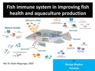
  • 1. Fish immune system in improving fish health and aquaculture production By: Bhukya Bhaskar Fisheries Ref: Dr. Ekaitz Maguregui, 2020
  • 2. Introduction • Traditionally, the immune system is divided into the innate or nonspecific immune system and the adaptive or specific immune system. • innate (non-specific) and adaptive (specific). • Cellular immune responses consist of innate and adaptive cell- mediated immune mechanisms, where all leukocyte subpopulations are included. • Among these are vital processes such as cell-mediated cytotoxicity and phagocytosis. • The main cellular constituents of the fish immune system are macrophages, granulocytes, dendritic cells, NK cells, and cytotoxic T cells. • The fish immune system has both types of immunity, innate immunity, and adaptive immunity. • fish immune system responsible for defending the body from external threats, such as viruses, bacteria, or protozoa and, therefore, it allows to prevent infections.
  • 3. • The adaptive immune system is highly specific to a specific antigen and can provide long- standing immunity. • Several ingredients of the adaptive system, including T cell receptors (TCR), immunoglobulins (Igs), and major histocompatibility complex (MHC), are presumed to have arisen in the first jawed vertebrates. • Teleost antibodies are found in the skin, intestine, bile, gill mucus, and systemically in the plasma. • IL-1β and TNF-α are cytokines implicated in inflammatory response induction in fish. • IL-6 has been reported to be involved in the cascade that induces an inflammatory response • The components of the innate immunity respond to pathogens via identifying pathogen- associated molecular patterns (PAMPs) that demonstrate no expression in host cells. • Among these PAMPs recognized by the innate immune receptors are lipopolysaccharides (LPS), lipoteichoic acid (LTA), phospholipomannan, beta-glucan, chitin, and hemagglutinin. • pathogen recognition, the pathogens are destructed by phagocytosis, one of the effector mechanisms of innate immunity. • Adaptive immunity recognizes pathogens through highly specific receptors, and consequently induces specific B and T cell clone proliferation and differentiation • The immune system of teleost fish is constituted by two receptor categories: the antigen- specific receptors and germline-encoded pattern recognition receptors (PRRs). Numerous other receptors or molecules can participate in the innate immunity
  • 4. Types of immunity • Like upper vertebrates, fish have two types of immunity, innate or nonspecific immunity and adaptive or specific immunity (table 1). • Innate or nonspecific immunity • Innate or non-specific immunity is the first line of defense that the fish’s immune system has to deal with the different pathogens that threaten their homeostasis. This defensive system can be divided into three main components: mucosal immunity, humoral components, and cellular components. • Mucosal immunity plays a key role in the fish immune system, acting as a barrier and preventing pathogens from reaching the body. In addition to the mechanical and physical protection provided by the mucus that coats the body surface of these animals, as well as gills and other tissues, it contains a large number of immune components such as antimicrobial peptides, complement factors and immunoglobulins. • The main difference between innate immunity and adaptive immunity is the specificity with which they are able to recognize different pathogens. Such specificity is determined by the type of receptors they can recognize. • The cells and humoral components that constitute nonspecific immunity recognize these types of molecular patterns: • Pathogen Associated Molecular Patterns (PAMPs): which constitute a set of highly conserved substances/structures in different types of pathogens that are not present in eukaryotic cells, such as cell wall lipopolysaccharides (LPS). In the case of teleostswei, TLR (Toll-like receptors), of which more than 20 different types have been described, play a primary role. • Damage/Danger Associated Molecular Patterns (DAMPs): signals emitted by cells of the body itself, for example, in case of thermal stress. • As in mammals, the innate fish immune system has different humoral and cellular components. Humoral components include antimicrobial peptides, the complement system, lectins, TNF-α and interleukins. • Antimicrobial peptides have been described in multiple aquatic species and are present in the mucus, covering the skin and gills. For example, in the case of the Atlantic cod (Gadus morhua), polypeptides present in the mucus have been described to be effective against Gram + and Gram – bacteria. • One of the defensive mechanisms of the innate immunity is the complement system. This system consists of a set of serum proteins that circulate inactively and are activated in the form of a cascade. As in the case of upper vertebrates, the complement system is activated by the 3-way, the classic, the alternative, and the lectins pathways. All of them lead to the formation of pores in the cell membrane of the pathogen and the consequent death by osmotic shock. One noteworthy difference between the mammalian and the fish immune system is the optimal temperature for this immune complex. In the case of teleost, its activity is maximum between 15-25°C and can remain active at temperatures between 0-4C°, while, in mammals, its optimal temperature is 35°C. • The innate immune system of fish has, at the same time, different cell types that participate in the nonspecific cellular response, including cells with phagocytic activity, such as monocytes and macrophages, and cells with cytotoxic activity, such as granulocytes and non-specific cytotoxic cells. • In the presence of an antigen, neutrophils are mobilized first from the anterior kidney and then the macrophages that mobilize from nearby tissues are attended. Thanks to the phagocytic and cytotoxic activity of these cells, pathogens are eliminated.
  • 5. Fish immune system characteristics, differential characteristics between the innate immune system and the adaptive immune system
  • 6. Cellular Immune Components (source: Doaa M. Mokhtar et al, 2023) • Immune cells in teleosts include natural killer cells, non-specific cytotoxic cells, macrophages, granular leucocytes, thrombocytes, monocytes, dendritic cells, lymphocytes, mast cells, and eosinophilic granule cells. In addition, fish also possess rodlet cells and melanomacrophage centers.
  • 7. 1. Natural Killer (NK) Cells • In teleost fish, there are two different types of NK cell homologs: non-specific cytotoxic cells (NCCs) and NK-like cells . • In contrast to NK cells, which have a large and granular morphology, NCCs are tiny, agranular cells that resemble catfish monocytes. • NCCs in seabream are highly variable in morphology. • The discovery of NK-like cells in teleosts suggests that NCCs are not the same as NK cells in fish. Accordingly, NK-like cells destroy allogeneic and virus-infected cells, according to a few investigations conducted on rainbow trout and catfish . • From teleost NK-like cells, the gene that acts as a cell marker, NK cell enhancement factor (NKEF), has been discovered. After viral and bacterial infection, NKEF gene expression is elevated in tissues such as the skin, gills, and other organs. • Interestingly, a recombinant NKEF protein enhances the cytotoxicity of NCC from the kidney in Nile tilapia. • The NCCs were the first recognized and are the most thoroughly investigated killer cell population in teleosts. The NCCs serve functions like those of the higher vertebrates, acting on various target cells, including virus-infected cells, tumor cells, and protozoan parasites. • NCCs may also be involved in antibacterial immunity by triggering the production and secretion of cytokines . • The NCCs of tilapia and catfish express components of the granule exocytosis pathway of cell-mediated cytotoxicity (CMC) analogous to cytotoxic lymphocytes of mammals. • Non-specific CMC reactions in mammals are mainly performed by NK cells. Non-specific cytotoxic cells (NCCs) and NK-like cells, two classes of NK cell homologues, are responsible for non-specific CMC mechanisms in fish. Fish NK-like cells have been isolated from blood leukocytes and have been shown to kill virus-infected, allogeneic, and xenogeneic target cells on their own. • NCCs, on the other hand, tend to target a variety of cells, including some protozoa and tumor cells, and are particularly active in the spleen and head kidney. NCCs are capable of spontaneously killing the affected cells through necrotic and apoptotic mechanisms. • These cells show variable morphological features, varying from tiny agranular monocyte-like cells in catfish to a mixture of acidophilic granulocytes, monocyte-macrophages, and lymphocytes in seabream. • Moreover, a population of circulating lymphocytes that resemble mammalian natural killer (NK) cells in terms of morphology and functionality has also been reported. • Additionally, NCCs express the NCC receptor protein 1 (NCCRP-1) on their cell surface and present a vimentin-like surface molecule. • It has been shown that catfish NCCs recognize and kill various human cell lines. • Further research reported that granulysin, perforin, and serglycin, gene-encoding molecules with lytic capacity, are expressed by NCCs. • Finally, the fish possess different NCC subsets in several of their immune compartments
  • 8. 2. Macrophages • Macrophages have a pivotal role in specific immune responses because of their function in lymphocyte activation and phagocytosis. Macrophages possess specific receptors capable of recognizing β-glucan, so that the immunostimulants augment leukocytes’ respiratory burst, which produces reactive oxygen species with bactericidal activity. • Another bactericidal mechanism is represented by the production of nitric oxide (NO), which is catalyzed by an NO synthase. Using enzyme histochemical techniques, Schoor and Plumb demonstrated inducible NO production from the head kidney of channel catfish (Ictalurus punctatus) infected with Edwardsiella ictaluri. In addition, Stafford et al. • distinguished the molecules found in crude leukocyte supernatants that stimulate NO production in macrophages of goldfish, indicating that transferrin is an essential mediator for the activation of both fish granulocytes and macrophages. • In fish, neutrophils and macrophages play an essential role in controlling the spread of infectious agents and are accountable for the destruction of phagocytosed pathogens. • Macrophages act as antigen-presenting cells in the distal intestine, enabling antigens to interact with the adaptive immune system for identification. Other granulocytes can also be identified in the intestinal segments for innate clearance, in addition to the resident macrophages. • The lamina propria and epithelial linings of the intestine contain these innate populations, allowing a close vicinity to the digested pathogens. • Macrophages express several receptors on their cell surface, including TLRs, PRRs, and CLRs, in addition to complement and scavenger receptors. Macrophages are also an essential source of chemokines and cytokines, linking innate and adaptive immunity, which mediates an efficient immune response. • Moreover, macrophages are important for antigen presentation to T cells. • Circulating monocytes are typically CD14+, and express chemokine receptors, TLRs, adhesion molecules, and surface molecules that are involved in the pathogen-associated molecular pattern recognition at sites of inflammation and/or infection
  • 9. 3. Neutrophils • In the blood, peritoneal cavity, and lymphoid organs, neutrophils are polymorphonuclear cells that have the ability to phagocytose cells or foreign particles and manufacture superoxide anions, a bactericidal component. • Neutrophils play an essential role in the inflammatory immune response against various viral, bacterial, fungal, and protozoan pathogens. • Neutrophils are the first granulocytes to be detected at the injury site, followed by macrophages, both of which are directed by chemotactic factors produced by wounded tissue. At the site of injury, neutrophils use antimicrobial peptides, proteolytic enzymes, and reactive oxygen species (ROS) to phagocytose microorganisms and destroy them. • Additionally, fish neutrophils have the ability to release extracellular fibers known as neutrophil extracellular traps (NETs) that include DNA, histones, and proteins that can bind, kill, and inactivate viruses, bacteria, fungi, and parasites. • Furthermore, the granular leukocyte-derived myeloperoxidase enzyme can interact with hydrogen peroxide to create hypochlorite, which in turn can be used to produce cloramins, oxidative substances that can attack microorganism membranes
  • 10. 4. Eosinophils, Basophils, Thrombocytes, and Monocytes • Eosinophils are widely distributed cells in connective tissue, particularly in the gastrointestinal tract, bloodstream, ovaries, and gills, and offer degranulation when parasite infections are present. • Basophils are large polymorphonuclear granular leucocytes that are rarely observed in teleost species. • Their cytoplasmic granules have an inflammatory mediator, histamine. These cells are involved in allergy and anaphylaxis. • Thrombocytes are oval-shaped, nucleated, and agranular cells. • They possess both a coagulation function and phagocytic ability. • They show acid phosphatase activity that causes cells to gather at the inflammatory site. • The monocytes show phagocytosis and non-specific cytotoxic activities. • These cells are thought to be transient blood cells because they migrate to the connective tissue during the inflammatory phase and change into macrophages. • Professional phagocytes that can be deployed to the site of inflammation include monocytes, macrophages, granulocytes, and dendritic cells
  • 11. 5. Dendritic Cells (DCs) • The DCs are among the antigen presenting cells found within several tissues and can initiate both innate and acquired immune responses. • DCs are characterized by their small cell bodies with many cytoplasmic processes (dendrites) and have been found in gills, gut, skin, and ovaries. Cell populations with DC-like functions and morphology have been demonstrated in many fish species. • With the assistance of MHC class 2 receptors, these cells deliver the processed antigens to T lymphocytes, triggering the cell-mediated acquired response. These cells appear to activate T cells in an antigen-dependent manner in zebrafish by expressing genes associated with antigen presentation, such as MHC class II invariant chain iclp1, csf1r, and il12. • The trout gut has been shown to include putative CD8 + DC-like cells that appear to have phagocytic activity and the capacity to excite naive T cells. • Recently, Alesci, et al. identified the intestinal DCs in several fish species including Scyliorhinus canicula (Chondrichthyes), Eptatretus cirrhatus (myxines), Polypterus senegalus (Osteichthyes, Brachiopterygii), Clarias batrachus (Osteichthyes, Teleostei), and Lepisosteus oculatus (Osteichthyes, Holostei). • These findings showed that intestinal DCs and DC-like cells were positive to TLR-2, Langerin/CD207, and MHC II.
  • 12. 6. Lymphocytes • B lymphocytes, which are professional antigen-presenting cells (APCs) produce antibodies against antigens and mediate humoral immunity. They also develop immunological memory by differentiating into memory B cells. • A key location for B cell production and development into mature naive B cells is the fish head kidney. Immunoglobulins are secreted into the plasma by B cells to form an antigen-binding complex. • These secretory cells, also known as antibody secreting cells (ASCs) or plasmablasts, resemble mammalian plasma cells. • When B cells come into contact with antigens, activation occurs, which can be either T cell-independent (for polysaccharides- and unmethylated CpG DNA-derived antigens) or T cell-dependent (for protein-derived antigens). • Once an antigen is bound by the BCR on an antigen-presenting B cell, the antigen is processed and delivered to T helper cells by MHC class II on the surface of B cells. T helper cells are then activated and differentiated as a result, and they subsequently express the surface protein cluster of differentiation 40 ligand (CD40L). When CD40L interacts with the B cell surface protein CD40, it causes naive B cells to activate and differentiate. • Furthermore, T cell activation is not necessary for T cell-independent B cell activation, which is triggered by the direct binding of an antigen to the BCR. • Activated B cells can either differentiate into memory B cells, which circulate throughout the body and are in charge of secondary antibody response, or into plasmablasts, which are proliferating, short-lived, and low affinity antibody-producing cells that later differentiate into plasma cells, which are not- proliferating, long-lived, or high-affinity antibody-producing cells • When memory B cells come into contact with the same antigen, which activates their parent B cells since these two share the same surface BCR, a secondary response is generated. • In the cell-mediated adaptive immune system, T cells play an inherent role in directing immune activity after the early non-specific defenses and offer an immunological response after antigen identification by APCs. The T cell may then direct the clearance of the foreign material by phagocytosis or cytokine stimulation after pattern recognition through the TCR. As a result, specific antigen–receptor interactions and the MHC pairing stimulate the cellular responses of the two main groups of T cells, T helper cells and cytotoxic T cells. • T cell differentiation and control are influenced in distinct ways by surrounding immunological molecules. While cytotoxic T cells are controlled by cytokine concentrations and surrounding helper T cells. T helper cells are affected by antigen binding affinity, antigen concentrations, and signaling from APCs Moreover, the release of IL-2 acts as a growth promoter for the overall development of T cells . • After differentiation, helper T cells produce lymphotoxin (LT) to interact with intracellular pathogens and release IFN-γ to help in resisting viral infections . • The helper T cells can be further classified into Th1 and Th2 according to their functions and target pathogens. The Th1 subset offers cell-mediated removal of microbes. On the other hand, the Th2 cell tends to target particular pathogens, such as parasites • In teleosts, T cells are correlated to the mammalian subset and possess complementarity-determining region 3 (CD3) binding domains within the TCR • The head kidney and thymus nurse the developing T cells and act as a base for plasma circulating T cells. Salmonid CD3 was proven by Koppang et al.to be an effective T cell marker to assess the anatomical distributions of T cells and expression regions • Numerous T cells have been reported to populate the thymus before migrating to the kidney and spleen after development . • The antigen presentation of both MHC class I and II has demonstrated the directional activation of a T cell response, in addition to T cell characterization. MHC class I interacts with CD8 + cytotoxic T cells, whereas MHC class II helps with complex formation in T helper cells
  • 13. 7. Mast Cells • In most teleost species, mast cells are a constituent of innate immunity and are located near the blood vessels in the ovaries, skin, intestine, and gills. • The mast cell granules express S100 protein, desmin, and CD117. • Moreover, their granules are stained positively with bromophenol blue and Safranin O. Using electron microscopy, these cells are characterized by the presence of numerous cytoplasmic electron-dense granules. • Several stress conditions, such as exposure to herbicide and heavy metals, chronic inflammation, and parasitic infections, cause an increased number of mast cells in the organs and tissues of teleosts. • Teleost fish have also demonstrated mast cell degranulation, cytokine release, and the ensuing inflammatory response after bacterial infection. • Piscidins, host-defense peptides, were initially discovered in hybrid striped bass. It is interesting to note that piscidins are found in mast cells, highly prevalent granulocytes that are present in all vertebrate classes • The recent localization of ACh in mast cells and the pavement cells, as well as the interaction of the mast cells with eosinophils and the innervation of the latter by nicotinic acetylcholine receptor (nAChR) alpha 2 subunits, were correlated with the modulation of the immune function in fish gill. • Both muscarinic and nicotinic cholinergic receptors have been identified on mast cells
  • 14. 8. Eosinophilic Granule Cells (EGCs) • Salmonids and cyprinids are among many species that frequently possess EGCs. EGCs are typically present in the skin’s epidermis, gills, connective tissue surrounding the spinal cord, and intestinal mucosa. • In the connective tissue stroma surrounding the bile duct, EGCs can also be identified. • They have spherical granules in their cytoplasm that are brightly stained red by hematoxylin and eosin (HE) because they contain basic proteins. • Additionally, their granules are stained deep magenta with PAS and alcian blue, due to their content of neutral and sulfated glycosaminoglycans. • They exhibit a positive reaction to bromophenol blue, which shows that they contain protein, and they show a metachromatic affinity to toluidine blue. • Many functions are reported for these cells, including the release of oxygen radicals and toxic proteins onto the surface of multicellular parasites in inflammatory areas. • Additionally, acute tissue damage results in EGC degranulation and release of inflammatory mediators, but chronically inflamed tissues frequently exhibit an increase in the number of these cells
  • 15. 9. Rodlet Cells • Fish only have rodlet cells, which are distinguished by their thicker capsule-like cell borders and rodlet cytoplasmic inclusions. • The rodlet cells of the grass carp head kidney expresses α- SMA. • Rodlet cells possess an actin cytoskeleton, which may contribute to the contraction that ejects their contents. During the expulsion phase, they go through a one-way contraction that eventually destroys the cell; the rodlets and all the contents of the cell are evacuated. • The rodlet cells are frequently distributed in the heart, thymus, kidney, spleen, gills, skin, gall bladder, liver, blood vessel endothelium, and pancreas. • Several functions have been reported for these cells, including a secretory function, which is a defensive role, especially against parasitic helminths. • They may play a role in water or electrolyte transport or lubrication, pH control, and antibiotic reactions. They may be modified goblet cells or may play a regulatory role in osmoregulation and transportation of ions. They can also be viewed as non-specific immune cells that are implicated in immunity, as they frequently occur in parasitic infestation. Rodlet cells, on the other hand, may be triggered because of tissue damage, simulating leukocyte reactions to various chemotactic stimuli. • Moreover, rodlet cells can be regarded as a type of eosinophilic granule cell or mast cell. ]The nature of their contents must be determined to throw additional light on the functions of RCs. • The molecular structure of RCs has been investigated using lectin histochemistry and immunohistochemistry to clearly distinguish rodlet cells from mucous cells, macrophages, and granulocytes. • Complex carbohydrates were detected in the RCs of fish, primarily glucosidic residues of N- acetyl-galactosamine and, less commonly, N-acetyl-glucosamine and sialic acid. • Sialic acid residues in the carbohydrate backbone help the mucosae to defend themselves against infections
  • 16. Mucosal Barriers • Fish can interact with their surroundings through mucosal barriers while still maintaining homeostasis. Mucosal barriers are extremely complex tissues with special adaptations that enhance a variety of physiological functions, including immunity, gas exchange, and nutrition. • The principal mucosa-associated lymphoid tissues (MALTs) of teleosts are the gut-associated lymphoid tissue (GALT), gill-associated lymphoid tissue (GIALT), skin-associated lymphoid tissue (SALT),) and nasopharynx-associated lymphoid tissue (NALT). • These component mucosal barriers serve as the host’s local immune system and protect it from pathogens and other threats. The fish MALT possesses the major populations of innate immune cells, as well as the lymphocyte populations of CD4 and CD8 T cells, and B cells. • However, only three immunoglobulins’ (Igs) isotypes have been discovered in teleost to date (IgM, IgD, and IgT/Z), each of which consists of two identical heavy and two identical light polypeptide chains. • IgT/Z is thought to be specialized in the mucosal immunity. In mammalian mucosal surfaces, the main immunoglobulin is IgA, which is mostly produced by plasma cells present in the gut lamina propria. In a similar way, the teleost IgA homologue, IgT/IgZ, has a dominant role in gut mucosal immunity. • The transport of IgM and IgT through mucosal barriers is carried out by the polymeric immunoglobulin receptor (pIgR), which is also expressed in the gut and skin of teleosts. • In zebrafish and rainbow trout, dendritic-like cells have recently been identified. • It is intriguing that DCs only make up around 15% of the mononuclear phagocytes in the skin of adult zebrafish but are sparse in the gut, spleen, and kidney. • It has been hypothesized that zebrafish skin has DCs comparable to mammalian Langerhans cells. • Additionally, innate immune genes such as TNF have been found in the mucosal tissues, indicating that innate immunity has a role in the antiviral process
  • 17. Schematic representation of the fish mucosal immunity (source: Doaa M. Mokhtar et al, 2023)
  • 18. • 1. Fish Mucus • Fish mucus that covers the mucosal surface is produced by many types of secretory cells, mainly the goblet cells (GCs), sacciform cells, and club cells. Goblet cells, which produce mucus granules and contain glycoproteins, are prevalent on all external surfaces, as well as the surface of the gills. • The initial line of defense against pathogen invasion is fish mucus. • Mucins are the major molecules found in mucus and are responsible for the mucus viscosity, pathogen entrapment, physical protection of the skin’s surface, and signaling at the cell surface. • Mucins are a heterogeneous family of high molecular weight glycoproteins with single or many protein domains that have numerous locations where O-glycan attachment can be formed. • Mucins contribute to the innate immune system in two ways that are significant. • First, they prevent the adhesion of pathogens, sustained colonization of potentially infectious bacteria, and invasion of parasites by being continuously generated and routinely shed. • Second, they contain a variety of innate immunity-related proteins and enzymes, including lysozyme, esterases, complement proteins, lectins, C-reactive protein (CRP), transferring, alkaline phosphatise (ALP), proteases, immunoglobulins, and antimicrobial peptides (AMPs), and various other antibacterial proteins and peptides, which are frequently responsible for deteriorating, inactivating, and controlling infections. • In addition, the fish skin mucus has been reported to have few metabolites with antibacterial properties such as N-acetylneuraminic acid, azelaic acid, and hydroxyisocaproic acid
  • 19. Cont... • According to reports, fish skin mucus contains a variety of saturated fatty acids (SFAs), monounsaturated fatty acids (MUFAs), and polyunsaturated fatty acids (PUFAs). Palmitic acid and stearic acid are SFAs that are present in mucus. Oleic acid is an example of a monosaturated fatty acid. • Moroctic and alpha-linolenic acids are polyunsaturated fatty acids. These fatty acids are thought to perform a significant defensive function against pathogens • Skin transcriptome analysis of S. trutta, M. anguillicaudatus, and C. striatus demonstrated that several genes are implicated in immunology and epidermal mucus secretion. • Mucins are divided into two structurally separate families: membrane-bound and large secreted gel forming (SGFM) forms, and, to date, more than 20 mucin genes have been identified in higher vertebrates. Cystein- rich (C8) and trypsin inhibitor-like cysteine-rich (TIL) domains are two of the domain structures that distinguish the secreted gel-forming mucins from other types. • These domains aid in the disulfide bond formation that leads to the oligomerization of the mucin proteins, which gives mucus its ability to gel. • Additionally, long repetitive sequences rich in proline, threonine, and serine residues, known as the PTS- domain, are present in the released gel-forming mucins. • MUC2, MUC5AC, MUC5B, MUC6, and MUC19 are examples of SGFM. However, the membrane-bound mucins include MUC1, MUC3, MUC4, MUC12, MUC13, MUC14, MUC15, MUC16, MUC17, and MUC18 • The mucin genes of a few teleost fish species, such as zebrafish (Danio rerio), gilthead sea bream (Sparus aurata), common carp (Cyprinus carpio), and sea trout (Salmo trutta), have been discovered. • Seven secreted gel-forming mucin genes were discovered in the Atlantic salmon based on many levels of evidence, including annotation, transcription, phylogeny, and domain structure. • Five genes were labelled as muc5 and two as muc2. • The muc5 genes were largely expressed on the epidermis and gills, while the muc2 genes were primarily expressed in the intestinal region. • The mucus cells and the composition of the mucus are influenced by several exogenous (e.g., hyperosmolarity, stress, infections, and pH) and endogenous (e.g., developmental stage, sex) factors.
  • 20. • The properties of the mucus vary among fish species, and they are subject to change in response to disease and environmental factors. Amoebic gill disease (AGD) is one of the main disorders affecting the mariculture of Atlantic salmon (Salmo salar L.). • AGD is characterized by increased gill mucus production and hyperplasia of the lamellar epithelium. • The regulation of two mucins (muc5, muc18) changed in response to illness. The secreted muc5 was significantly upregulated in AGD, but the membrane-bound muc18 showed the reverse pattern. • Surprisingly, the properties of the skin mucus of carp appear to alter in response to increases in the total amount of bacteria in the water. Increases in acidic glycoconjugates and overall glycosylation can be noticed as changes in the terminal presence of certain sugars. • Similar to this, myxozoan parasite infection alters the characteristics of seabream (Sparus aurata) gut mucus, resulting in higher levels of glycosylation and terminal glycosylation of mucus proteins in the posterior gut. • The fish mucus has a wide range of functions, including protection, respiration, disease resistance, as well as osmotic regulation, excretion, reproduction, communication, nest building, and feeding. • Fish mucus extract is increasingly known to possess a wide range of biological qualities, for instance, antiviral, antibacterial, antiparasitic, and antifungal qualities, which may be employed in both human medicine and fish farming. • Sacciform cells and acidophilic granular cells (orserous goblet cells) have also been demonstrated in fish skin whose secretions mix with the secretions of goblet cells to form mucus. Sacciform cells may be analogous to granular glands of amphibians, which produce crinotoxic and repellent substances, but secretions of these cells also have a protective and regulatory role. • On the other hand, the adaptive immune components include Igs, which are expressed on the B cells’ surface as B cell receptors or in a soluble form in body fluids. • |These mucosal Igs (secretory Igs “sIgs”) are widespread in the mucosal barriers of teleosts, including the gut, olfactory organ , gills , skin, and buccal and pharyngeal cavities • Recently, a bursa of Fabricius-like structure in Atlantic salmon (Salmo salar) cloaca was identified where IgT+ and IgM+ B cells could be distinguished, which indicates that sIgs may be secreted into the mucus around this structure. • The expressions of constituents involved in the cholinergic system, including acetylcholinesterase (ChAT) and vesicular ACh transporter (VAChT), have been confirmed in various immune cells including macrophages and epithelial cells. • Several studies have demonstrated that most epithelial cells do not concentrate ACh into vesicles; instead, they release it directly from the cytoplasm
  • 21. • 2. The Intestinal Mucosa • The intestinal mucosa of teleosts is a saturated substrate for leukocytes, and they are housed within the lamina propria. • These strata contain proliferating granulocytes, lysozyme, and NK cells, which together promote the production of lysozyme and superoxides. • The epithelial cells also play the role of a secondary barrier, securing the passage while permitting proper nutrient flows, signaling permeability, and benefiting gut microflora to improve digestion. • The intestinal regions possess an essential population of immunoglobulins and isolated populations of both T and B cells, called intra-epithelial lymphocytes (IELs). • Dezfuli et al. have found that mast-cell analogues have a role in the indirect propagation of immunoglobulins following viral infection. Inami et al. found a larger number of IgM-positive cells in the rectum of Atlantic cod (Gadus morhua), compared with the stomach regions and foregut. • This result agrees with the familiar adaptive immunocompetency of the posterior intestine and demonstrates the capability of Igs to aggregate in specific regions away from common GALT. • As fish lack IgA, serum or secretory immunoglobulin, IgM is rapidly degraded within the harsh environment
  • 22. Immunoglobulins • In bony fish and other jawed vertebrates, the adaptive immune system relies on B and T cells and on the tremendous variety and specificity of their antigen receptors, the immunoglobulins (IG) or antibodies, and the T cell receptors, respectively. • Immunoglobulins are the main constituents of the immune response against pathogens. Among the mechanisms implying humoral immunity mediated by IGs are pathogen elimination via phagocytosis, toxin and virus neutralization, and complement cascade activation. • In bony fish, three isotypes of IGs are produced by B cells: IgD, IgM, and IgT/Z. Therefore, three major B cell lineages have been characterized in teleosts. • The predominant and most ancient immunoglobulin in fish is the IgM class tetramer, which contains eight antigenic combining sites. • Although IgM can be expressed at the surface of B cells, secreted tetrameric IgM is considered the most predominant IG in fish serum. • Many studies have reported that mucosal and serum noncovalently accompanies IgM monomers in diverse fish species • In rainbow trout, a new teleost fish Ig was detected and was named IgT. This new IG was identified as IgZ in zebrafish • This IgT has been distinguished in most studied teleost fish, except for medaka, channel catfish, and turquoise killifish • In rainbow trout, at least three subclasses of IgT are expressed. Mucosal and systemic lymphoid tissues have been reported to express the IgT1 subclass, while IgT2 has been documented to be mainly expressed in systemic lymphoid organs. Furthermore, IgT3 protein is reported in rainbow trout serum. • IgD is the second immunoglobulin isotype characterized in fish, specifically channel catfish. As a result of its sequence similarity with mammalian IgD, its immediate location under the IgM gene, and its expression on B cells, IgD has been suggested to be as old as IgM • The secreted form of IgD has been interestingly identified in trout and catfish, with a different structure
  • 23. Cytokines • Cytokines play an essential role in the immune system through their specific receptors binding to the cellular membrane, providing a cascade-enhancing induction, stimulation, or suppression of nuclear cytokine-regulated genes. Several critical cytokines have been recognized in teleost fish, including tumor necrosis factor-α (TNF-α), interleukin-1β (IL-1β), transforming growth factor-β (TGF-β), interferon (IFN), and many chemokines , recently numerous other cytokines have been identified in teleosts . • The interferons (IFNs) are the key cytokines present in all jawed vertebrates and are involved in antiviral immunity. • In vertebrate host cells, INFs play a critical role in the defense mechanism against viral infection through INFα/β secretion upon the recognition of viral nucleic acid • These INFs keep other cells away from viral infection via binding to different receptors, resulting in the induction of multiple genes, where some of these genes encode antiviral proteins. • The IFNs have been categorized into three broad groups, where type I and III are originally embroiled in antiviral defense, while type II IFN has a broader function and shares in cell-mediated immunity in response to a wide variety of pathogens during adaptive immune responses. • IL-1β and TNFα are considered the proximal cytokines that are first released and boost response through enhancing the secretion of distal cytokines such as IL-6 and IL-8. All these cytokines have been detected in fish, where IL-1β, IL-6, and TNFα, with their family members, have been reported in several fish studies investigating inflammatory responses against infection . • IL-1β was one of the first characterized cytokines in teleosts. • As reported in zebrafish, IL-1β is produced as a precursor molecule that is cleaved to release a bioactive mature protein, which is linked to the activation of the inflammasome complex that is accountable for the secretion of IL-1β • IL-1β is produced by various cell types following the stimulation of host pattern recognition receptors through PAMPs or DAMPs. • IL-1β improves vaccine efficacy as it boosts antibody production when administered with bacterial vaccines. • In primary leucocytes and macrophages, IL-1β action has been extensively analyzed, where induced expression of pro- inflammatory genes such as TNF-α, IL-1β, and IL-6 is evident . • Furthermore, IL-1β activates the expression of genes that are considered to be immune response suppressors IL-1β genes have revealed wide variations among fish species. In carp, two IL-1β genes were found: IL-β1 and IL-1β2. Three functional IL-1β genes (IL-1β1–3), in addition to pseudogene (IL-1β4), have been identified in salmonids , while only a single type II IL-1β was detected in gadoids.
  • 24. Cont.. • IL-18 is an essential cytokine responsible for the induction of IFN-γ production and promotion of TH1 immunity in vertebrates. It exerts an essential role in regulating inflammation within mucosal tissues. • IL-18 has been described in a few fish species including elephant shark, pufferfish, rainbow trout, and seabream . • IL-18 is fundamentally expressed in fish immune or non-immune tissues, unlike IL-1β, which is induced by inflammatory stimuli • A recent study has suggested that IL-18 is likely developed in an evolutionary-independent event to IL-1β. • TNFα is a member of the TNF superfamily, which primarily comprises type II transmembrane proteins, which can be cleaved to release a soluble cytokine. TNF-α presence has been reported in teleosts • In carp, many TNF-α copies have been detected • In catfish cell lines, TNF-α expression has been observed in T cells and macrophages; however, there are no expressions in B cells or fibroblasts • n salmonids, four copies of TNF-α have been reported. • Several fish studies have suggested that TNF-α and -β are essential macrophage activators. Previous studies in some fish species, including goldfish, rainbow trout, sea bream, turbot, and catfish, have revealed that TNF induces macrophage activation, resulting in increased respiratory activity, phagocytosis, and nitric oxide production. • IL-6 was first discovered in teleost fish. • It is a member of the IL-6 family, and is considered a pivotal acute phase response cytokine. Moreover, it is implicated in the differentiation of B cells into antibody-secreting cells. It is highly expressed in the kidney and spleen, and during caudal fin regeneration following amputation. IL-6 has been used as a vaccine adjuvant, to achieve higher serum antibody levels for protection. In salmonids, two IL-6 genes have been detected (IL-6A and IL-6B) • IL-11 is one of the other members of the IL-6 cytokine family related to inflammatory responses. It was first detected in teleosts in the bacterial-challenged fish. Two IL-11 types (IL-11a, IL-11b) have been reported in teleosts, where IL-11a is highly expressed in the intestine and gills of trout. • TGF-β is one of the TGF-β protein family. It plays a fundamental role in various functions, including cell proliferation, differentiation, and regulation. In fish, three TGF-β proteins have been detected (TGF-β1, TGF-β2, TGF-β3). TGF-β1 is the main immune active isoform, and two TGF-β1 paralogues (TGF-β1a and TGF-β1b) have been found in zebrafish. In carp, TGF-β1 is highly expressed in the spleen, and it can induce expressions of pro inflammatory cytokine and IL-10. In rainbow trout, three paralogues of TGF-β1 have been reported, with expression highest in lymphoid tissues. The highest levels of TGF-β1 expression have been found in macrophages and the gills of seabream
  • 25. Immune Cells—Nerve Interaction • To monitor infections and inflammation and to take appropriate action, the neurological and immune systems work in concert as an integrated physiological system. The nervous system, including the brain and peripheral divisions, can stimulate or inhibit a variety of immune system functions, including those of the innate and adaptive immune systems. It has been established that the anti-bacterial peptides (AMPs), which are signaling molecules involved in the antimicrobial immunity, are synthesized by the enzymatic processing of neuropeptide precursors such as proenkephalin A (PEA) and chromogranin B. • It has been shown that some peptides involved in neuronal or neuroendocrine signaling also have potent antibacterial activities. Additionally, they are widely distributed across immunological, endocrine, neuroendocrine, and neuronal cells. • The microbiota–gut–brain axis (MGB axis) is based on the reciprocal interactions between the CNS and gut microbiota that involve endocrine, immunological, and neurological pathways. • in the MGB axis, lymphocytes stimulate the gut lumen and internally release cytokines, which are then activated by gut peptides released by enteroendocrine cells from sensory nerve terminals, such as those of the vagus nerve. To create the link between the immune system and the central nervous system (CNS), which results in tissue homeostasis, communication pathways based on neurotransmitters, neuropeptides, cytokines, hormones, and growth factors are required . • The innervation of lymphoid tissue has been investigated in the Coho salmon (Oncorhynchus kisutch) spleen, where nerve fibers are accompanied by the vascular supply and MMCs. Additionally, adrenergic receptors are among the neurotransmitter receptors that are expressed by immune cells. Adrenergic receptors have so far been sequenced in the leukocytes of zebrafish, trout, catfish, and goldfish. • The activation of the sympathetic nervous system can modulate the production and circulation of leucocytes, through activation of hematopoietic stem cells and NK cells, upregulation of myelopoiesis, and production of monocytes and neutrophils by the spleen. • Gut neuropeptides, such as gut antimicrobial neuropeptides, constitute a complex network between the neurological and immunological systems, where they play a crucial modulatory role. I • n response to various stimuli, both enteroendocrine cells and enteric neurons release gut neuropeptides. Most neuropeptides created by neurons during an immune response have neuroendocrine properties that can affect both the stomach and the brain. • The role of the mast cells, mucous cells, and neuroepithelial endocrine cells (NECs) of teleosts’ gills in innate and adaptative immunity has been examined in several studies. These substances, such as immunoglobulins (Igs), antibody-secreting cells, and piscidins, are produced by these molecules or cells through immune-related pathways. • Piscidin 1, an antimicrobial peptide, and enkephalin, were found to be co-localized in fish gill NECs and the neurological system of the gill filaments, suggesting a potential role in the bidirectional relationship between endocrine, immune cells, and their interaction with the filament nervous system.
  • 26. Cont... • Investigations have been conducted into the possibility of tight communication between immune cells and afferent nerve terminals in the gill. The eosinophils in the gill lamellae of Boleophthalmus are immunostained with antibodies against Pis 1 and 5HT (serotonin), and they come into contact with neurons that are also immunolabeled with antibodies against acetylated tubulin. • Many neuroactive substances and AMPs, including 5-HT, piscidin 1, nitric oxide (NO), tyrosine hydroxylase (TH), and the nicotinic acetylcholine receptor, are found in the terminals of afferent nerves. These substances are released by mast cells and eosinophils. Additionally, it has been discovered that eosinophils and NECs include neuropeptide receptors, such as the GABA B R1 receptor, which suggests that the immune and neurological systems interact closely. • During lung infections, neuro-immune interactions also take place at the surface of the lung air barrier. Bacterial pathogens are killed by the neuropeptide calcitonin-gene related peptide (CGRP). In addition to sensory nerves, the pulmonary neuroepithelial endocrine cells (PNECs) also express CGRP. PNECs have been identified to be co-localized with innate lymphoid cells (ILC2). • Cytokines or other mediator molecules stimulated by cytokines would be accessible to the fish brain. However, the preoptic region of the carp brain contains the cytokine receptor IL-1RI mRNA • Prostaglandins, which are smaller, neuroactive, and lipophilic, can be the mediators. • There is evidence that immune mechanisms that signal to the brain in fish are similar to those in mammals. It has been suggested that the vagus nerve could act as a channel for cytokines to reach the brain. • However, because stresses can increase peripheral cytokines by stimulating pathways such as the noradrenergic pathway, these, in turn, may cause a rise in brain IL-1 via one of the immune to brain pathways. • The role of cytokines in the activation of the HPI and sympatho-chromaffin (SC) axis and the central regulation of the stress reaction in fish is not well understood because there have been few studies that have demonstrated the signaling pathways from peripheral blood molecules to the brain. Adrenaline has been demonstrated to modulate cytokines in fish, which can cause the signaling pathways to the brain to activate. Pro-inflammatory cytokines in the brain decrease the activity of neurotransmitters such as noradrenaline, serotonin, and dopamine, which will have a substantial impact on how behavioral patterns are modulated. • To further integrate peripheral pro-inflammatory and antigenic signals with physiological and behavioral response, cytokines also activate a number of other signaling pathways in the brain. • Under stressful conditions, all these processes will cooperate to combine immune cells’ necessary protective response with the physiological and behavioral response. • Finally, other forms of indirect control, such as those connected to the relationship between the gut and the brain, can also exist in fish. It has been demonstrated that information is transmitted between the two organs via chemical signals in both fish and mammals. As a result, when the neuro- immuno-endocrine relationship is effectively initiated, other processes such as reproduction or growth will also be impacted. The Sockeye salmon (Oncorhynchus nerka) provides a clear case study, as degeneration of various glands and organs has been linked to extremely high steroid levels and the loss of immunocompetence, particularly innate responses, during the migratory period. • In order to gather energy that ensures the fish can spawn, cortisol and other reproductive steroids redirect energy from numerous biological processes, including immunological functions. Sex steroids have also been found to modulate the cortisol production in the internal tissues of salmonids
  • 27. Adaptive or specific immunity • The specific or adaptive immune system is present in cartilaginous fish and has been described in all jawed fish. • Like mammals, this specific response in fish starts from the detection of the molecules of the major histocompatibility complex (MHC) by T cells. • The MHC molecules are a superfamily of immunoglobulins that act as receptors. • Unlike higher mammals, where MHC genes are joined into a single chromosome, in the fish immune system these genes are not connected and can be found in different chromosomes. • The two types of MHC receptors described in upper vertebrates are present in teleosts. • The MHC I molecules, which are related to endogenous antigens, are present in all nucleated cells and are recognized by CD8 or cytotoxic T cells. • The MHC II molecules are related to exogenous antigens and are only found in antigen-presenting cells (dendritic cells, macrophages, and B lymphocytes). • B lymphocytes are responsible for the production of immunoglobulins, which allow this immune system to have a certain “memory”, causing a more intense and lasting response in a second exposure to the same antigen. • Activation of B lymphocytes requires two steps. On the one hand, they must recognize external antigens through type II MHC, and, at the same time, such antigens have to be presented by a CD4 T lymphocyte or helper. • Once activated, B lymphocytes are transformed into plasma cells capable of secreting the different types of immunoglobulins.
  • 28. Process of activation of B lymphocytes in plasma cells
  • 29. Cont... • Teleosts are able to synthesize 3 types of immunoglobulins: IgM, IgD and IgT/IgZ, unlike upper vertebrates, where 5 types of immunoglobulins are described. • IgM is the most abundant immunoglobulin in the fish immune system and, as in mammals, is able to pass through epithelial membranes. • The IgM of teleosts is a tetramer consisting of two heavy chains and two light chains (2H:2L). • This immunoglobulin plays a key role in the innate immune system against bacteria and viruses, as it is responsible for fixing the complement, opsonization and activation of the cytotoxic response, among other functions. • The location of immunoglobulins is specific in certain parasitic infections, for example, in trouts affected by Ceratomixa shasta, a greater increase in IgT/IgZ was observed in the intestinal mucosa, while the IgM title increased significantly in the serum. • These assays seem to indicate some affinity in the location of immunoglobulins, as IgM are more abundant in serum and IgT/IgZ are the most abundant in mucous- associated immunity.
  • 30. Stressors and stress response • Stress, especially chronic stress, is known to have a negative effect on different productive species, including aquaculture species, because the immune response and disease resistance decrease. • Stressors in fish can be divided into three large groups, environmental factors, social and reproductive factors and physical or management factors. • Environmental factors include sudden changes in temperature and oxygen, as well as changes in water quality and high culture densities. • Social factors refer to the establishment of a dominance hierarchy within cages or ponds. The reproductive season has also proven to be a very stressful period for aquaculture species and should certainly be taken into account in production systems. • There are physical or handling factors that can affect fish immune system, including the transport of animals, both within the same facility and between different facilities, and tasks such as tank cleaning, vaccinations, and animal classifications. • The stress state can be divided into three stages: initial stage, endurance stage and exhaustion stage. When an animal is exposed to a stressful agent, different signals are sent from the sensitive organs that reach the central nervous system, activating the hypothalamus- hypophysis axis. • Activation of this nervous axis causes the release of catecholamines and cortisol, secreted by chromaffin cells and interrenal cells, respectively, located in the kidney of these aquatic animals. • Catecholamines and cortisol are responsible for the immune system’s response to stress. • The secretion of these molecules prepares the animal to face a possible challenge by increasing heart rate, blood flow, energy availability (glucose levels) and stimulating the immune response. • If the stressor persists, we enter the resistance phase, during which changes occur in metabolism and enzymatic secretion that allow to maintain the alarm state. • However, if the stressful stimulus continues for a long time, the animal is not able to maintain this stage, and its immune response decreases, which is known as the exhaustion stage. • At this stage, the animal is not able to continue producing high levels of the molecules involved in defensive mechanisms (lysozyme, complement system, IgM, leukocytes, etc.). • In Table 2, we can see how the number of leukocytes increases when animals are subjected to acute stress and, if stress is sustained, the number of white blood cells decreases, making animals more susceptible to pathogens. •
  • 31. Effect of stress on immune cells. ( Source: Barcellos et al., Aquaculture 237 (2004) 229–236) • These stressors have a negative impact on aquaculture production. On one hand, they decrease animal growth rate, because stressed animals ingest a smaller amount of food. • On the other hand, stress states generate immunosuppression in fish, making them more susceptible to the microorganisms present in the environment.
  • 32. Example: Immune system of Tilapia • Innate immune defences of tilapia include physical barriers and various cellular and humoral components. • Skin, scales and the epithelial layers of gills and the gastrointestinal tract act as physical barriers, helping to prevent the entry of pathogens into the fish. • The mucus covering these surfaces also acts as a physical barrier by trapping pathogens and contains various antimicrobial substances such as lectins, lysozymes, complement proteins and antimicrobial peptides (AMPs) that can neutralize and kill the pathogen
  • 33. Cont... • If the pathogen is able to breach the physical barriers and enter the host, humoral and cellular components of the innate immune system respond in an attempt to prevent the infection from progressing. • The humoral response uses a diverse range of antimicrobial components (e.g., AMPs, lysozyme, complement proteins and acute phase proteins) to directly destroy the pathogen, or to promote inflammation and phagocytosis by cellular components of the innate immune response. • Several studies report on the functional characterization and activity of complement in tilapia. • Measurement of lysozyme activity is often used as an indicator of the innate immune response in tilapia to various stimuli. • Over 90 AMPs have been identified in teleost fish, including molecules like β-defensins, cathelicidins, hepcidins, histone-derived peptides and fish-specific piscidins. • Nile tilapia β-defensin has been shown to have an inhibitory effect on the growth of Escherichia coli and Streptococcus agalactiae. • The therapeutic potential of piscidins,including tilapia piscidins, has recently been reviewed by Hazam and Chen and Raju et al. • The cellular components of the tilapia's innate immune response consist of monocytes/macrophages and granulocytes, that is, neutrophils and eosinophilic granule cells. Initiation of the cellular response is mediated through the binding of pattern recognition receptors (PRRs) on these cells to pathogen- associated molecular patterns (PAMPs) located on different microbial pathogens, including viruses, bacteria, fungi and parasites, or to danger-associated molecular patterns (DAMP) found on proteins or other molecules released from stressed or injured cells. • Toll-like receptors (TLRs) are a group of PPRs responsible for monitoring the extracellular environment for pathogens.
  • 34. Cont... • TLRs detected in tilapia include TLR3, 7, 21 and 22, with high expression of TLR3 found in tilapia liver, brain and spleen. • Retinoic acid-inducible gene-I (RIG-I)-like receptors (RLRs) on the other hand, detect viral RNA. Tilapia appear to lack RLR genes RIG-I and LGP2, but possess MDA5 and RLR adaptor IPS-1 genes. • The nucleotide-binding oligomerization domain (NOD)-like receptors (NLRs) that have been detected in tilapia include NOD1, NOD2 and NRL3. • These molecules are responsible for detecting host cell membrane damage and intracellular pathogens, and the stimulation of inflammatory cytokine expression, such as interleukin-1β (IL-1β), leading to the formation of inflammasomes. • The process of inflammation is the host's response to infection, and is aimed at eliminating the pathogen and initiating tissue healing.
  • 35. • Various inflammatory cytokines have been identified in tilapia, and their expression profiles investigated in response to various pathogens, for example, upregulation of IL-1β and tumour necrosis factor alpha (TNFα) during infection by Lactobacillus rhamnosus or S. Agalactiae showed that IL- 10 was up-regulated, and TNFα and IL-6 down-regulated during an infection by S. agalactiae, indicating antagonistic pro- and anti-inflammatory cytokines responses. • The main phagocytes in fish are monocytes/macrophages and neutrophils. • Monocyte/macrophages, neutrophils and eosinophils have been shown to have phagocytic activity in fish, with many reports available on the phagocytic activity of tilapia macrophages, for example, phagocytosis of Aeromonas hydrophila in vivo. • During phagocytosis, the pathogen is taken up by the phagocyte and is enclosed into a phagosome, which then combines with a lysosome to form a phagolysosome, where the pathogen is killed by antimicrobial substances such as reactive oxygen species (ROS) released during respiratory burst and nitric oxide (NO).
  • 36. Cont... • If a pathogen persists within the host, the adaptive immune system becomes activated. Adaptive immunity in tilapia, like other teleosts, is divided into cell-mediated and humoral immune responses, with B and T lymphocyte cells responsible for delivering cellular immunity. Antigen presenting cells (APCs) from the innate response, including dendritic cells, monocyte/macrophages and B cells present processed phagocytosed materials to the T cells of the adaptive immune system in a process known as antigen presentation, linking the innate and adaptive immune responses. • Genes associated with pathogen recognition, antigen presentation and activation of the adaptive response have been detected in tilapia through transcriptomic analysis. • Presentation occurs in association with major histocompatibility complex (MHC) antigens on the surface of the APC and the T cell receptor (TCR) to stimulate T cell differentiation. Types of T cells include helper (CD4+) T cells, cytotoxic (CD8+) T cells and regulatory T cells (Treg). • The protein structure of the TCR β chain has been characterized in Nile tilapia. • CD4 is the T-cell co-receptor found on APCs, and is associated with the recognition of processed antigens presented by MHC-II. CD8 is the T-cell co-receptor associated with binding to antigens presented via the MHC-I molecule. • MHC class Iα gene, and two MHC-II molecules, MHC-IIa and MHC-IIb have been described for Nile tilapia. • Both CD4 and CD8 genes have also been identified in tilapia. • The CD8+ T cells (also referred to as cytotoxic T lymphocytes or CTLs) are an important immune defence against intracellular pathogens (viruses and bacteria) or tumour cells. • The CD4+ T cells, also referred to as T helper cells (Th cells), play a major role in initiating and regulating adaptive immune responses.
  • 37. Cont... • The humoral adaptive immune response is delivered by B cells, which produce high-affinity immunoglobulins (Ig) specific for their target antigen. • Three classes of Ig have been identified in teleosts, IgM, IgD and IgT, all of which have been detected in tilapia. • gM is the major Ig found in serum and the systemic response of tilapia, and is found in all immune tissues. Although its function remains unclear, IgT plays a major role in mucosal immunity and is found predominantly in the mucus on mucosal surfaces of gills, skin and intestines. • gM is frequently used as a measure of systemic antibody responses, while IgT is often used as a measure of a mucosal antibody response, including in tilapia. • The mucosal-associated lymphoid tissues (MALTs) are important in preventing pathogen invasion into the fish during the early stages of infection and are associated with skin (skin-associated lymphoid tissue [SALT]), gut (gut-associated lymphoid tissue [GALT]), gills (gill associated lymphoid tissues [GIALT]) and nares (nasopharynx- associated lymphoid tissue [NALT]). • While the contribution of MALTs in tilapia immunity has not yet been fully elucidated, mucosal vaccination of tilapia with a nanoparticle vaccine against F. columnare, delivered by immersion, was shown to trigger upregulation of IgT, IgM, TNF α, IL1-β and MHC-1 in the gills (mucosal response)and of IgT, IgM, TNF α and IL1-β in the blood (systemic response)of vaccinated fish. • Since the products discussed in this review tend to be delivered orally to fish, we need a better understanding of how they influence the GALT as well as other MALT responses. For example, alternations to the intestinal microbial community of the host after feeding probiotics are known to influence growth, digestion, immunity and disease resistance of the fish and development and function of the fish's immune response. • Understanding how these products influence different components of the tilapia's immune response may provide insight into how they can be used against specific types of pathogens, for example, bacteria, viruses or parasites. Several reviews are available describing the effects of dietary immunostimulants on various immune responses of fish. • However, these responses depend on the type and dose of immunostimulant used and how it is administered (route and duration). The overuse of immunostimulants can actually lead to immunosuppression. • These aspects need further investigation to optimize the use of immunostimulants in tilapia.
  • 38. Use of immunostimulants in aquaculture • Fish are subjected to different stressors during their production. • Even if we can prevent some of them, especially environmental factors such as sudden changes in temperature or poor water quality, and maintain appropriate culture densities, other factors, especially those associated with management practices, are difficult to prevent. • There are routine tasks, such as cleaning tanks, fish classification, vaccination, and transportation, necessary within farms, but, at the same time, they generate great stress in animals and affect their productive performance. • There are immunostimulant products capable of improving the immune response of fish during stressful periods. • They are products based on active molecules of a botanical origin, such as immunostimulant pronutrients that, added in feed, are able to improve the immune response, both innate and adaptive, and reduce the effects of stress. • Results in hematological parameters in Nile tilapia (Oreochromis niloticus) supplemented with immunostimulant pronutrients. T0. Control, T1. Pronutrients 2 ml/kg, T2. Pronutrients 4 ml/kg, T3. Pronutrients 6 ml/kg. Trial conducted in Lima, Peru (2018).
  • 39. Example of Immunostimulant: pronutrients • promote the activity of macrophages and neutrophils, which increase the elimination of pathogens and, at the same time, stimulate the secretion of various molecules involved in the immune response, such as the gamma-interferon. • Improving the fish immune system has a direct effect on the productive parameters, because they are more prepared to fight infections and, thus, avoid their negative effect on growth and feed utilization. • The use of immunostimulant pronutrients allows to improve feed conversion rate by 31% and increase weight gain by 61%, thanks to their direct effect on the immune system • Results in the productive parameters in Nile tilapia (Oreochromis niloticus) supplemented with immunostimulant pronutrients. T0. Control, T1. Pronutrients 2 ml/kg, T2. Pronutrients 4 ml /kg, T3. Pronutrients 6 ml/kg. Trial conducted in Lima, Peru (2018).
  • 40. References • Dr. Ekaitz Maguregui, 2020. https://www.veterinariadigital.com/en/articulos/fish-immune-system-how-to-improve- aquaculture-production-through-immunity/ • 50Riera Romo M, Pérez-Martínez D, Castillo FC. Innate immunity in vertebrates: an overview. Immunology. 2016; 148(2): 125- 139. doi:10.1111/imm.12597 – View – https://onlinelibrary.wiley.com/doi/full/10.1111/raq.12731 – CAS PubMed Web of Science® Google Scholar • 51Smith NC, Rise ML, Christian SL. A comparison of the innate and adaptive immune systems in cartilaginous fish, ray-finned fish, and lobe-finned fish. Front Immunol. 2019; 10: 2292. doi:10.3389/fimmu.2019.02292 – View – CAS PubMed Web of Science® Google Scholar • 52Ellis AE. Innate host defense mechanisms of fish against viruses and bacteria. Dev Comp Immunol. 2001; 25(8): 827-839. doi:10.1016/S0145-305X(01)00038-6 – View – CAS PubMed Web of Science® Google Scholar • 53Ángeles EM. An overview of the immunological defenses in fish skin. ISRN Immunol. 2012; 2012:853470. doi:10.5402/2012/853470 – View – Google Scholar • 54Van Doan H, Hoseinifar SH, Sringarm K, et al. Effects of Assam tea extract on growth, skin mucus, serum immunity and disease resistance of Nile tilapia (Oreochromis niloticus) against Streptococcus agalactiae. Fish Shellfish Immunol. 2019; 93: 428- 435. doi:10.1016/j.fsi.2019.07.077 – View – CAS PubMed Web of Science® Google Scholar • 55Gomez D, Sunyer JO, Salinas I. The mucosal immune system of fish: the evolution of tolerating commensals while fighting pathogens. Fish Shellfish Immunol. 2013; 35(6): 1729-1739. doi:10.1016/j.fsi.2013.09.032 – View – CAS PubMed Web of Science® Google Scholar • 56Wangkahart E, Secombes CJ, Wang T. Dissecting the immune pathways stimulated following injection vaccination of rainbow trout (Oncorhynchus mykiss) against enteric redmouth disease (ERM). Fish Shellfish Immunol. 2019; 85: 18-30. doi:10.1016/j.fsi.2017.07.056 – View – CAS PubMed Web of Science® Google Scholar • 57Rakers S, Niklasson L, Steinhagen D, et al. Antimicrobial peptides (AMPs) from fish epidermis: perspectives for investigative dermatology. J Invest Dermatol. 2013; 133(5): 1140-1149. doi:10.1038/jid.2012.503 – View – CAS PubMed Web of Science® Google Scholar • 58Salinas I. The mucosal immune system of teleost fish. Biology (Basel). 2015; 4(3): 525-539. doi:10.3390/biology4030525 – View – CAS PubMed Google Scholar • 59Abdel-Salam SGR, Tsujikura M, Kondo M, Somamoto T, Nakao M. Purification and functional characterization of complement C3 and a novel zymosan-binding protein in tilapia serum. Fish Sci. 2014; 80(2): 301-310. doi:10.1007/s12562-014-0700-7 – View – CAS Web of Science® Google Scholar • 60Valladão GMR, Gallani SU, Pala G, et al. Practical diets with essential oils of plants activate the complement system and alter the intestinal morphology of Nile tilapia. Aquacult Res. 2017; 48(11): 5640-5649. doi:10.1111/are.13386 – View – CAS Web of Science® Google Scholar • 61Chen M, Ding M, Li Y, et al. The complement component 1 q (C1q) in Nile tilapia (Oreochromis niloticus): functional characterization in host defense against bacterial infection and effect on cytokine response in macrophages. Dev Comp Immunol. 2018; 87: 98-108. doi:10.1016/j.dci.2018.05.023 – View – CAS PubMed Web of Science® Google Scholar • 62Galagarza OA, Smith SA, Drahos DJ, Eifert JD, Williams RC, Kuhn DD. Modulation of innate immunity in Nile tilapia (Oreochromis niloticus) by dietary supplementation of Bacillus subtilis endospores. Fish Shellfish Immunol. 2018; 83: 171-179. doi:10.1016/j.fsi.2018.08.062 – View – CAS PubMed Web of Science® Google Scholar – CAS PubMed Web of Science® Google Scholar • 79Dong J, Wei Y, Sun C, et al. Interaction of Group B Streptococcus sialylated capsular polysaccharides with host Siglec-like molecules dampens the inflammatory response in tilapia. Mol Immunol. 2018; 103: 182-190. doi:10.1016/j.molimm.2018.09.014 – View – CAS PubMed Web of Science® Google Scholar • 92Pang JC, Gao FY, Lu MX, Ye X, Zhu HP, Ke XL. Major histocompatibility complex class IIA and IIB genes of Nile tilapia Oreochromis niloticus: genomic structure, molecular polymorphism and expression patterns. Fish Shellfish Immunol. 2013; 34(2): 486-496. doi:10.1016/j.fsi.2012.11.048 – View – CAS PubMed Web of Science® Google Scholar • 93Wei X, Ai K, Li H, Zhang Y, Li K, Yang J. Ancestral T cells in fish require mTORC1-coupled immune signals and metabolic programming for proper activation and function. The. J Immunol. 2019; 207: 1172-1188. doi:10.4049/jimmunol.1900008 – View – Web of Science® Google Scholar • 94Mai TT, Kayansamruaj P, Taengphu S, et al. Efficacy of heat-killed and formalin-killed vaccines against Tilapia tilapinevirus in juvenile Nile tilapia (Oreochromis niloticus). J Fish Dis. 2021; 44(12): 2097-2109. doi:10.1111/jfd.13523 – View – CAS PubMed Web of Science® Google Scholar • 95Secombes JC, Belmonte R. Overview of the fish adaptive immune system. In: A Adams, ed. Fish Vaccines. Birkhäuser Advances in Infectious Diseases. Springer; 2016: 35-52. – View – Google Scholar • 96Velázquez J, Acosta J, Lugo JM, et al. Discovery of immunoglobulin T in Nile tilapia (Oreochromis niloticus): a potential molecular marker to understand mucosal immunity in this species. Dev Comp Immunol. 2018; 88: 124- 136. doi:10.1016/j.dci.2018.07.013 – View – CAS PubMed Web of Science® Google Scholar • 97Phuyindee C, Unajak S, Srisapoome P. Diversity analysis of the immunoglobulin M heavy chain gene in Nile tilapia, Oreochromis niloticus (Linnaeus). Afr J Biotechnol. 2015; 14: 2282-2299. doi:10.5897/AJB2014.14001 – View – CAS Google Scholar • 98Wang B, Wang P, Wu Z-H, Lu Y-S, Wang Z-L, Jian J-C. Molecular cloning and expression analysis of IgD in Nile tilapia (Oreochromis niloticus) in response to Streptococcus agalactiae stimulus. Int J Mol Sci. 2016; 17(3): 348. doi:10.3390/ijms17030348 – View – PubMed Web of Science® Google Scholar • 99Zhang Y-A, Salinas I, Li J, et al. IgT, a primitive immunoglobulin class specialized in mucosal immunity. Nat Immunol. 2010; 11: 827-835. doi:10.1038/ni.1913 – View – CAS PubMed Web of Science® Google Scholar • 100Tellez-Bañuelos MC, Santerre A, Casas-Solis J, Zaitseva G. Endosulfan increases seric interleukin-2 like (IL-2L) factor and immunoglobulin M (IgM) of Nile tilapia (Oreochromis niloticus) challenged with Aeromona hydrophila. Fish Shellfish Immunol. 2010; 28(2): 401-405. doi:10.1016/j.fsi.2009.11.017 – View – CAS PubMed Web of Science® Google Scholar • 101Rombout JHWM, Yang G, Kiron V. Adaptive immune responses at mucosal surfaces of teleost fish. Fish Shellfish Immunol. 2014; 40(2): 634-643. doi:10.1016/j.fsi.2014.08.020 – View
  • 41. • 64Dhanarso P, Yunissa H, Istiqomah I, Isnansetyo A. Complement system activation in red tilapia (Oreochromis sp.) orally administered with probiotics SEAL. IOP Conf Ser Earth Environ Sci. 2021; 718(1):012055. doi:10.1088/1755-1315/718/1/012055 – View – Google Scholar • 65Sankaran K, Gurnani S. On the variation in the catalytic activity of lysozyme in fishes. Indian J Biochem Biophys. 1972; 9(2): 162-165. – CAS PubMed Web of Science® Google Scholar • 66Taoka Y, Maeda H, Jo J-Y, et al. Use of live and dead probiotic cells in tilapia Oreochromis niloticus. Fish Sci. 2006; 72(4): 755-766. doi:10.1111/j.1444- 2906.2006.01215.x – View – CAS Web of Science® Google Scholar • 67Welker TL, Lim C, Yildirim-Aksoy M, Klesius PH. Growth, immune function, and disease and stress resistance of juvenile Nile tilapia (Oreochromis niloticus) fed graded levels of bovine lactoferrin. Aquaculture. 2007; 262(1): 156-162. doi:10.1016/j.aquaculture.2006.09.036 – View – CAS Web of Science® Google Scholar • 68Nurani F, Sukenda S, Nuryati S. Maternal immunity of tilapia broodstock vaccinated with polyvalent vaccine and resistance of their offspring against Streptococcus agalactiae. Aquacult Res. 2020; 51(4): 1513-1522. doi:10.1111/are.14499 – View – CAS Web of Science® Google Scholar • 69Dong HT, Nguyen VV, Le HD, et al. Naturally concurrent infections of bacterial and viral pathogens in disease outbreaks in cultured Nile tilapia (Oreochromis niloticus) farms. Aquaculture. 2015; 448: 427-435. doi:10.1016/j.aquaculture.2015.06.027 – View – Web of Science® Google Scholar • 70Zaccone G, Capillo G, Fernandes JMO, et al. Expression of the antimicrobial peptide Piscidin 1 and neuropeptides in fish gill and skin: a potential participation in neuro- immune interaction. Mar Drugs. 2022; 20(2):145. doi:10.3390/md20020145 – View – PubMed Web of Science® Google Scholar • 71Hazam P, Chen J-Y. Therapeutic utility of the antimicrobial peptide tilapia Piscidin 4 (TP4). Aquac Rep. 2020; 17:100409. doi:10.1016/j.aqrep.2020.100409 – View – Web of Science® Google Scholar • 72Raju SV, Sarkar P, Kumar P, Arockiaraj J. Piscidin, fish antimicrobial peptide: structure, classification, properties, mechanism, gene regulation and therapeutical importance. Int J Pept Res Ther. 2021; 27(1): 91-107. doi:10.1007/s10989-020-10068-w – View – CAS Web of Science® Google Scholar • 73Abouelmaatti RR, Algammal AM, Li X, Ma J, Abdelnaby EA, Elfeil WMK. Experimental immunology cloning and analysis of Nile tilapia toll-like receptors type-3 mRNA. Cent Eur J Immunol. 2013; 38(3): 277-282. doi:10.5114/ceji.2013.37740 – View – CAS Web of Science® Google Scholar • 74Pang J-C, Gao F-Y, Wang M, Zhao J-L, Lu M-X. Isolation and characterization of toll-like receptor 21 and 22 genes from Nile tilapia, Oreochromis niloticus (Linnaeus). Aquacult Res. 2017; 48(7): 3528- 3544. doi:10.1111/are.13179 – View – CAS Web of Science® Google Scholar • 75Gao FY, Pang JC, Lu MX, et al. Molecular characterization, expression and functional analysis of NOD1, NOD2 and NLRC3 in Nile tilapia (Oreochromis niloticus). Fish Shellfish Immunol. 2018; 73: 207-219. doi:10.1016/j.fsi.2017.12.012 – View – CAS PubMed Web of Science® Google Scholar • 76Mogensen TH. Pathogen recognition and inflammatory signaling in innate immune defenses. Clin Microbiol Rev. 2009; 22(2): 240-273. doi:10.1128/CMR.00046-08 – View – CAS PubMed Web of Science® Google Scholar • 77Xia Y, Lu M, Chen G, et al. Effects of dietary Lactobacillus rhamnosus JCM1136 and Lactococcus lactis subsp. lactis JCM5805 on the growth, intestinal microbiota, morphology, immune response and disease resistance of juvenile Nile tilapia, Oreochromis niloticus. Fish Shellfish Immunol. 2018; 76: 368-379. doi:10.1016/j.fsi.2018.03.020 – View – CAS PubMed Web of Science® Google Scholar • 78Zhu J, Fu Q, Ao Q, et al. Transcriptomic profiling analysis of tilapia (Oreochromis niloticus) following Streptococcus agalactiae challenge. Fish Shellfish Immunol. 2017; 62: 202-212. doi:10.1016/j.fsi.2017.01.023 – View 102Kitiyodom S, Trullàs C, Rodkhum C, et al. Modulation of the mucosal immune response of red tilapia (Oreochromis sp.) against columnaris disease using a biomimetic-mucoadhesive nanovaccine. Fish Shellfish Immunol. 2021; 112: 81- 91. doi:10.1016/j.fsi.2021.02.017 • 63Chen J, Zhi T, Xu X, Zhang S, Zheng Y, Yang T. Molecular characterization and dynamic expressions of three Nile tilapia (Oreochromis niloticus) complement genes after Gyrodactylus cichlidarum (Monogenea) infection. Aquaculture. 2019; 502: 176- 188. doi:10.1016/j.aquaculture.2018.12.018 – View – CAS Web of Science® Google Scholar – CAS PubMed Web of Science® Google Scholar –
  • 42. • 80Esteban MÁ, Cuesta A, Chaves-Pozo E, Meseguer J. Phagocytosis in teleosts. Implications of the new cells involved. Biology. 2015; 4(4): 907-922. doi:10.3390/biology4040907 – View – CAS PubMed Google Scholar • 81Suzuki K. Morphological and phagocytic characteristics of peritoneal exudate cells in tilapia, Oreochromis niloticus (Trewavas), and carp, Cyprinus carpio L. J Fish Biol. 1986; 29(3): 349-364. doi:10.1111/j.1095- 8649.1986.tb04951.x – View – Web of Science® Google Scholar • 82Fernandes DC, Eto SF, Funnicelli MIG, et al. Immunoglobulin Y in the diagnosis of Aeromonas hydrophila infection in Nile tilapia (Oreochromis niloticus). Aquaculture. 2019; 500: 576-585. doi:10.1016/j.aquaculture.2018.10.045 – View – CAS Web of Science® Google Scholar • 83Zhang L, Wang C, Liu H, Fu P. The important role of phagocytosis and interleukins for nile tilapia (Oreochromis niloticus) to defense infection of Aeromonas hydrophila based on transcriptome analysis. Fish Shellfish Immunol. 2019; 92: 54-63. doi:10.1016/j.fsi.2019.05.041 – View – CAS PubMed Web of Science® Google Scholar • 84Hodgkinson JW, Grayfer L, Belosevic M. Biology of bony fish macrophages. Biology (Basel). 2015; 4(4): 881- 906. doi:10.3390/biology4040881 – View – CAS PubMed Google Scholar • 85Lu X-J, Chen J. Specific function and modulation of teleost monocytes/macrophages: polarization and phagocytosis. Zool Res. 2019; 40(3): 146-150. doi:10.24272/j.issn.2095-8137.2019.035 – View – PubMed Web of Science® Google Scholar • 86Uribe C, Folch H, Enríquez R, Moran G. Innate and adaptive immunity in teleost fish: a review. Vet Med (Praha). 2011; 56: 486-503. doi:10.17221/3294-VETMED – View – CAS Web of Science® Google Scholar • 87Sayeed S. A comparison of immune responses and disease resistance in clonal lines of nile tilapia Oreochromis niloticus L. PhD thesis. University of Sterling; 2003. – Google Scholar • 88Zhu J, Gan X, Ao Q, et al. Basal polarization of the immune responses to Streptococcus agalactiae susceptible and resistant tilapia (Oreochromis niloticus). Fish Shellfish Immunol. 2018; 75: 336-345. doi:10.1016/j.fsi.2018.01.022 – View – CAS PubMed Web of Science® Google Scholar • 89Fischer U, Koppang EO, Nakanishi T. Teleost T and NK cell immunity. Fish Shellfish Immunol. 2013; 35(2): 197- 206. doi:10.1016/j.fsi.2013.04.018 – View – CAS PubMed Web of Science® Google Scholar • 90Nithikulworawong N, Yakupitiyage A, Rakshit S, Srisapoome P. Molecular characterization and increased expression of the Nile tilapia, Oreochromis niloticus (L.), T-cell receptor beta chain in response to Streptococcus agalactiae infection. J Fish Dis. 2012; 35(5): 343-358. doi:10.1111/j.1365-2761.2012.01353.x – View – CAS PubMed Web of Science® Google Scholar • 91Poonsawat S, Areechon N, Srisapoome P, Maita M, Endo M. Polymorphism of major histocompatibility complex class I alpha cDNA and resistance against Streptococcosis of six strains of Nile tilapia (Oreochromis niloticus Linnaeus). Kasetsart J Nat Sci. 2009; 43(2): 348- 357. http://kasetsartjournal.ku.ac.th/kuj_files/2009/A0912091415557500.pdf – CAS Google Scholar • 92Pang JC, Gao FY, Lu MX, Ye X, Zhu HP, Ke XL. Major histocompatibility complex class IIA and IIB genes of Nile tilapia Oreochromis niloticus: genomic structure, molecular polymorphism and expression patterns. Fish Shellfish Immunol. 2013; 34(2): 486-496. doi:10.1016/j.fsi.2012.11.048 – View – CAS PubMed Web of Science® Google Scholar • 93Wei X, Ai K, Li H, Zhang Y, Li K, Yang J. Ancestral T cells in fish require mTORC1-coupled immune signals and metabolic programming for proper activation and function. The. J Immunol. 2019; 207: 1172-1188. doi:10.4049/jimmunol.1900008 – View – Web of Science® Google Scholar • 94Mai TT, Kayansamruaj P, Taengphu S, et al. Efficacy of heat-killed and formalin-killed vaccines against Tilapia tilapinevirus in juvenile Nile tilapia (Oreochromis niloticus). J Fish Dis. 2021; 44(12): 2097-2109. doi:10.1111/jfd.13523 – View – CAS PubMed Web of Science® Google Scholar • 95Secombes JC, Belmonte R. Overview of the fish adaptive immune system. In: A Adams, ed. Fish Vaccines. Birkhäuser Advances in Infectious Diseases. Springer; 2016: 35-52. – View – Google Scholar • 96Velázquez J, Acosta J, Lugo JM, et al. Discovery of immunoglobulin T in Nile tilapia (Oreochromis niloticus): a potential molecular marker to understand mucosal immunity in this species. Dev Comp Immunol. 2018; 88: 124-136. doi:10.1016/j.dci.2018.07.013 – View – CAS PubMed Web of Science® Google Scholar • 97Phuyindee C, Unajak S, Srisapoome P. Diversity analysis of the immunoglobulin M heavy chain gene in Nile tilapia, Oreochromis niloticus (Linnaeus). Afr J Biotechnol. 2015; 14: 2282-2299. doi:10.5897/AJB2014.14001 – View – CAS Google Scholar • 98Wang B, Wang P, Wu Z-H, Lu Y-S, Wang Z-L, Jian J-C. Molecular cloning and expression analysis of IgD in Nile tilapia (Oreochromis niloticus) in response to Streptococcus agalactiae stimulus. Int J Mol Sci. 2016; 17(3): 348. doi:10.3390/ijms17030348 – View – PubMed Web of Science® Google Scholar • 99Zhang Y-A, Salinas I, Li J, et al. IgT, a primitive immunoglobulin class specialized in mucosal immunity. Nat Immunol. 2010; 11: 827-835. doi:10.1038/ni.1913 – View – CAS PubMed Web of Science® Google Scholar • 100Tellez-Bañuelos MC, Santerre A, Casas-Solis J, Zaitseva G. Endosulfan increases seric interleukin-2 like (IL- 2L) factor and immunoglobulin M (IgM) of Nile tilapia (Oreochromis niloticus) challenged with Aeromona hydrophila. Fish Shellfish Immunol. 2010; 28(2): 401-405. doi:10.1016/j.fsi.2009.11.017 – View – CAS PubMed Web of Science® Google Scholar • 101Rombout JHWM, Yang G, Kiron V. Adaptive immune responses at mucosal surfaces of teleost fish. Fish Shellfish Immunol. 2014; 40(2): 634-643. doi:10.1016/j.fsi.2014.08.020 – View – CAS PubMed Web of Science® Google Scholar • 102Kitiyodom S, Trullàs C, Rodkhum C, et al. Modulation of the mucosal immune response of red tilapia (Oreochromis sp.) against columnaris disease using a biomimetic-mucoadhesive nanovaccine. Fish Shellfish Immunol. 2021; 112: 81-91. doi:10.1016/j.fsi.2021.02.017