
Recommended
More Related Content
What's hot
What's hot (20)
Similar to Endocrine Organs and their physiological functions in fishes by B.pptx
Similar to Endocrine Organs and their physiological functions in fishes by B.pptx (20)
More from B. BHASKAR
More from B. BHASKAR (20)
Recently uploaded
Recently uploaded (20)
Endocrine Organs and their physiological functions in fishes by B.pptx
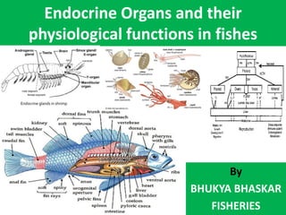
- 1. Endocrine Organs and their physiological functions in fishes By BHUKYA BHASKAR FISHERIES
- 2. Introduction • The endocrine system mainly controls the relatively slow metabolism of carbohydrates and water through the adrenal cortical tissue, the metabolism of nitrogen through the adrenal cortical tissue and thyroid gland, and the maturation and sexual behavior of germ cells through the pituitary and sex hormones. • The pituitary gland is called the master gland, because the hormones secreted by these glands directly through the help of different biochemical reactions and reveals various characteristics, as well as play a role in controlling the secretion of other endocrine glands. • The pituitary gland is mainly divided into two parts based on the cell structure, relative position, presence of secretory cells and pigmentation capacity of different parts of the pituitary gland, viz. • (1) Adenohypophysis • (2) Neurohypophysis • Adenohypophysis): It is divided into three types: These are: • Pro-adenohypophysis • Meso-adenohypophysis • Meta-adenohypophysis
- 3. Endocrine glands in Fin fishes • There are 12 types of endocrine glands in fish. Their names are mentioned below: 1. Pituitary gland 2. Thyroid gland 3. Interrenal tissue 4. Chromaffin tissue 5. Corpuscles of stannius 6. Ultimobranchial gland 7. Islets of Langerhans 8. Gonads 9. Intestinal mucosa 10. Thymus gland 11. Pineal body 12. Urophysis
- 4. • All hormones are made up of proteins or polypeptides. The hormones are- • Prolactin Growth hormone (somatotropin; STH) • Gonadotropins (GTH) – These are two types: Follicle stimulating hormone (FSH) and luteinizing hormone (LH) • Thyroid stimulating hormone (TSH) • Adrenocorticotropin (ACTH) • Melanocyte stimulating hormone (MSH) or intermedine • Vasopressin and • Oxytocin.
- 9. Classification of Hormones secreted from the endocrine glands • Different types of hormones are secreted from the endocrine glands. Hormones are generally of four types, viz • 1. Protein and Polypeptide Hormone: These hormones are made up of proteins or peptides. Parathyroid hormone secreted from the anterior and posterior pituitary glands, insulin and glucagon are secreted from the pancreas, calcitonin is secreted from the ultimobronchial gland, relaxin hormones are secreted by the ovarian and hormones of other glands. • 2. Steroid Hormone: This type of hormone is made up of steroid substances. It is usually a hormone secreted by the adrenal cortex (cortisol, aldosterone), ovaries (estrogen and progesterone), testis (testosterone) and placenta (estrogen and progesterone). • 3. Amino acid derivatives or biogenic amines: These hormones are derived from amino acids such as melanin and adrenal medullary adrenaline. • 4. Iodinated amino acids: Iodine is combined with amino acids to produce iodinated amino acids such as thyroxine.
- 10. Based on the origin the components of the endocrine system • Hormones are important regulators that often regulate the metabolism of different types of living cells. • The endocrine gland is unanimously referred to as the ductless gland whose secretory substance is released directly into the blood or lymph. • Based on the origin, the components of the endocrine system are divided into the following: • (1) Discrete endocrine glands: Such glands are pituitary (hypophysis), thyroid and pineal. • (2) Organs containing exocrine and endocrine functions: In fish, kidneys, gonads and intestines perform such functions. Heterotropic thyroid follicles, interenal and stannius corpuscles are present in the kidneys. • (3) Scattered cells with endocrine function: These are called expanded neuroendocrine. These cells are found in the digestive tract. These are usually called paracrine cells (such as somatostatin). These are gastrointestinal peptides that are classified as specific hormones or paracrine agents. They are also called Pupative Hormones.
- 11. • Prolactin cells: These cells are mainly located in pro-adenohypophysis and meso- adenohypophysis or pars distalis and form dense mass. In some species, such cells have a follicular structure. Such cells contain red pigments in erythrocytes or azocarmine and the acid fuchsin. However, they are not pigmented with Periodic acid-Schiff (PAS), Alkan Blue or Aldehyde fuchsin (AF). Studies have shown that these cells secrete prolactin-like hormones. • Function: Prolactin affects milk production in mammals, but in fish it plays a role in controlling circulation. • Corticotropes (ACTH cells): Such cells are also located in pro-adenohypophysis and neurohypophysis (NH). They are thought to secrete adrenocorticotropic hormones. In fish such as Poecilia, these cells are granular long chromophobic types. Sometimes these cells are lightly pigmented with erythrocyte or azocarmine and turn gray with lead hematoxylin. In eel fish, these cells contain more grains which are more pigmented in alizarin and lead hematoxylin. • Function: The hormone produced from these cells stimulates the adrenal cortical tissue to produce ACH and metabolize stored fats, water, carbohydrates and proteins. These cells also contain sodium ions and help in the metabolism of electrolytes. In Carassius fish, it helps in cell melanogenesis. • Somatropes (STH cells): It is formed from acidophils cells of pro-adenohypophysis and meso- adenohypophysis and is pigmented by orange G. Some species of teleost have two types of acidophil cells. One type of cell is pigmented by orange G (somatotropes). Other types of cells are stained with azacarmine or erythrosine (prolactin cells). In some species, such as Anguilla and Poecilia, two types of acidophils cells have been distinguished based on location, morphology, and pigmentation capacity. In many teleosts, such as Anguilla and Poecilia, the somatotropes are acidophils type and are located in the meso-adenohypophysis and are mixed with thyrotropes. • Function: Such cells help to increase body size.
- 12. Cont... • Gonadotropes (FSH and LH cells): These cells are usually present in meso-adenohypophysis, but sometimes, especially during the sexual maturity of eel (Olivereau 1967) and trout (Baker 1968), these cells enter pro-adenohypophysis. These cells contain glycoproteins and are stained by PAS, aldehyde fuchsin (FA), alcian blue and aniline blue. Various studies have shown that two types of gonadotropes are found in fish which are known as FSH and LH in mammals. Specific physiological and biochemical tests have shown that these two types of hormones are secreted by teleosts but not readily available (Olivereau 1967). The relationship of gonadotropes with sexual perfection has been proven experimentally on Indian teleost. • Function: These cells control the secretion of sex hormones.
- 13. • Thyrotropes (TSH cells): • Thyrotropes and gonadotropes are types of basophils (mucoid cells). • They are found in meso-adenohypophysis. Thyrotropes cannot be distinguished from gonadotropes in most teleosts. • In some teleosts such as Phoxinus, Rutilus and Astyanax, thyrotrophs aldehydes are used to stain fuchsin (AF). • Gonadotropes, however, are not stained by AF. Moreover, their positions vary. • Thyroid hormone (TSH) is present on the surface of meso-adenohypophysis and in some species of such as Poecilia, gonadotropes are found in the ventral side of meso-adenohypophysis. • Function of Thyrotropes : Controls the secretion of thyroid gland. • The presence of two distinct cells in meta-adenohypophysis suggests that the region secretes two types of hormones. • One of these is the melaminophore stimulating hormone (Intermedin- MSH) and the other is the melanophore concentrating hormone (MCH- Melanophore Concentrating Hormone). • Neurohypophysis • It releases two types of hormones, such as arginine vasotocin and oxytocin. • Functions: Arginine plays an important role in controlling vasotocin secretion and maintaining salt-water balance, and oxytocin helps in sexual intercourse and egg laying.
- 15. Thyroid Gland • Thyroid Gland • In most teleosts, the thyroid gland is composed of numerous follicles surrounding the arterial artery and the afferent bronchial blood vessel. However, in some fish such as Parrotfish (Spariidae), Swordfish (Xiphias gladius), Gymnarchus niloticus and Thynnus thynnus it is densely packed and covered. Some aerial fish such as Channa punctatus, C. gachua, C. marulius, C. striatus and Heteroneustes fossilis have a dense and covered thyroid gland near the aortic artery (Belsare 1959). Intermediate conditions are observed in Clarias batrachus. In this case, the gland does not have a capsule, the gland seems to be three condensed grains. • Histologically, the thyroid gland is made up of numerous follicles. The follicle, like a hollow ball full of fluid, contains a single layer of epithelial cells. These follicles vary in size and shape and are joined by connective tissue. As this gland is more rich in red blood cells, the blood supply system is well developed in it. The epithelium around the follicle may be thin or thick, and the height of the cells depends on their excretory activity. Normally less active follicles contain thinner epithelium. There are two main types of cells in the epithelium. • (1) Chief cells – These cells are dense and columnar in shape and have transparent cytoplasm attached. • (2) Colloid cells (Colloid cells or Benstay`s cells) – These cells contain granules of excretory material. • In some teleosts, the presence of thyroid follicles in various abnormal locations such as the main kidney, brain, esophagus and pleura has been reported. The idea is that all these heterotropic thyroid follicles migrate from the pharyngeal region to different parts of the body. The thyroid follicle collects inorganic iodine from the blood and stores it in its cells. It then combines iodine with tyrosine to make thyroid hormone. These hormones are secreted from the thyroid gland under the influence of thyrotropic hormones secreted from the pituitary gland. There are three types of thyroid hormones in fish, viz • Mono-iodothyrosine • Diiodotyrosine and • Thyroxine. • Functions • (1) Thyroid hormone plays an important role in oxygen uptake. However, the current study has found the opposite result. In Goldfish (Carassius auratus), Muller (1953) has been shown to increase their oxygen uptake by applying the hormone thyroxine, but Matty (1954) has shown that removal of the thyroid gland in the elasmobranch (surgical thyroidectomy) has no effect on respiration. In the case of teleost, the application of thiouria has been shown to reduce the need for oxygen uptake. • (2) Thyroid glands affect the control of osmosis (salmon and Gasterosteus).
- 16. ENDOCRINE CONTROL IN THE CRUSTACEA • 1The hormones of crustaceans may be grouped into those which control effector organs (chromatophores, muscles) and those which control the more gradual sequences of growth, development and reproduction; they have been termed energetic and metabolic hormones respectively. Most of the known crustacean hormones originate in neurosecretory centres in the central nervous system. • 2The sinus gland is an important part of an elaborate neurosecretory system in the eyestalk of stalk-eyed crustaceans and in the head of sessile-eyed forms. It is largely composed of the swollen terminations of neurosecretory fibres. Some cellular elements have also been detected, but it is not yet known whether they have a secretory function. It is generally accepted that the greater part of the secretory material in the sinus gland has been transported thither along axon fibres from cell bodies in the central nervous system, though it has been suggested that the staining reactions of the sinus gland indicate also the probability of chemical transformation, if not autochthonous secretion. After-sinus gland ablation the cut stump of the nerve leading to it continues to accumulate secretory material. • 3An indiscriminate use of the term X organ in the eyestalk has led to confusion. The X organ, originally described in detail by Hanstrom, is characterized by its association with a sensory papilla and by its contained concentric-layered structures, here called ‘onion bodies’. The term X organ has been used by some authors to denote a group of neurosecretory cell bodies, the fibres of which terminate in the sinus gland. It is suggested that the sensory papilla X organ (Hanstrom's X organ) should be distinguished from the other X organs as it differs from them morphologically and physiologically. • 4The post-commissure organs comprise enlargements of the epineurium of two post-commissure nerves, containing the terminations of neurosecretory fibres. They have been shown to contain chromactivating hormones. • 5The pericardial organs comprise epineurial enlargements containing fine-fibre terminations. They lie in the pericardial spaces and have been shown to contain substances active on the heart. • 6The Y organ, a glandular structure which contains hormones affecting the rate of development, is located in the antennary segment of those forms which have a maxillary excretory organ and in the second maxillary segment of those which have an antennary excretory organ. • 7Various groups of neurosecretory cells have been detected in the brain and in thoracic ganglia.
- 17. ENDOCRINE CONTROL IN THE CRUSTACEA • 8. The term neurohaemal organ has been proposed to denote those tissues, through which substances produced in neurosecretory cells gain ready access to the blood. • 9The pigment pattern of any one crustacean species is fairly constant, but patterns differ in the different groups. The pigments are contained in monochromatic, dichromatic, trichromatic and tetrachromatic chromatophores. The chromactivating substances which have so far been separated from tissue extracts seem to act differentially on these chromatophore types. • 10The sinus glands and the post-commissure organs contain a substance, found also in the corpora cardiaca of insects, which concentrates the red pigments in the large red and small red chromatophores of Leander serratus; it has been called the A-substance. When tissue extracts containing the A-substance have been allowed to stand for some time the A-substance disappears and is replaced by one or more substances which affect the red pigment in the small chromatophores but not that in the large chromatophores. • 11The post-commissure organs contain, in addition to the A-substance, a substance B which disperses red pigments, and also an A'-substance which concentrates white chromatophore pigments and at the same time the red pigments of the large red chromatophores. • 12A substance which disperses black pigments in the chromatophores of crabs has been found in a great number of different crustacean species. It is present in the Natantia, but its function in these forms is not yet known. • 13There is some evidence that light may affect chromatophore and eye pigments directly, possibly by sensitizing them to chromactivating hormones. • 14Chromatophore and eye pigments continue to migrate rhythmically even if animals are kept in constant darkness or under constant illumination. One such rhythm in Uca has been shown to be in synchrony with the solar cycle, another with the lunar cycle. The rhythms seemed to be largely independent of temperature, and of the eyes and sinus glands. • 15The migration of distal retinal pigment appears to be controlled by one or two substances originating in the sinus glands; there is some evidence that the proximal and reflecting pigments may be influenced by hormones originating outside the sinus glands. • 16A heart-accelerating substance found in the pericardial organs is without effect on the chromatophores.
- 18. ENDOCRINE CONTROL IN THE CRUSTACEA • 17. The sinus glands release a diabetogenic principle which maintains the blood-sugar level above a certain minimum. This substance appears to be released in response to stressing agents. In crabs, centres outside the eyestalk may also produce the hormone. The eyestalks seem to exert some measure of endocrine influence over glycogen metabolism, though this may be indirect. • 18. There is some evidence that the eyestalks are implicated in an endocrine control of chitin formation and the decomposition of lipoids and carotenoids. The evidence for the hormonal control of protein metabolism is suggestive but incomplete. • 19Removal of the eyestalks has been shown to affect respiratory metabolism, and tissue homogenates in vitro can be affected by eyestalk extracts and by extracts of the sinus glands or of the central nervous system. Respiratory data in crabs deprived of their sinus glands suggest that a hormone controlling oxygen consumption is released at the sinus gland, though formed elsewhere. • 20. There is evidence that a sinus-gland principle influences calcium metabolism, but that this may only be effective in the winter non-moulting season in those animals which moult annually. The phosphate content of the tissues and the blood changes cyclically with the calcium content and there is some evidence of endocrine control, though the substance appears to be different from that which affects calcium metabolism. • 21. The eyestalk appears to contain an antidiuretic and water-balance regulating hormone. It seems likely that this differs from the ‘moulting hormone’, although their actions seem to be very closely co-ordinated. • 22. The moulting cycle is under the control of at least three hormones and possibly as many as six. The moult-inhibiting hormone of the eyestalk and brain inhibits the onset of the premoult, the period of preparation for moulting; the moult- accelerating hormone accelerates and correlates the processes of the premoult once this has begun. Both these hormones may act upon the Y organ, which itself produces a hormone necessary for the moult process. The amount of water taken in at moulting is regulated by the water-balance hormone of the eyestalk. • 23. Ovarian growth is controlled, in both Natantia and Reptantia, by an ovarian-inhibiting hormone emanating from the eyestalk. • 2. 4Testicular growth is controlled in Brachyura, but not in those Natantia so far investigated, by a testis-inhibiting hormone emanating from the eyestalk. • 25. The ovary of some Malacostraca may secrete a progestational hormone responsible for promoting the development of the brooding characteristics. • 26. The vas deferens gland in Amphipoda secretes a hormone controlling the development of the male secondary and accessory sexual characteristics. • 27. Some colour-change hormones and substances which affect the heart beat have recently been separated by chromatography and electrophoresis. Preliminary studies indicate that at least one of the colour-change hormones contains peptide bonds which are necessary for its activity; one of the active substances in pericardial organ extracts is closely related to 5-hydroxytryptamine. It is suggested that the ovarian-inhibiting hormone is steroid in nature.
- 19. Endocrine glands in shrimp • X-organ are located in the medulla terminalis. • The X-organ secretes several neurohormones such as Ovary (gonad) Inhibiting Hormones (OIH/GIH) Moult Inhibiting Hormone (MIH) Light adapting hormone or distal retinal pigment hormone Hyper Glycemic Factor (CHH- Crustacean Hyperglycaemic Hormone) Erythrophore concentrating hormone & Neuro depressing hormone. • Out of these, OIH and MIH are directly involved in reproduction GSH : neuropeptide Growth Stimulating Hormone(GSH) is also known as Moult or Gonad Stimulating Hormone. It is secreted by theThoracic ganglia.
- 20. Y-Organ: • Y-Organ: • Located in the antennary / maxillary segment of the body anteriorly. It secretes a moulting hormone (ecdysone) which is a steroid hormone. The secretary activity of this gland is controlled by moult inhibiting hormone secreted by Xorgan and secretion of mandibular organ. MIH inhibits Y- organ whereas secretion of mandibular organ will stimulate the gland • Molting: Regular phenomenon in crustaceans as it grows. Molt cycle: Divided into 4 phases- Premolt: Inorganic constituents of old exoskeleton reabsorbed & stored in gastroliths (constrictions in the stomach wall) or hepatopancrease + glycogen deposited in hypodermis + increased oxygen concentration indicating accelerated metabolic activity. Molt: Old cuticle sloughed off + increase in size of body due to rapid absorption of water /hydration. Postmolt: New exoskeleton formed due to redeposition of chitin and inorganic salts. • Premolt & molt stages: Molting hormone (ecdysone) secreted byY-organ. Post molt & intermolt stages: Ecdysone secretion stopped by molt inhibiting hormone of X-organ. During anecdysis (A prolonged period without ecdysis)(period between molts) Y-organ is greatly reduced (indicating dysfunction) resulting in lack of molting.
- 21. Mandibular organ • The mandibular organ in the crustaceans is similar to that of an endocrine gland in higher vertebrates. It secretes hormones or hormone like substances, that will have the influence on the reporducitve organs or reproductive process. It has been involved in ovarian growth and vitellogenesis. Mandibular organ synthesizes a substance called methyl farnesoate (MF) which is responsible for juvenile characteristics. MF has been shown to have stimulatory effect on Y-organ to secrete ecdysone
- 22. Cont... • Vitellogenesis (also known as yolk deposition) is the process of yolk formation via nutrients being deposited in the oocyte, or female germ cell involved in reproduction of lecithotrophic organisms. In insects, it starts when the fat body stimulates the release of juvenile hormones and produces vitellogenin protein. Endocrine glands are glands of the endocrinesystem that secrete their products, hormones, directly into the blood rather than through a duct. The majorglands of the endocrine system include the pinealgland, pituitary gland, pancreas, ovaries, testes, thyroid gland, parathyroid gland, hypothalamus and adrenal glands