Lymphatic system

Lymphatic system It was studied for the first time by Thomas Bartholin in 1703. It is a sub-system of circulatory system in vertebrate body that consists of a complex network of lymph vessels or lymphatic’s lymph tissues (nodes) and organs such as tonsils, thymus and spleen which carries a milky fluid Called lymph. LYMPH Colorless clearly watery fluid contains lymphocytes similar in composition to with the exception of some proteins. The Main functions of lymph are as under: Carries plasma proteins which seep out of the capillary beds from the blood stream. It also carries larger particles such as bacteria and other waste product, cell debris from the damaged tissues which is then filtered out and destroyed by the lymph node. In lacteals fats absorbed into blood plasma called (chyle) which has a milky appearance.

Recommended
More Related Content
What's hot
What's hot (20)
Similar to Lymphatic system
Similar to Lymphatic system (20)
Recently uploaded
Recently uploaded (20)
Lymphatic system
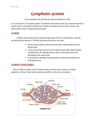
- 1. 1 | P a g e Lymphatic system It was studied for the first time by Thomas Bartholin in 1703. It is a sub-system of circulatory system in vertebrate body that consists of a complex network of lymph vessels or lymphatic’s lymph tissues (nodes) and organs such as tonsils, thymus and spleen which carries a milky fluid Called lymph. LYMPH Colorless clearly watery fluid contains lymphocytes similar in composition to with the exception of some proteins. The Main functions of lymph are as under: Carries plasma proteins which seep out of the capillary beds from the blood stream. It also carries larger particles such as bacteria and other waste product, cell debris from the damaged tissues which is then filtered out and destroyed by the lymph node. In lacteals fats absorbed into blood plasma called (chyle) which has a milky appearance. LYMPH CAPILLARIES They are blind end tubes in the interstitial spaces and have same structure as blood capillaries. All tissues have lymph vessels except CNS, cornea of eye, and bones.
- 2. 2 | P a g e LYMPH VESSELS Lymph capillaries join up to form larger lymph vessels. Lymph vessels are found running alongside the arteries and veins. Wall of lymph vessels are consists of three layers as in the blood vessels. LYMPHATIC DUCTS Lymph vessels are joined together to form a larger vessels called lymphatic ducts. They are of two types which are: THORACIC DUCT It drains lymph from both legs, pelvic and abdominal cavity, left half of the thorax, head, neck and left arm and open into the left subclavian vein in the root of neck. RIGHT LYMPHATIC DUCT It drains lymph from the right half of the thorax, head, neck, and right arm and open into the right subclavian vein.
- 3. 3 | P a g e LYMPH NODE Nodes are oval or bean shaped organs covered by capsule. Invagination of the capsule forms many cavities called trabeculae which consist of lymphocytes and macrophages. They also consist of two types of tissues called the lymphatic and reticular tissues. Lymph enters into the nodes through three or four vessels called afferent vessels while their exit occurs through another vessel called efferent vessel. Nodes help in phagocytosis of pathogens and thus help in filtration. There are more than hundred nodes present in human body some of them are: Cervical node located in neck region. Axillary node located in armpits. Inguinal node located in groin. Thoracic region. Abdominal region. Pelvic region.
- 4. 4 | P a g e TONSILS Pair of soft masses located in the pharynx composed of tissues similar to lymph nodes covered by pink mucosa. Tonsils trap pathogens present in the inhaled air and hence help in the filtration of air. Sometimes tonsils are itself infected by pathogens (TONSILITIS) and inflammation occurs due to whichtonsils become swell up and are called HYPERTROPHIC due to which breathlessness and sleep apnea (choking and snoring sound during sleeping) occur. SPLEEN Spleen is the largest organ of lymphatic system 12 cm long and 7cm wide, purplish in color, weight 200 g consists of lymphocytes and macrophages. FUNCTIONS OF SPLEEN Phagocytosis: spleen helps in the destruction of old and abnormal RBC’s and their breakdown products (bilirubin and iron) are transported to liver. Storage of blood: spleen store about 350 ml of blood every time it releases it in hemorrhagic condition and during the stimulation of sympathetic nervous system. Erythropoiesis: spleen is the best site for the production of blood cells in fetal life. THYMUS GLAND Thymus gland is located in the chest cavity. At the time of birth its size is about from 10 to 15g and grows until puberty to about 30 to 40g then it is returned to approximately its weigh at birth. T lymphocytes are produced in this gland. Thymus gland also provides each lymphocyte with the ability to react to only one specific antigen. T lymphocytes are then transported to the blood stream.
- 5. 5 | P a g e EXCHANGE ACROSS CAPILLARY WALLS There are two types of pressure involved in this process which are: Blood pressure /capillary hydrostatic pressure: Pressure that is exerted by blood on the walls of the blood vessels is called blood pressure. Due to this pressure water, some proteins and small solutes squeezed out of the capillary into the interstitial spaces. Osmotic pressure: Pressure exerted by proteins (mainly Albumin) in blood vessels plasma that usually tends to pull water into the capillary from interstitial spaces. CREATED BY: AZIZ KHAN BS ZOOLOGY: 3RD SEMESTER UNIVERSITY OF SWAT