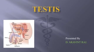
The testis is the male gonad located in the scrotum. It has two poles, two borders, and two surfaces. The testis is covered by three layers: the tunica vaginalis, tunica albuginea, and tunica vasculosa. Internally, the testis contains 200-300 lobules with seminiferous tubules that produce sperm. The testis receives blood supply from the abdominal aorta and drains into veins that lead to the inferior vena cava or left renal vein. Lymphatic drainage is to the pre-aortic and para-aortic lymph nodes. The testis has both sensory and motor nerve supply. Abnormalities can
2. Introduction.
Shape and size.
External features.
Coverings of the testis.
Structure of the testis.
Arterial supply.
Venous supply.
Lymphatic drainage.
Nerve supply.
Clinical anatomy.
3. MALE GENITAL ORGANS :
They are situated both outside the pelvic cavity. As lower
temperature is required for spermatogenesis. The testis are placed outside
the pelvic cavity in the scrotal sac.
EXTERNAL GENITAL ORGANS:
1.Penis.
2.scrotum.
3.Testes.
4.Epididymes.
5.Spermatic cords.
4. The testis is a male gonad.
It is homologous with the ovary of the female.
Suspended in the scrotum by the spermatic cord.
Upper pole is tiled forwards and laterally.
The left testis is slightly lower than the right.
5. Two poles - Upper pole and Lower pole .
Upper and lower poles are convex and smooth .
Upper pole provides attachment to spermatic cord.
Two borders - Anterior Border and Posterior Border.
Anterior border is convex and smooth. Fully covered by tunica
vaginalis.
Posterior border is straight .partially covered by tunica vaginalis .
Two surfaces - Medial Surface and Lateral Surface.
Medial surface is separated by extension of cavity of tunica vaginalis
extension called sinus of epididymis.
Medial and lateral surfaces are convex and smooth.
6.
7. The testis is covered by layers of the scrotum.
1.Tunica Vaginalis.
2.Tunica Albuginea.
3.Tunica Vasculosa.
1.Tunica vaginalis:
It represents the lower persistent portion of the
processus vaginalis.
Parietal and visceral layer with a cavity in between.It
covers the whole testis
8. 2.Tunica Albuginea:
Tunica albuginea “ Latin White” fibrous coat covering the
testis all around
It is covered by the visceral layer of the tunica vaginalis.
The posterior border is thickened to form an incomplete
vertical septum called Mediastinum testis.
They incompletely divide the testis into 200 to 300 lobules.
3.TunicaVasculosa:
Tunica Vasculosa is the innermost ,vascular coat of the testis
lining its lobules.
9.
10. Testis consists of 200 to 300 lobules .
Each lobule contains two to three seminiferous tubules.
Each tubule measure about 60 cm length, and it’s about 0.2 mm
in diameter.
The tubule is lined by cells represent stages formation of
spermatozoa.
The seminiferous tubules lobules to from 20 to 30 straight
tubules of mediastinum.
The rete testis give rise to 12 to 30 efferent ductless.
11. Branch of the abdominal aorta
given off the level off vertebra
12.
It descends on posterior
abdominal wall reach deep
inguinal ring .
Small branches enter the
posterior border.
Larger branches medial and
lateral ,the tuica albuginea.
12. It drain into the inferior vena cava on the right side
And into the left renal vein on the left side.
13. It drain into the pre-
aortic and para-
aortic groups of
lymph nodes at the
level of second
lumbar vertebra.
14. Sympathetic nerves arises from segment T10 of the
spinal cord .
They passes through the renal and aortic plexuses.
The nerves are both afferent for testicular sensation
and efferent to the blood vessels (vasomotor).
15. Unilateral Absence – Monorchism.
Bilateral Absence - Anorchism.
Undescended testis or cryptorchidism : The organ may lie in the
lumbar, iliac, inguinal, or upper scrotal region.
Testis and epididymis may be site of various infection and
tumors .
Testis may be palpated to check any nodules ,or irregularity or
size consistency.
Varicocele is produced by dilatation of the pampiniform plexus
of veins.