
Recommended
More Related Content
Similar to ENT.pptx
Similar to ENT.pptx (20)
More from 9459654457
More from 9459654457 (20)
Recently uploaded
Recently uploaded (20)
ENT.pptx
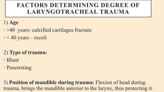
- 1. FACTORS DETERMINING DEGREE OF LARYNGOTRACHEAL TRAUMA 1) Age • >40 years- calcified cartilages fracture • < 40 years – recoil 2) Type of trauma: • Blunt • Penetrating 3) Position of mandible during trauma: Flexion of head during trauma, brings the mandible anterior to the larynx, thus protecting it.
- 2. 4) Force of impact: severity of injury is directly proportional to the force of impact. 5) Angle of impact: • If force is from the front- larynx is pressed against the cervical vertebrae and causes more damage. • If there is lateral force- larynx moves to certain extent and the damage is less.
- 3. Ossification of the laryngeal cartilages occurs after 40 years of age. If age is more than 40 years , the cartilage fracture occurs in multiple areas and incidence of obstructed airway is high. If the age is less than 40 years, the commonest site of fracture is in the mid line and elastic recoil tends to bring the fracture site forwards reducing the incidence of airway obstruction.
- 4. CLINICAL FEATURES SYMPTOMS :- Symptoms of laryngotracheal injury would vary , greatly depending on the structures damaged and the severity of damage. They include: • Respiratory distress • Change in voice- hoarseness or aphonia • Painful or difficult swallowing- This is accompanied by aspiration of the food.
- 5. • Local pain in the larynx – More marked on speaking or swallowing. • Hemoptysis, usually the result of tears in laryngeal or tracheal mucosa. • Otalgia • Swelling or open wound • Cough
- 6. EXTERNAL SIGNS:- • Bruises or abrasions over the skin. • Palpation of the laryngeal area is painful. • Subcutaneous emphysema due to mucosal tears. It may increase on coughing. • Flattening of thyroid prominence and contour of anterior cervical region. Thyroid notch may not be palpable.
- 7. • Fracture displacements of thyroid or cricoid cartilage or hyoid bone. Gap may be felt between the fractured fragments. • Bony crepitus between fragments of hyoid bone, thyroid or cricoid bone may sometimes be elicited. • Separation of cricoid cartilage from larynx or trachea.
- 8. • Tenderness on springing of bone or cartilage. • Step deformity may be present in the frature of laryngeal cartilage. • Stridor • Open wound with / without larynx / trachea exposed. • Swelling
- 9. • INTERNAL FEATURES:– • Edema / hematoma/ mucosal tears will be seen. • Airway obstruction • Dislocation of cricoarytenoid joint • Vocal cord palsy • Falling back of epiglottis or avulsion • Disrupted vocal cord or ventricular bands • Exposed cartilage and laryngeotracheal lumen • Asymmetrical laryngeal inlet and laryngeotracheal lumen.
- 10. • Laryngeal trauma may be part of multitrauma involving other parts of the body and therefore must be assessed in a systematic manner. • Urgent review of patients is vital as patients initially exhibiting only subtle symptoms and signs of upper aerodigestive tract dysfunction can progress to complete airway obstruction from worsening oedema.
- 11. INVESTIGATIONS 1) INDIRECT LARYNGOSCOPY OR RIGID ENDOSCOPY OF THE LARYNX: It is the most valuable examination. Indirect laryngoscopy reveals:- Edema, its location and degree Hematoma Mucosal lacerations Displacement of epiglottis Fragments of cartilage Asymmetry of larynx
- 12. 2) FLEXIBLE LARYNGOSCOPY THROUGH THE NOSE:- It provides better visualisation of:- Hematoma Mucosal oedema Arytenoid avulsion or dislocation Exposure of cartilages Vocal cord paralysis Direct laryngoscopy is relatively contraindicated as it may precipitate respiratory distress and need for tracheostomy.
- 13. 3) X- Ray:- soft tissue lateral view of the neck may show: Subcutaneous emphysema Mucosal swelling Fracture / displacement of epiglottis Thyroid and cricoid cartilages Hyoid bone Changes in the air column
- 14. 4) CT Scan :- It is the most preferred imaging modality these days as it provides three dimensional reconstructions. It reveals injuries of laryngeal cartilages better than x- rays. • Additionally, it also gives information about injuries to cervical spine and vascular structures. • It is very useful in the investigation of :- Mucosal oedema Fractures of thyroid and cricoid cartilages Dislocation of joints
- 15. 5) ASSOCIATED INJURIES:– It is essential to examine for other injuries to head , cervical spine, chest, abdomen and extremities. • X- Ray chest for pneumothorax and gastrograffin swallow for oesophageal tears may be required. 6) VIDEOSTROBOSCOPY OR LARYNGEAL ELECTROMYOGRAPHY: • It is valuable in determining conditions of the vocal folds.