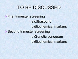
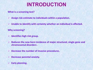
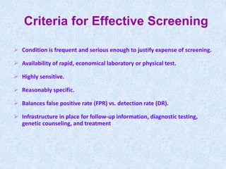
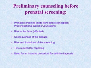
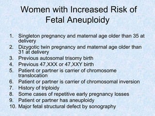
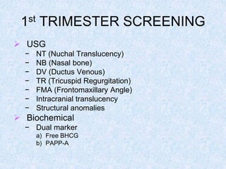
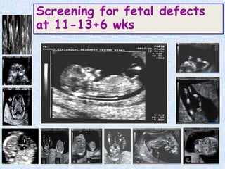
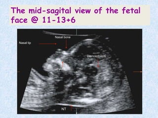
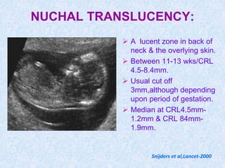
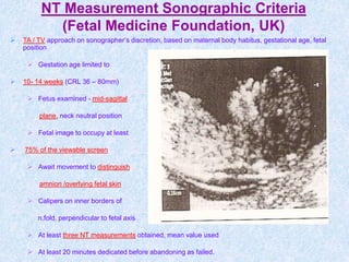
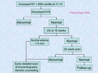
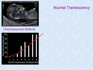
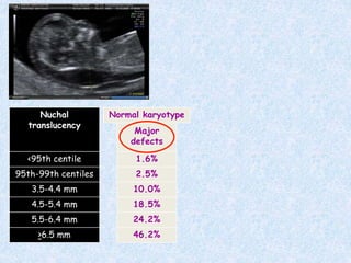
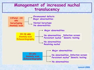
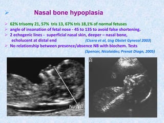
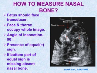
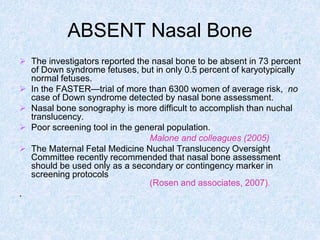
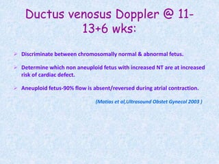
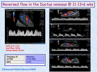
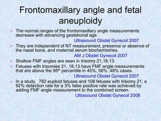
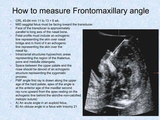
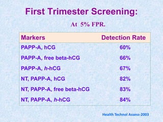
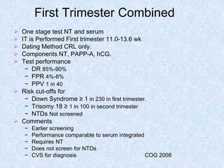
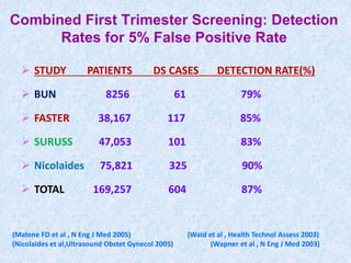
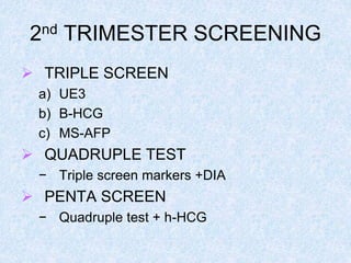
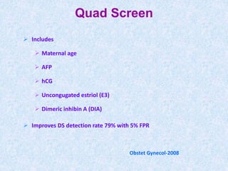
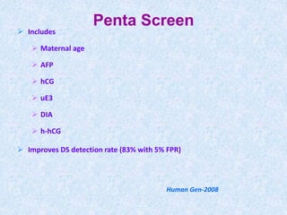
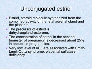
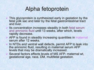
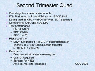
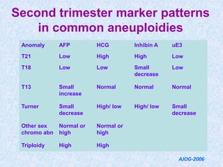
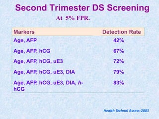
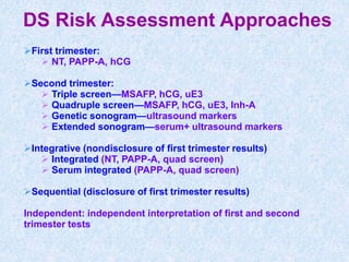
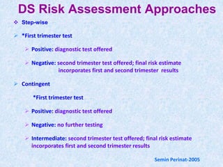
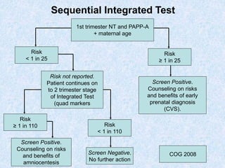
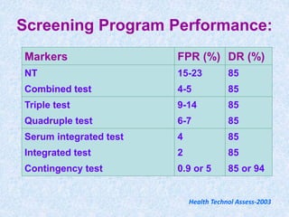
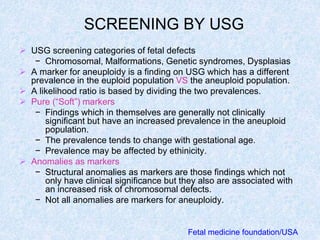
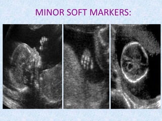
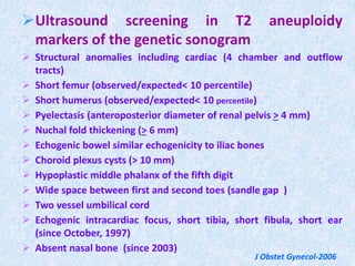
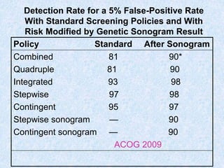
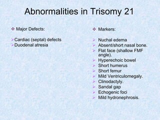
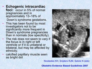
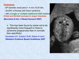
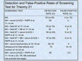
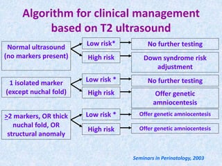
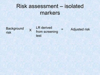
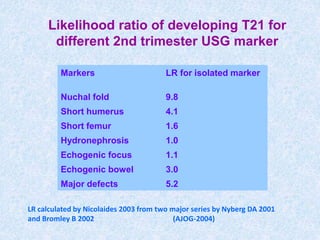
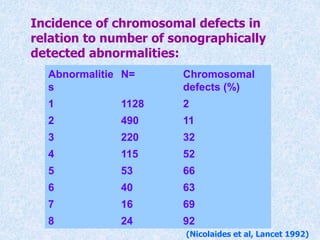
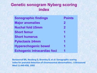
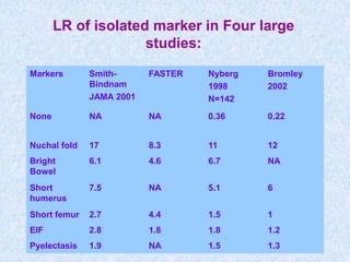
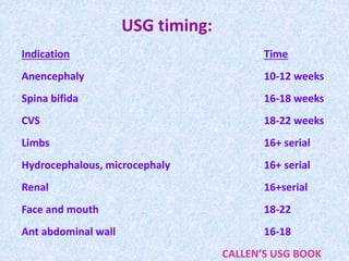
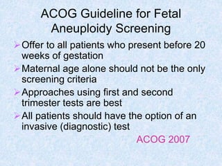
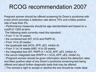
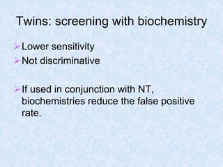
SlideShare a Scribd company logo
1 of 80
ANTENATAL SCREENING IN FIRST
AND SECOND TRIMESTER
Candidate : Dr Rajnish
Guide : Dr Vatsla Dadhwal
Sr-guide : Dr Shankar
TO BE DISCUSSED
First trimester screening
a)Ultrasound
b)Biochemical markers
Second trimester screening
a)Genetic sonogram
b)Biochemical markers
INTRODUCTION
What is a screening test?
 Assign risk estimate to individuals within a population.
 Unable to identify with certainty whether an individual is affected.
Why screening?
 Identifies high risk group.
 Reduces the new born incidence of major structural, single gene and
chromosomal disorders .
 Decrease the number of invasive procedures.
 Decrease parental anxiety.
 Early planning.
Criteria for Effective Screening
 Condition is frequent and serious enough to justify expense of screening.
 Availability of rapid, economical laboratory or physical test.
 Highly sensitive.
 Reasonably specific.
 Balances false positive rate (FPR) vs. detection rate (DR).
 Infrastructure in place for follow-up information, diagnostic testing,
genetic counseling, and treatment
Preliminary counseling before
prenatal screening:
 Prenatal screening starts from before conception:-
Preconceptional Genetic Counselling
 Risk to the fetus (affected)
 Consequences of the disease
 Risk and limitations of the screening
 Time required for reporting
 Need for an invasive procedure for definite diagnosis
PRECONCEPTION GENETIC COUNSELLING:
 Indications :-
 Previous pregnancy history
 Fetal demise
 Previous child with a genetic disorder
 Recurrent pregnancy loss
 Disorder
 Chromosomal: Down syndrome
 Structural: dwarfism, neural tube defect
 Metabolic disorder
 Hematological: anemia, bleeding disorder
 Mental retardation
 Family history of a genetic disorder
 Bleeding disorders: hemophilia
 Neurological disease: muscular dystrophy
 Mental retardation: fragile (X) syndrome
PRECONCEPTION GENETIC COUNSELLING (contd…)
 Ethnic origin from population at high risk of genetic disorder
 Ashkenazi Jewish: Tay-Sachs disease
 French-Canadian: Tay-Sachs disease
 African-American: Sickle cell anemia
 Mediterranean: Beta thalassemia
 Oriental: Alpha thalassemia and beta thalassemia
 Maternal medical disorder
 Diabetes
 Phenylketonuria
 Maternal medications
 Anticonvulsants
 Lithium
 Socially used drugs
 Alcohol ,cocaine COG-2008
Women with Increased Risk of
Fetal Aneuploidy
1. Singleton pregnancy and maternal age older than 35 at
delivery
2. Dizygotic twin pregnancy and maternal age older than
31 at delivery
3. Previous autosomal trisomy birth
4. Previous 47,XXX or 47,XXY birth
5. Patient or partner is carrier of chromosome
translocation
6. Patient or partner is carrier of chromosomal inversion
7. History of triploidy
8. Some cases of repetitive early pregnancy losses
9. Patient or partner has aneuploidy
10. Major fetal structural defect by sonography
1st TRIMESTER SCREENING
 USG
− NT (Nuchal Translucency)
− NB (Nasal bone)
− DV (Ductus Venous)
− TR (Tricuspid Regurgitation)
− FMA (Frontomaxillary Angle)
− Intracranial translucency
− Structural anomalies
 Biochemical
− Dual marker
a) Free BHCG
b) PAPP-A
Maternal Serum Screening
• Photograph of Brahms Kryptor Immunoassay analyzer.
Normal anatomy
Screening for fetal defects
at 11-13+6 wks
NT
Nasal bone
Nasal tip
Diencephalon
Palate
The mid-sagital view of the fetal
face @ 11-13+6
NUCHAL TRANSLUCENCY:
 A lucent zone in back of
neck & the overlying skin.
 Between 11-13 wks/CRL
4.5-8.4mm.
 Usual cut off
3mm,although depending
upon period of gestation.
 Median at CRL4.5mm-
1.2mm & CRL 84mm-
1.9mm.
Snijders et al,Lancet-2000
NT Measurement Sonographic Criteria
(Fetal Medicine Foundation, UK)
 TA / TV approach on sonographer’s discretion, based on maternal body habitus, gestational age, fetal
position
 Gestation age limited to
 10- 14 weeks (CRL 36 – 80mm)
 Fetus examined - mid-sagittal
plane, neck neutral position
 Fetal image to occupy at least
 75% of the viewable screen
 Await movement to distinguish
amnion /overlying fetal skin
 Calipers on inner borders of
n.fold, perpendicular to fetal axis
 At least three NT measurements obtained, mean value used
 At least 20 minutes dedicated before abandoning as failed.
Increased NT > 95th centile at 11-14
Karyotype/CVS
Early detailed scan
Echocardiography
Genetic counseling
Abnormal Normal
20 week scan
Nuchal edema
> 5 mm
US at 16 weeks
Normal
Abnormal
Normal
Follow-up
Prenat Diagn 2002
ASSOCIATION OF NT WITH
TRISOMY:
 Fetal Medicine Foundation in England:
 total no patients-96,127
 detection rate:82%
 FPR-8% (Snijders et al,Lancet-1998)
 SURUSS Trial:
 detection rate-85%
 FPR-21%
 FASTER Trial: -
 detection rate-86%
 FPR-20% ( Malone F,N Eng J Med-2005)
Increased NT, Normal Karyotype
and Major Heart Defects
NT N CHD
3 mm 1102 5 /
1,000
4 mm 188 27 /
1,000
5 mm 56 54 /
1,000
>6 mm 43 233 /
1,000
Total 1,389 17 /
1,000
3mm 4mm 5mm >6mm
Lancet-2006
Nuchal Translucency
0
20
40
60
80
100
1 2 3 4 5 6 7 8
Nuchal translucency thickness (mm)
Abnormal
karyotype
(%)
Chromosomal Defects
46.2%
>6.5 mm
24.2%
5.5-6.4 mm
18.5%
4.5-5.4 mm
10.0%
3.5-4.4 mm
2.5%
95th-99th centiles
1.6%
<95th centile
Major
defects
Nuchal
translucency
Normal karyotype
11Fetal -14
wks
karyotyping
Anomaly scan
Chromosomal defects
Major abnormalities
Normal karyotype
No abnormalities
14-16 wks
Anomaly scan
Echocardiography
No abnormalities
Resolving nuchal
Major abnormalities
No abnormalities
Persistent nuchal
Infection screen
Genetic testing
20 wks
Anomaly scan
Echocardiography
No abnormalities
Major abnormalities
No abnormalities
Persistent nuchal
Infection screen
Genetic testing
Management of increased nuchal
translucency
Lancet-2006
 Nasal bone hypoplasia
 62% trisomy 21, 57% tris 13, 67% tris 18,1% of normal fetuses
 angle of insonation of fetal nose - 45 to 135 to avoid false shortening.
 2 echogenic lines - superficial nasal skin, deeper – nasal bone,
echolucent at distal end (Cicero et al, Usg Obstet Gynecol 2003)
 No relationship between presence/absence NB with biochem. Tests
(Spencer, Nicolaides; Prenat Diagn, 2005)
HOW TO MEASURE NASAL
BONE?
 Fetus should face
transducer.
 Face & thorax
occupy whole image.
 Angle of insonation-
90`.
 Presence of equal(=)
sign.
 If bottom part of
equal sign is
missing-absent
nasal bone.
NT
Nasal bone
Nasal tip
Diencephalon
Palate
Sonek et al , AJOG-2006
ABSENT Nasal Bone
 The investigators reported the nasal bone to be absent in 73 percent
of Down syndrome fetuses, but in only 0.5 percent of karyotypically
normal fetuses.
 In the FASTER—trial of more than 6300 women of average risk, no
case of Down syndrome detected by nasal bone assessment.
 Nasal bone sonography is more difficult to accomplish than nuchal
translucency.
 Poor screening tool in the general population.
Malone and colleagues (2005)
 The Maternal Fetal Medicine Nuchal Translucency Oversight
Committee recently recommended that nasal bone assessment
should be used only as a secondary or contingency marker in
screening protocols
(Rosen and associates, 2007).
.
Ductus venosus Doppler @ 11-
13+6 wks:
 Discriminate between chromosomally normal & abnormal fetus.
 Determine which non aneuploid fetus with increased NT are at increased
risk of cardiac defect.
 Aneuploid fetus-90% flow is absent/reversed during atrial contraction.
(Matias et al,Ultrasound Obstet Gynecol 2003 )
Reversed flow in the Ductus venosus @ 11-13+6 wks
Gate: 0.5-1 mm
Low filter: 50 Hz
Sweep speed: 2cm/s
Contamination from hepatic vein
Contamination from umbilical vein
Trisomy 21
n=125
65%
Normal
n=21,000
3.5%
(Ultrasound Obstet Gynecol 2003)
Frontomaxillary angle and fetal
aneuploidy
 The normal ranges of the frontomaxillary angle measurements
decrease with advancing gestational age.
Ultrasound Obstet Gynecol 2007
 They are independent of NT measurement, presence or absence of
the nasal bone, and maternal serum biochemistries.
AM J Obstet Gynecol 2007
 Shallow FMF angles are seen in trisomy 21,18,13.
 Fetuses with trisomies 21, 18,13 have FMF angle measurements
that are above the 95th percentile in 45%, 58% ,48% cases.
Ultrasound Obstet Gynecol 2007
 In a study, 782 euploid fetuses and 108 fetuses with trisomy 21, a
92% detection rate for a 3% false positive rate was achieved by
adding FMF angle measurement to the combined screen.
Ultrasound Obstet Gynecol 2008
How to measure Frontomaxillary angle
 CRL 45-84 mm 11 to 13 + 6 wk.
 MID saggital fetus must be facing toward the transducer.
 Face of the transducer is approximately
parallel to long axis of the nasal bone.
 Fetal profile must include an echogenic
line representing the skin over nasal
bridge and in front of it an echogenic
line representing the skin over the
nasal tip.
 Intracranial structures hypoechoic areas
representing the region of the thalamus,
pons and medulla oblangata.
 Space between the upper palate and the
nose should be devoid of an echogenic
structure representing the zygomatic
process.
 FMF angle first ray is drawn along the upper
age of the hard palate, apex of the angle is
at the anterior age of the maxillar second
ray runs upward from the apex resting on the
echogenic line behind the skin(the non-calcified
metopic suture)
 A) An acute angle in an euploid fetus.
 B) An obtuse angle in a fetus with trisomy 21
Tricuspid valve doppler and fetal aneuploidy
 The prevalence of tricuspid regurgitation varies with
gestational age and also increase as the NT
measurement increases.
 Tricuspid regurgitation is seen in trisomy 21,18,13 and
monosomy X.
 The prevalence of tricuspid regurgitation in fetuses with
trisomies 21, 18, 13 and monosomy X is 56%, 33%, 30%
and 38% respectively.
 The prevalence of tricuspid regurgitation in euploid
fetuses is 1%.
 In a study, 19 614 fetuses, a 96% detection rate for a 3%
false positive rate was achieved for trisomy 21 by adding
tricuspid valve evaluation to the combined screen.
 The detection rates for trisomies 18 ,13, and monosomy
X were 92%, 100% and 100% respectively.
Ultrasound Obstet Gynecol 2009
 Other fetal measurements potentially affected
in chromosomally abnormal fetuses in first
trimester USG scan
• Fetal heart rate.
• Maxillary length.
• Ear length.
• Femur length.
• Humeral length.
Free beta-hCG
Produced in the trophoblast cells.
Maternal serum concentration reaches a
peak at appr. 15 wks, followed by a rapid
decline until 17wks and more gradual
decrease between 17 and 22 wks.
Free beta-hCG are increased twice the
normal value during second trimester in
aneuploidy fetus.
PAPP-A (pregnancy associated
plasma protein- A)
 PAPP-A level normally increases with gestation.
 It is lower in pregnancies of trisomy 21.
 Used alone in the first trimester allows for
trisomy 21 DR 40% and FR 5%.
 Increase PAPP-A level in women were
associated with high risk of
preeclampsia,preterm birth.
Hypertens Pregnancy 2003
First Trimester Screening:
Markers Detection Rate
PAPP-A, hCG 60%
PAPP-A, free beta-hCG 66%
PAPP-A, h-hCG 67%
NT, PAPP-A, hCG 82%
NT, PAPP-A, free beta-hCG 83%
NT, PAPP-A, h-hCG 84%
At 5% FPR.
Health Technol Assess-2003
First Trimester Combined
 One stage test NT and serum
 IT is Performed First trimester 11.0-13.6 wk
 Dating Method CRL only.
 Components NT, PAPP-A, hCG.
 Test performance
− DR 85%-90%
− FPR 4%-6%
− PPV 1 in 40
 Risk cut-offs for
− Down Syndrome ≥ 1 in 230 in first trimester.
− Trisomy 18 ≥ 1 in 100 in second trimester
− NTDs Not screened
 Comments
− Earlier screening
− Performance comparable to serum integrated
− Requires NT
− Does not screen for NTDs
− CVS for diagnosis COG 2008
Combined First Trimester Screening: Detection
Rates for 5% False Positive Rate
 STUDY PATIENTS DS CASES DETECTION RATE(%)
 BUN 8256 61 79%
 FASTER 38,167 117 85%
 SURUSS 47,053 101 83%
 Nicolaides 75,821 325 90%
 TOTAL 169,257 604 87%
(Malone FD et al , N Eng J Med 2005) (Wald et al , Health Technol Assess 2003)
(Nicolaides et al,Ultrasound Obstet Gynecol 2005) (Wapner et al , N Eng J Med 2003)
2nd TRIMESTER SCREENING
 TRIPLE SCREEN
a) UE3
b) B-HCG
c) MS-AFP
 QUADRUPLE TEST
− Triple screen markers +DIA
 PENTA SCREEN
− Quadruple test + h-HCG
Obstet Gynecol-2008
Quad Screen
 Includes
 Maternal age
 AFP
 hCG
 Uncongugated estriol (E3)
 Dimeric inhibin A (DIA)
 Improves DS detection rate 79% with 5% FPR
Penta Screen
 Includes
 Maternal age
 AFP
 hCG
 uE3
 DIA
 h-hCG
 Improves DS detection rate (83% with 5% FPR)
Human Gen-2008
Unconjugated estriol
 Estriol, steroid molecule synthesized from the
combined activity of the fetal adrenal gland and
the placenta.
 The precursor of estriol is
dehydroepiandrosterone.
 The concentration of estriol in the second
trimester of pregnancy is decreased about 25%
in aneuploid pregnancies.
 Very low level of uE3 are associated with Smith-
Lemli-Optiz syndrome, placental sulfatase
deficiency.
Alpha fetoprotein
 This glycoprotein is synthesized early in gestation by the
fetal yolk sac and later by the fetal gastrointestinal tract
and liver.
 Its concentration increases steadily in both fetal serum
and amnionic fluid until 13 weeks, after which, levels
rapidly decrease.
 AFP is found in steadily increasing quantities in maternal
serum after 12 weeks.
 In NTDs and ventral wall defects, permit AFP to leak into
the amnionic fluid, resulting in maternal serum AFP
levels that may be dramatically increased.
 Several factors effects levels of MS AFP- maternal wt,
gestational age, race, DM, multifetal gestation.
Prenat Diag-2000
Dimeric Inhibin A (DIA)
 Hormone synthesized by the gonads and placenta
 Levels vary the least with increasing gestational age in second trimester
 Elevated maternal serum levels associated with DS
 Increase levels in down syndrome about 1.77 MoMs
 The concentration of inhibin A is not used to calculate the risk for T18
 1996: addition to triple screen could improve DS detection rate
 Quad screen proposed
 Test at 15-20 weeks gestation
Prenat Diag-2008
Hyperglycosylated hCG (h-hCG):
 Produced very early in pregnancy by placenta
 Elevated levels associated with DS
 Low levels associated with trisomy 18, trisomy 13, triploidy, and
preeclampsia
 In 2000, h-hCG reported to add value to second trimester screening
 Penta screen proposed.
(BJOG-2007)
AFP, hCG, uE3, and DIA vs
Gestational Age:
15 20
Gestational Age (weeks)
AFP
UE3
HCG
Screening Window
DIA
Second Trimester Quad
 One stage test maternal serum only
 IT is Performed in Second Trimester 15.0-22.6 wk.
 Dating Method CRL or BPD Preferred. LMP accepted.
 Components AFP, uE3,hCG,DIA.
 Test performance
− DR 80%-85%
− FPR 5%-8%
− PPV 1 in 50
 Risk cut-offs for
− Down Syndrome ≥ 1 in 270 in Second trimester.
− Trisomy 18 ≥ 1 in 100 in Second trimester
− NTDs AFP ≥ 2.0 MoM.
 Comments
− Best second trimester screening test
− U/S not Required
− Screens for NTDs
− Amniocentesis for diagnosis COG 2008
Second trimester marker patterns
in common aneuploidies
Anomaly AFP HCG Inhibin A uE3
T21 Low High High Low
T18 Low Low Small
decrease
Low
T13 Small
increase
Normal Normal Normal
Turner Small
decrease
High/ low High/ low Small
decrease
Other sex
chromo abn
Normal or
high
Normal or
high
Triploidy High High
AJOG-2006
Health Technol Assess-2003
Second Trimester DS Screening
Markers Detection Rate
Age, AFP 42%
Age, AFP, hCG 67%
Age, AFP, hCG, uE3 72%
Age, AFP, hCG, uE3, DIA 79%
Age, AFP, hCG, uE3, DIA, h-
hCG
83%
At 5% FPR.
First trimester:
 NT, PAPP-A, hCG
Second trimester:
 Triple screen—MSAFP, hCG, uE3
 Quadruple screen—MSAFP, hCG, uE3, Inh-A
 Genetic sonogram—ultrasound markers
 Extended sonogram—serum+ ultrasound markers
Integrative (nondisclosure of first trimester results)
 Integrated (NT, PAPP-A, quad screen)
 Serum integrated (PAPP-A, quad screen)
Sequential (disclosure of first trimester results)
Independent: independent interpretation of first and second
trimester tests
DS Risk Assessment Approaches
 Step-wise
 *First trimester test
 Positive: diagnostic test offered
 Negative: second trimester test offered; final risk estimate
incorporates first and second trimester results
 Contingent
*First trimester test
 Positive: diagnostic test offered
 Negative: no further testing
 Intermediate: second trimester test offered; final risk estimate
incorporates first and second trimester results
Semin Perinat-2005
Serum Integrated
 Two stage test serum only
 IT is Performed in First and second trimester10.3-13.6+15.0-22.6wk
 Dating Method CRL or BPD preferred. LMP accepted.
 Components PAPP-A in first trimester. Quad markers in second
trimester
 Test performance
− DR 85%-90%
− FPR 4%-6%
− PPV 1 in 40
 Risk cut-offs for
– Down Syndrome ≥ 1 in 110 in second trimester.
– Trisomy 18 ≥ 1 in 100 in second trimester
− NTDs AFP ≥ 2.0 MoM
 Comments
– Fewer false positives than Quad
− NT not required
− Screens for NTDs
− Amniocentesis for diagnosis
− CVS for diagnosis COG 2008
Full Integrated
 Two stage test NT and Serum
 IT is Performed in First and second trimester 10.3-13.6+15.0-22.6wk
 Dating Method CRL only.
 Components NT and PAPP-A in first trimester Quad markers in
second trimester
 Test performance
− DR 85%-90%
− FPR 1%-2%
− PPV 1 in 10
 Risk cut-offs for
– Down Syndrome ≥ 1 in 110 in second trimester.
– Trisomy 18 ≥ 1 in 100 in second trimester
− NTDs AFP ≥ 2.0 MoM
 Comments
– Fewer false positives
− Safest test by far
− Requires NT
− Screens for NTDs
− Amniocentesis for diagnosis COG 2008
1st trimester NT and PAPP-A
+ maternal age
2 trimester quad markers
(AFP, uE3, hCG, inhA)
Screen Positive.
Counseling on risks
and benefits of
amniocentesis
Risk
≥ 1 in 110 Risk
< 1 in 110
Screen Negative.
No further action
Integrated Test
COG 2008
Sequential Integrated
 Two stage test NT and Serum very high risks reported in first
trimester
 IT is Performed First and second trimester10.3-13.6+15.0-22.6wk
 Dating Method CRL only.
 Components NT and PAPP-A in first trimester. Quad markers in
second trimester
 Test performance
− DR 85%-90%
− FPR 1.5%-2.5%
− PPV 1 in 10
 Risk cut-offs for
– Down Syndrome ≥ 1 in 25 (first); ≥ 1 in 110 (second)
– Trisomy 18 ≥ 1 in 100 in second trimester
− NTDs AFP ≥ 2.0 MoM
 Comments
– Fewer false positives
− Requires NT
− Screens for NTDs in most
– First trimester diagnosis for some
– CVS/amniocentesis for diagnosis
1st trimester NT and PAPP-A
+ maternal age
Screen Positive.
Counseling on risks
and benefits of early
prenatal diagnosis
(CVS).
Risk not reported.
Patient continues on
to 2 trimester stage
of Integrated Test
(quad markers
Screen Positive.
Counseling on risks
and benefits of
amniocentesis
Risk
≥ 1 in 110 Risk
< 1 in 110
Risk
≥ 1 in 25
Risk
< 1 in 25
Screen Negative.
No further action
Sequential Integrated Test
COG 2008
Screening Program Performance:
Markers FPR (%) DR (%)
NT 15-23 85
Combined test 4-5 85
Triple test 9-14 85
Quadruple test 6-7 85
Serum integrated test 4 85
Integrated test 2 85
Contingency test 0.9 or 5 85 or 94
Health Technol Assess-2003
Ultrasound
SCREENING BY USG
 USG screening categories of fetal defects
− Chromosomal, Malformations, Genetic syndromes, Dysplasias
 A marker for aneuploidy is a finding on USG which has a different
prevalence in the euploid population VS the aneuploid population.
 A likelihood ratio is based by dividing the two prevalences.
 Pure (“Soft”) markers
− Findings which in themselves are generally not clinically
significant but have an increased prevalence in the aneuploid
population.
− The prevalence tends to change with gestational age.
− Prevalence may be affected by ethinicity.
 Anomalies as markers
− Structural anomalies as markers are those findings which not
only have clinical significance but they also are associated with
an increased risk of chromosomal defects.
− Not all anomalies are markers for aneuploidy.
Fetal medicine foundation/USA
Continue…
 Modify the a priori risk of aneuploidy (maternal age, gestational age,
history, serum biochemistries, prior screening)
 The more markers are present in a single fetus, the more likely it is
that aneuploidy is present.
 The more anomalies are present in a fetus, the more likely it is that
aneuploidy is present.
 Most markers are either reported as present or absent.
 Even if a measurement is used, most markers are reported as either
yes or no rather than a continuum.
 Notable exceptions
− Nuchal translucency measurement (11-136/7 wks)
− Nasal bone measurement(2nd trimester only)
− Frontomaxillary facial angles(1st and 2nd trimesters)
Genetic sonogram
 Major anomalies
− Congenital heart defects
− Duodenal atresia
 Major markers
− Increased nuchal thickness
− Hyperechoic bowel
− Shortened humerus
− Shortened femur
− Echogenic intracardiac focus
− Renal Pyelectasis
 Minor markers
− Shortened or absent nasal bone
− Foot length
− ‘Sandal gap’ of the foot
− Widened ischial spine angle
− Hypoplasia of the mid-phalynx of the fifth digit
− Brachycephaly
Obstetric Evidence Based Guidelines 2007
Most Common Structural Defects Diagnosed
Prenatally during Screening at 11 to 14 Weeks
 Atrloventrlcular septal defects and
hypoplastic left heart syndrome
Acrania and alobar holoprosencephaly
Omphalocele and gastroschlsis
Megacystis
Lethal skeletal defects
MINOR SOFT MARKERS:
Ultrasound screening in T2 aneuploidy
markers of the genetic sonogram
 Structural anomalies including cardiac (4 chamber and outflow
tracts)
 Short femur (observed/expected< 10 percentile)
 Short humerus (observed/expected< 10 percentile)
 Pyelectasis (anteroposterior diameter of renal pelvis > 4 mm)
 Nuchal fold thickening (> 6 mm)
 Echogenic bowel similar echogenicity to iliac bones
 Choroid plexus cysts (> 10 mm)
 Hypoplastic middle phalanx of the fifth digit
 Wide space between first and second toes (sandle gap )
 Two vessel umbilical cord
 Echogenic intracardiac focus, short tibia, short fibula, short ear
(since October, 1997)
 Absent nasal bone (since 2003)
J Obstet Gynecol-2006
Detection Rate for a 5% False-Positive Rate
With Standard Screening Policies and With
Risk Modified by Genetic Sonogram Result
Policy Standard After Sonogram
Combined 81 90*
Quadruple 81 90
Integrated 93 98
Stepwise 97 98
Contingent 95 97
Stepwise sonogram — 90
Contingent sonogram — 90
ACOG 2009
Abnormalities in Trisomy 21
 Markers:
 Nuchal edema
 Absent/short nasal bone.
 Flat face (shallow FMF
angle).
 Hyperechoic bowel
 Short humerus
 Short femur
 Mild Ventriculomegaly.
 Clinodactyly.
 Sandal gap
 Echogenic foci
 Mild hydronephrosis.
 Major Defects:
Cardiac (septal) defects
Duodenal atresia
• Echogenic intracardiac
foci: occur in 5% of normal
pregnancies and in
approximately 13–18% of
Down’s syndrome gestations.
• This has been found by most
investigators not to be
significantly more frequent in
Down’s syndrome pregnancies
than in normals (low specificity).
• The risk does not seem to vary if
the focus is in right or left
ventricle or if it is unilateral or
bilateral, but may be affected by
ethnicity.
• calcified papillary muscle seen
as bright dot
Nicolaides KH, Snijders RJM, Sebire N (eds)
Obstetric Evidence Based Guidelines 2007
Pyelectasis
- AP diameter renal pelvis > 4 mm,15-20 wks
- 20-25% of fetuses with Down syndrome
- with a single or multiple additional USG markers :
8-fold and 62-fold increase in major trisomies
(Bornstein E.Am J Obstet Gynecol 2007)
 This has been found by some not to be
significantly more frequent in Down’s
syndrome pregnancies than in normals
(low specificity)
Nicolaides KH, Snijders RJM, Sebire N (eds)
Obstetric Evidence Based Guidelines 2007
Detection and False-Positive Rates of Screening
Test for Trisomy 21
SCREENING TEST DETECTION FALSE-POSITIVE
RATE (%) RATE (%)
MA 30 (or 50) 5 (or 15)
MA + serum β-hCG + PAPP-A at 60 5
11–14 wk
MA + fetal NT at 11–14 wk 75 (or 70) 5 (or 2)
MA + fetal NT + NB at 11–14 wk 90 5
MA + fetal NT + serum β-hCG + 90 (or 80) 5 (or 2)
PAPP-A at 11–14 wk
MA + fetal NT + NB + serum β-hCG + 97 (or 95) 5 (or 2)
PAPP-A at 11–14 wk
MA + serum biochemistry at 15–18 wk 60–70 5
Ultrasound for fetal defects and 75 10–15
markers at 16–23 wk
MA + serum β-hCG + PAPP-A at 90 2-3
11–14 wk ± DV, TR, NB (individual
risk-oriented two-stage)
Abnormalities in Trisomy 18
 Markers:
 Ventriculomegaly.
 Choroid plexus cysts
 Microgynathia.
 Nuchal oedema.
 Talipes
 Rocker bottom feet
 2 vessel cord
 IUGR
 Major Defects:
Strawberry shaped head
Posterior fossa cyst
Facial cleft
Cardiac defects
Diaphragmatic hernia
Omphalocoele
Renal anomalies
Radial anomalies
Clenched fists
Abnormalities in Trisomy 13
 Markers:
 Ventriculomegaly.
 Nuchal oedema.
 Echogenic foci
 Postaxial polydactyly
 IUGR
 Major Defects:
Holoprosencephaly
Microcephaly
Anophthalmia
Facial cleft
Cardiac defects
Renal anomalies
Omphalocoele
Myelomeningocele
Algorithm for clinical management
based on T2 ultrasound
Normal ultrasound
(no markers present)
1 isolated marker
(except nuchal fold)
>2 markers, OR thick
nuchal fold, OR
structural anomaly
Low risk*
High risk
Low risk *
High risk
Low risk *
High risk
No further testing
Down syndrome risk
adjustment
No further testing
Offer genetic
amniocentesis
Offer genetic amniocentesis
Seminars in Perinatology, 2003
Risk assessment – isolated
markers
Background
risk
LR derived
from screening
test
Adjusted risk
=
X
Likelihood ratio of developing T21 for
different 2nd trimester USG marker
Markers LR for isolated marker
Nuchal fold 9.8
Short humerus 4.1
Short femur 1.6
Hydronephrosis 1.0
Echogenic focus 1.1
Echogenic bowel 3.0
Major defects 5.2
LR calculated by Nicolaides 2003 from two major series by Nyberg DA 2001
and Bromley B 2002 (AJOG-2004)
Incidence of chromosomal defects in
relation to number of sonographically
detected abnormalities:
Abnormalitie
s
N= Chromosomal
defects (%)
1 1128 2
2 490 11
3 220 32
4 115 52
5 53 66
6 40 63
7 16 69
8 24 92
(Nicolaides et al, Lancet 1992)
Genetic sonogram Nyberg scoring
index
Sonographic findings Points
Major anomalies 2
Nuchal fold ≥5mm 2
Short femur 1
Short humerus 1
Pylectasis ≥4mm 1
Hyperechogenic bowel 1
Echogenic intracardiac foci 1
Benacerraf BR, Neuberg D, Bromley B, et al: Sonographic scoring
index for prenatal detection of chromosomal abnormalities. J Ultrasound
Med 11:449-458, 1992
LR of isolated marker in Four large
studies:
Markers Smith-
Bindnam
JAMA 2001
FASTER Nyberg
1998
N=142
Bromley
2002
None NA NA 0.36 0.22
Nuchal fold 17 8.3 11 12
Bright
Bowel
6.1 4.6 6.7 NA
Short
humerus
7.5 NA 5.1 6
Short femur 2.7 4.4 1.5 1
EIF 2.8 1.8 1.8 1.2
Pyelectasis 1.9 NA 1.5 1.3
Indication Time
Anencephaly 10-12 weeks
Spina bifida 16-18 weeks
CVS 18-22 weeks
Limbs 16+ serial
Hydrocephalous, microcephaly 16+ serial
Renal 16+serial
Face and mouth 18-22
Ant abdominal wall 16-18
CALLEN’S USG BOOK
USG timing:
ACOG Guideline for Fetal
Aneuploidy Screening
Offer to all patients who present before 20
weeks of gestation
Maternal age alone should not be the only
screening criteria
Approaches using first and second
trimester tests are best
All patients should have the option of an
invasive (diagnostic) test
ACOG 2007
RCOG recommendation 2007
Pregnant women should be offered screening for Down’s syndrome with
a test which provides a detection rate above 75% and a false positive
rate of less than 3%.
Performance measures should be age standardised and based on a
cutoff of 1/250 at term.
The following tests currently meet this standard:
• From 11 to 14 weeks:
• the combined test (NT, hCG and PAPP-A)
• From 14 to 20 weeks:
• the quadruple test (hCG, AFP, uE3, inhibin A)
• From 11 to 14 weeks AND 14 to 20 weeks:
• the integrated test (NT, PAPP-A + hCG, AFP, uE3, inhibin A)
• the serum integrated test (PAPP-A + hCG, AFP, uE3, inhibin A).
Pregnant women should be given information about the detection rates
and false positive rates of any Down’s syndrome screening test being
offered and about further diagnostic tests that may be offered.
The woman’s right to accept or decline the test should be made clear.
Advantages of the 11-14 week
screening scan in multiple gestation
Identification of all multiple gestations
Determination of chorionicity
Evaluation of anatomy
Nuchal translucency screening is the best
screening tool for aneuploidy available.
Chorionicity in decision making
− Calculation of risks based on NT screening.
Assessing risk for trisomy 21 in
twins
 Monochorionic twins
 Measure NT in each fetus
 Calculate risks for each
fetus
 Counsel by the
AVERAGE risk
 Invasive technique: CVS
Dichorionic twins
 Measure NT in each fetus
Calculate risks for each fetus
Give risks for each fetus
Select invasive technique
depending on the risk level.
Assessing risk for trisomy 21in twins
Monozygotic:
− Risk one fetus has trisomy 21 = 1 in 200
− Risk both fetuses have trisomy 21 = 1 in 200
Dizygotic:
− Risk one fetus has trisomy 21 = 1 in 100
− Risk both fetuses have trisomy 21 = 1 in
40000
Twins: screening with biochemistry
Lower sensitivity
Not discriminative
If used in conjunction with NT,
biochemistries reduce the false positive
rate.

More Related Content

What's hot

Fetal brain anomalies
Fetal brain anomaliesFetal brain anomalies
Fetal brain anomaliesBatnasan Kh
 
ACOG : Indications for NIPT
ACOG : Indications for NIPT ACOG : Indications for NIPT
ACOG : Indications for NIPT Asha Reddy
 
Doppler in gyneacology Dr. Muhammad Bin Zulfiqar
Doppler in gyneacology Dr. Muhammad Bin ZulfiqarDoppler in gyneacology Dr. Muhammad Bin Zulfiqar
Doppler in gyneacology Dr. Muhammad Bin ZulfiqarDr. Muhammad Bin Zulfiqar
 
Ultrasound in Obstetric Emergencies by Dr Wannanee Meennuch
Ultrasound in Obstetric Emergencies by Dr Wannanee MeennuchUltrasound in Obstetric Emergencies by Dr Wannanee Meennuch
Ultrasound in Obstetric Emergencies by Dr Wannanee MeennuchRathachai Kaewlai
 
Focused approach to antenatal care - First trimester screening
Focused approach to antenatal care - First trimester screeningFocused approach to antenatal care - First trimester screening
Focused approach to antenatal care - First trimester screeningBharti Gahtori
 
Ultrasound in infertility
Ultrasound in infertilityUltrasound in infertility
Ultrasound in infertilityRupal Shah
 
First trimester ultrasound
First trimester ultrasoundFirst trimester ultrasound
First trimester ultrasoundRoshan Valentine
 
Role of ultrasound in the Infertility management
Role of ultrasound in the Infertility  management Role of ultrasound in the Infertility  management
Role of ultrasound in the Infertility management Bharti Gahtori
 
Retained products of conception dr.mohamed Soliman
Retained products of conception dr.mohamed SolimanRetained products of conception dr.mohamed Soliman
Retained products of conception dr.mohamed SolimanMohamed Soliman
 
Obstetric ultrasound - Radiology
Obstetric ultrasound - Radiology Obstetric ultrasound - Radiology
Obstetric ultrasound - Radiology Birhanu Melese
 
Ultrasound of Fetal anomalies in first trimester
Ultrasound of Fetal anomalies in first trimesterUltrasound of Fetal anomalies in first trimester
Ultrasound of Fetal anomalies in first trimesterSyed Yousaf Gilani
 
NIPT, Dr. Sharda Jain, Life Care centre
NIPT, Dr. Sharda Jain, Life Care centre NIPT, Dr. Sharda Jain, Life Care centre
NIPT, Dr. Sharda Jain, Life Care centre Lifecare Centre
 
First trimester ultrasound Dr. Muhammad Bin Zulfiqar
First trimester ultrasound Dr. Muhammad Bin ZulfiqarFirst trimester ultrasound Dr. Muhammad Bin Zulfiqar
First trimester ultrasound Dr. Muhammad Bin ZulfiqarDr. Muhammad Bin Zulfiqar
 

What's hot (20)

Fetal brain usg 1
Fetal brain usg   1Fetal brain usg   1
Fetal brain usg 1
 
Fetal brain anomalies
Fetal brain anomaliesFetal brain anomalies
Fetal brain anomalies
 
ACOG : Indications for NIPT
ACOG : Indications for NIPT ACOG : Indications for NIPT
ACOG : Indications for NIPT
 
Second trimestric soft markers of aneuploidy
Second trimestric soft markers of aneuploidySecond trimestric soft markers of aneuploidy
Second trimestric soft markers of aneuploidy
 
Approach to infertility
Approach to infertilityApproach to infertility
Approach to infertility
 
Doppler in gyneacology Dr. Muhammad Bin Zulfiqar
Doppler in gyneacology Dr. Muhammad Bin ZulfiqarDoppler in gyneacology Dr. Muhammad Bin Zulfiqar
Doppler in gyneacology Dr. Muhammad Bin Zulfiqar
 
Isuog fetal cns usg guidelines
Isuog fetal cns usg guidelinesIsuog fetal cns usg guidelines
Isuog fetal cns usg guidelines
 
Ultrasound in Obstetric Emergencies by Dr Wannanee Meennuch
Ultrasound in Obstetric Emergencies by Dr Wannanee MeennuchUltrasound in Obstetric Emergencies by Dr Wannanee Meennuch
Ultrasound in Obstetric Emergencies by Dr Wannanee Meennuch
 
Focused approach to antenatal care - First trimester screening
Focused approach to antenatal care - First trimester screeningFocused approach to antenatal care - First trimester screening
Focused approach to antenatal care - First trimester screening
 
Early pregnancy ultrasonographic evaluation
Early pregnancy ultrasonographic evaluationEarly pregnancy ultrasonographic evaluation
Early pregnancy ultrasonographic evaluation
 
Ultrasound in infertility
Ultrasound in infertilityUltrasound in infertility
Ultrasound in infertility
 
Antenatal doppler
Antenatal dopplerAntenatal doppler
Antenatal doppler
 
First trimester ultrasound
First trimester ultrasoundFirst trimester ultrasound
First trimester ultrasound
 
Role of ultrasound in the Infertility management
Role of ultrasound in the Infertility  management Role of ultrasound in the Infertility  management
Role of ultrasound in the Infertility management
 
Retained products of conception dr.mohamed Soliman
Retained products of conception dr.mohamed SolimanRetained products of conception dr.mohamed Soliman
Retained products of conception dr.mohamed Soliman
 
Obstetric ultrasound - Radiology
Obstetric ultrasound - Radiology Obstetric ultrasound - Radiology
Obstetric ultrasound - Radiology
 
Ultrasound of Fetal anomalies in first trimester
Ultrasound of Fetal anomalies in first trimesterUltrasound of Fetal anomalies in first trimester
Ultrasound of Fetal anomalies in first trimester
 
NIPT, Dr. Sharda Jain, Life Care centre
NIPT, Dr. Sharda Jain, Life Care centre NIPT, Dr. Sharda Jain, Life Care centre
NIPT, Dr. Sharda Jain, Life Care centre
 
First trimester ultrasound Dr. Muhammad Bin Zulfiqar
First trimester ultrasound Dr. Muhammad Bin ZulfiqarFirst trimester ultrasound Dr. Muhammad Bin Zulfiqar
First trimester ultrasound Dr. Muhammad Bin Zulfiqar
 
Follicular study
Follicular studyFollicular study
Follicular study
 

Similar to ANTENATAL_SCREENING_IN_FIRST_AND_SECOND_TRIMESTER.ppt

Prenatal diagnosis and fetal therapy
Prenatal diagnosis and fetal therapyPrenatal diagnosis and fetal therapy
Prenatal diagnosis and fetal therapyMohit Satodia
 
Gyula Richard Nagy: Prenatal diagnostic methods
Gyula Richard Nagy: Prenatal diagnostic methodsGyula Richard Nagy: Prenatal diagnostic methods
Gyula Richard Nagy: Prenatal diagnostic methodsKatalin Cseh
 
exome sequencing improves genetic diagnosis of fetal increased nuchal translu...
exome sequencing improves genetic diagnosis of fetal increased nuchal translu...exome sequencing improves genetic diagnosis of fetal increased nuchal translu...
exome sequencing improves genetic diagnosis of fetal increased nuchal translu...Võ Tá Sơn
 
Aneuploidy screening Aneuploidy screening
Aneuploidy screening  Aneuploidy screening Aneuploidy screening  Aneuploidy screening
Aneuploidy screening Aneuploidy screening Dr Praman Kushwah
 
NT SCAN (11-13.6 weeks) & Serum BIOPROFILE
NT SCAN (11-13.6 weeks) & Serum BIOPROFILENT SCAN (11-13.6 weeks) & Serum BIOPROFILE
NT SCAN (11-13.6 weeks) & Serum BIOPROFILEDrNisheethOza
 
prenatal genet.pptx
prenatal genet.pptxprenatal genet.pptx
prenatal genet.pptxkrts131j
 
Primary Care direct access to thyroid ultrasound: Audit of clinical efficienc...
Primary Care direct access to thyroid ultrasound: Audit of clinical efficienc...Primary Care direct access to thyroid ultrasound: Audit of clinical efficienc...
Primary Care direct access to thyroid ultrasound: Audit of clinical efficienc...u.surgery
 
Manegement of adenexal masses in pregnancy
Manegement of adenexal masses in pregnancyManegement of adenexal masses in pregnancy
Manegement of adenexal masses in pregnancyWafaa Benjamin
 
Ultrasonography Scans in Pregnancy
Ultrasonography Scans in PregnancyUltrasonography Scans in Pregnancy
Ultrasonography Scans in PregnancyDrNisheethOza
 
Seminar prenatal genetic screening
Seminar prenatal genetic screeningSeminar prenatal genetic screening
Seminar prenatal genetic screeningSreelasya Kakarla
 
Sridhar prenatal diagnosis
Sridhar prenatal diagnosisSridhar prenatal diagnosis
Sridhar prenatal diagnosisSridhar Mulaka
 
Diagnóstico prenatal no invasivo en sangre materna
Diagnóstico prenatal no invasivo en sangre maternaDiagnóstico prenatal no invasivo en sangre materna
Diagnóstico prenatal no invasivo en sangre maternaGinecólogos Privados Ginep
 
Perinatal airway management haemangiomas and vascular malformations
Perinatal   airway   management  haemangiomas   and   vascular   malformationsPerinatal   airway   management  haemangiomas   and   vascular   malformations
Perinatal airway management haemangiomas and vascular malformationsArul Lakshmanaperumal
 
20150918 E. Pompilii - Microarray in diagnosi prenatale: la complessità della...
20150918 E. Pompilii - Microarray in diagnosi prenatale: la complessità della...20150918 E. Pompilii - Microarray in diagnosi prenatale: la complessità della...
20150918 E. Pompilii - Microarray in diagnosi prenatale: la complessità della...Roberto Scarafia
 
Hp mar06 rqectopic
Hp mar06 rqectopicHp mar06 rqectopic
Hp mar06 rqectopicAbeer Ahmed
 
Prenatal diagnosis
Prenatal diagnosisPrenatal diagnosis
Prenatal diagnosisobgymgmcri
 
screening for down syndrome
screening for down syndromescreening for down syndrome
screening for down syndromeSadaf Khan
 

Similar to ANTENATAL_SCREENING_IN_FIRST_AND_SECOND_TRIMESTER.ppt (20)

Prenatal diagnosis and fetal therapy
Prenatal diagnosis and fetal therapyPrenatal diagnosis and fetal therapy
Prenatal diagnosis and fetal therapy
 
Gyula Richard Nagy: Prenatal diagnostic methods
Gyula Richard Nagy: Prenatal diagnostic methodsGyula Richard Nagy: Prenatal diagnostic methods
Gyula Richard Nagy: Prenatal diagnostic methods
 
exome sequencing improves genetic diagnosis of fetal increased nuchal translu...
exome sequencing improves genetic diagnosis of fetal increased nuchal translu...exome sequencing improves genetic diagnosis of fetal increased nuchal translu...
exome sequencing improves genetic diagnosis of fetal increased nuchal translu...
 
Aneuploidy screening Aneuploidy screening
Aneuploidy screening  Aneuploidy screening Aneuploidy screening  Aneuploidy screening
Aneuploidy screening Aneuploidy screening
 
NT SCAN (11-13.6 weeks) & Serum BIOPROFILE
NT SCAN (11-13.6 weeks) & Serum BIOPROFILENT SCAN (11-13.6 weeks) & Serum BIOPROFILE
NT SCAN (11-13.6 weeks) & Serum BIOPROFILE
 
prenatal genet.pptx
prenatal genet.pptxprenatal genet.pptx
prenatal genet.pptx
 
Primary Care direct access to thyroid ultrasound: Audit of clinical efficienc...
Primary Care direct access to thyroid ultrasound: Audit of clinical efficienc...Primary Care direct access to thyroid ultrasound: Audit of clinical efficienc...
Primary Care direct access to thyroid ultrasound: Audit of clinical efficienc...
 
Manegement of adenexal masses in pregnancy
Manegement of adenexal masses in pregnancyManegement of adenexal masses in pregnancy
Manegement of adenexal masses in pregnancy
 
1 sk jain 2 rev
1 sk jain 2 rev1 sk jain 2 rev
1 sk jain 2 rev
 
Ultrasonography Scans in Pregnancy
Ultrasonography Scans in PregnancyUltrasonography Scans in Pregnancy
Ultrasonography Scans in Pregnancy
 
Seminar prenatal genetic screening
Seminar prenatal genetic screeningSeminar prenatal genetic screening
Seminar prenatal genetic screening
 
Morbidly adherent placenta
Morbidly adherent placentaMorbidly adherent placenta
Morbidly adherent placenta
 
Sridhar prenatal diagnosis
Sridhar prenatal diagnosisSridhar prenatal diagnosis
Sridhar prenatal diagnosis
 
Diagnóstico prenatal no invasivo en sangre materna
Diagnóstico prenatal no invasivo en sangre maternaDiagnóstico prenatal no invasivo en sangre materna
Diagnóstico prenatal no invasivo en sangre materna
 
Perinatal airway management haemangiomas and vascular malformations
Perinatal   airway   management  haemangiomas   and   vascular   malformationsPerinatal   airway   management  haemangiomas   and   vascular   malformations
Perinatal airway management haemangiomas and vascular malformations
 
20150918 E. Pompilii - Microarray in diagnosi prenatale: la complessità della...
20150918 E. Pompilii - Microarray in diagnosi prenatale: la complessità della...20150918 E. Pompilii - Microarray in diagnosi prenatale: la complessità della...
20150918 E. Pompilii - Microarray in diagnosi prenatale: la complessità della...
 
Hp mar06 rqectopic
Hp mar06 rqectopicHp mar06 rqectopic
Hp mar06 rqectopic
 
Prenatal diagnosis
Prenatal diagnosisPrenatal diagnosis
Prenatal diagnosis
 
Down Syndrome
Down SyndromeDown Syndrome
Down Syndrome
 
screening for down syndrome
screening for down syndromescreening for down syndrome
screening for down syndrome
 

More from 9459654457

investigation in pediatrics for a case of .pptx
investigation in pediatrics for a case of .pptxinvestigation in pediatrics for a case of .pptx
investigation in pediatrics for a case of .pptx9459654457
 
hip joint anatomy physiology and injuries.pptx
hip joint anatomy physiology and injuries.pptxhip joint anatomy physiology and injuries.pptx
hip joint anatomy physiology and injuries.pptx9459654457
 
EXPLORATORY LAPROTOMY indications and procedure.pptx
EXPLORATORY LAPROTOMY indications and procedure.pptxEXPLORATORY LAPROTOMY indications and procedure.pptx
EXPLORATORY LAPROTOMY indications and procedure.pptx9459654457
 
Vasculitis- Small, medium, large vessel vasculitis.pptx
Vasculitis- Small, medium, large vessel vasculitis.pptxVasculitis- Small, medium, large vessel vasculitis.pptx
Vasculitis- Small, medium, large vessel vasculitis.pptx9459654457
 
Lichtenstein hernioplasty surgery ppt.pptx
Lichtenstein hernioplasty surgery ppt.pptxLichtenstein hernioplasty surgery ppt.pptx
Lichtenstein hernioplasty surgery ppt.pptx9459654457
 
Steroidal contraceptives.pptx
Steroidal contraceptives.pptxSteroidal contraceptives.pptx
Steroidal contraceptives.pptx9459654457
 
ANTINEUTROPHIL CYTOPLASMIC ANTIBODY ASSOCIATED VASCULITIS.pptx
ANTINEUTROPHIL CYTOPLASMIC ANTIBODY ASSOCIATED  VASCULITIS.pptxANTINEUTROPHIL CYTOPLASMIC ANTIBODY ASSOCIATED  VASCULITIS.pptx
ANTINEUTROPHIL CYTOPLASMIC ANTIBODY ASSOCIATED VASCULITIS.pptx9459654457
 
old-age-faculty---opmh-personality-disorders-in-older-adults-tier-2---2021394...
old-age-faculty---opmh-personality-disorders-in-older-adults-tier-2---2021394...old-age-faculty---opmh-personality-disorders-in-older-adults-tier-2---2021394...
old-age-faculty---opmh-personality-disorders-in-older-adults-tier-2---2021394...9459654457
 
Rh negative case.pptx
Rh negative case.pptxRh negative case.pptx
Rh negative case.pptx9459654457
 
Anatomy of larynx
Anatomy of larynxAnatomy of larynx
Anatomy of larynx9459654457
 
CRITERIA FOR SCREENING.pptx
CRITERIA FOR SCREENING.pptxCRITERIA FOR SCREENING.pptx
CRITERIA FOR SCREENING.pptx9459654457
 
Antenatal care.pptx
Antenatal care.pptxAntenatal care.pptx
Antenatal care.pptx9459654457
 
hydatid cyst.pptx
hydatid cyst.pptxhydatid cyst.pptx
hydatid cyst.pptx9459654457
 
Presentation 1.pptx
Presentation 1.pptxPresentation 1.pptx
Presentation 1.pptx9459654457
 
ca gall bladder seminar.ppt
ca gall bladder seminar.pptca gall bladder seminar.ppt
ca gall bladder seminar.ppt9459654457
 
Metabolic Alkalosis .pptx
Metabolic Alkalosis .pptxMetabolic Alkalosis .pptx
Metabolic Alkalosis .pptx9459654457
 
friends Quiz.pptx
friends Quiz.pptxfriends Quiz.pptx
friends Quiz.pptx9459654457
 

More from 9459654457 (20)

investigation in pediatrics for a case of .pptx
investigation in pediatrics for a case of .pptxinvestigation in pediatrics for a case of .pptx
investigation in pediatrics for a case of .pptx
 
hip joint anatomy physiology and injuries.pptx
hip joint anatomy physiology and injuries.pptxhip joint anatomy physiology and injuries.pptx
hip joint anatomy physiology and injuries.pptx
 
EXPLORATORY LAPROTOMY indications and procedure.pptx
EXPLORATORY LAPROTOMY indications and procedure.pptxEXPLORATORY LAPROTOMY indications and procedure.pptx
EXPLORATORY LAPROTOMY indications and procedure.pptx
 
Vasculitis- Small, medium, large vessel vasculitis.pptx
Vasculitis- Small, medium, large vessel vasculitis.pptxVasculitis- Small, medium, large vessel vasculitis.pptx
Vasculitis- Small, medium, large vessel vasculitis.pptx
 
Lichtenstein hernioplasty surgery ppt.pptx
Lichtenstein hernioplasty surgery ppt.pptxLichtenstein hernioplasty surgery ppt.pptx
Lichtenstein hernioplasty surgery ppt.pptx
 
Steroidal contraceptives.pptx
Steroidal contraceptives.pptxSteroidal contraceptives.pptx
Steroidal contraceptives.pptx
 
ANTINEUTROPHIL CYTOPLASMIC ANTIBODY ASSOCIATED VASCULITIS.pptx
ANTINEUTROPHIL CYTOPLASMIC ANTIBODY ASSOCIATED  VASCULITIS.pptxANTINEUTROPHIL CYTOPLASMIC ANTIBODY ASSOCIATED  VASCULITIS.pptx
ANTINEUTROPHIL CYTOPLASMIC ANTIBODY ASSOCIATED VASCULITIS.pptx
 
old-age-faculty---opmh-personality-disorders-in-older-adults-tier-2---2021394...
old-age-faculty---opmh-personality-disorders-in-older-adults-tier-2---2021394...old-age-faculty---opmh-personality-disorders-in-older-adults-tier-2---2021394...
old-age-faculty---opmh-personality-disorders-in-older-adults-tier-2---2021394...
 
Rh negative case.pptx
Rh negative case.pptxRh negative case.pptx
Rh negative case.pptx
 
Anatomy of larynx
Anatomy of larynxAnatomy of larynx
Anatomy of larynx
 
IHD .pptx
IHD .pptxIHD .pptx
IHD .pptx
 
angina.pptx
angina.pptxangina.pptx
angina.pptx
 
CRITERIA FOR SCREENING.pptx
CRITERIA FOR SCREENING.pptxCRITERIA FOR SCREENING.pptx
CRITERIA FOR SCREENING.pptx
 
Antenatal care.pptx
Antenatal care.pptxAntenatal care.pptx
Antenatal care.pptx
 
hydatid cyst.pptx
hydatid cyst.pptxhydatid cyst.pptx
hydatid cyst.pptx
 
Presentation 1.pptx
Presentation 1.pptxPresentation 1.pptx
Presentation 1.pptx
 
ca gall bladder seminar.ppt
ca gall bladder seminar.pptca gall bladder seminar.ppt
ca gall bladder seminar.ppt
 
Metabolic Alkalosis .pptx
Metabolic Alkalosis .pptxMetabolic Alkalosis .pptx
Metabolic Alkalosis .pptx
 
friends Quiz.pptx
friends Quiz.pptxfriends Quiz.pptx
friends Quiz.pptx
 
ENT.pptx
ENT.pptxENT.pptx
ENT.pptx
 

Recently uploaded

ENGLISH 7_Q4_LESSON 2_ Employing a Variety of Strategies for Effective Interp...
ENGLISH 7_Q4_LESSON 2_ Employing a Variety of Strategies for Effective Interp...ENGLISH 7_Q4_LESSON 2_ Employing a Variety of Strategies for Effective Interp...
ENGLISH 7_Q4_LESSON 2_ Employing a Variety of Strategies for Effective Interp...JhezDiaz1
 
How to do quick user assign in kanban in Odoo 17 ERP
How to do quick user assign in kanban in Odoo 17 ERPHow to do quick user assign in kanban in Odoo 17 ERP
How to do quick user assign in kanban in Odoo 17 ERPCeline George
 
MULTIDISCIPLINRY NATURE OF THE ENVIRONMENTAL STUDIES.pptx
MULTIDISCIPLINRY NATURE OF THE ENVIRONMENTAL STUDIES.pptxMULTIDISCIPLINRY NATURE OF THE ENVIRONMENTAL STUDIES.pptx
MULTIDISCIPLINRY NATURE OF THE ENVIRONMENTAL STUDIES.pptxAnupkumar Sharma
 
HỌC TỐT TIẾNG ANH 11 THEO CHƯƠNG TRÌNH GLOBAL SUCCESS ĐÁP ÁN CHI TIẾT - CẢ NĂ...
HỌC TỐT TIẾNG ANH 11 THEO CHƯƠNG TRÌNH GLOBAL SUCCESS ĐÁP ÁN CHI TIẾT - CẢ NĂ...HỌC TỐT TIẾNG ANH 11 THEO CHƯƠNG TRÌNH GLOBAL SUCCESS ĐÁP ÁN CHI TIẾT - CẢ NĂ...
HỌC TỐT TIẾNG ANH 11 THEO CHƯƠNG TRÌNH GLOBAL SUCCESS ĐÁP ÁN CHI TIẾT - CẢ NĂ...Nguyen Thanh Tu Collection
 
AmericanHighSchoolsprezentacijaoskolama.
AmericanHighSchoolsprezentacijaoskolama.AmericanHighSchoolsprezentacijaoskolama.
AmericanHighSchoolsprezentacijaoskolama.arsicmarija21
 
ENGLISH6-Q4-W3.pptxqurter our high choom
ENGLISH6-Q4-W3.pptxqurter our high choomENGLISH6-Q4-W3.pptxqurter our high choom
ENGLISH6-Q4-W3.pptxqurter our high choomnelietumpap1
 
What is Model Inheritance in Odoo 17 ERP
What is Model Inheritance in Odoo 17 ERPWhat is Model Inheritance in Odoo 17 ERP
What is Model Inheritance in Odoo 17 ERPCeline George
 
Romantic Opera MUSIC FOR GRADE NINE pptx
Romantic Opera MUSIC FOR GRADE NINE pptxRomantic Opera MUSIC FOR GRADE NINE pptx
Romantic Opera MUSIC FOR GRADE NINE pptxsqpmdrvczh
 
ACC 2024 Chronicles. Cardiology. Exam.pdf
ACC 2024 Chronicles. Cardiology. Exam.pdfACC 2024 Chronicles. Cardiology. Exam.pdf
ACC 2024 Chronicles. Cardiology. Exam.pdfSpandanaRallapalli
 
Hierarchy of management that covers different levels of management
Hierarchy of management that covers different levels of managementHierarchy of management that covers different levels of management
Hierarchy of management that covers different levels of managementmkooblal
 
Gas measurement O2,Co2,& ph) 04/2024.pptx
Gas measurement O2,Co2,& ph) 04/2024.pptxGas measurement O2,Co2,& ph) 04/2024.pptx
Gas measurement O2,Co2,& ph) 04/2024.pptxDr.Ibrahim Hassaan
 
Computed Fields and api Depends in the Odoo 17
Computed Fields and api Depends in the Odoo 17Computed Fields and api Depends in the Odoo 17
Computed Fields and api Depends in the Odoo 17Celine George
 
call girls in Kamla Market (DELHI) 🔝 >༒9953330565🔝 genuine Escort Service 🔝✔️✔️
call girls in Kamla Market (DELHI) 🔝 >༒9953330565🔝 genuine Escort Service 🔝✔️✔️call girls in Kamla Market (DELHI) 🔝 >༒9953330565🔝 genuine Escort Service 🔝✔️✔️
call girls in Kamla Market (DELHI) 🔝 >༒9953330565🔝 genuine Escort Service 🔝✔️✔️9953056974 Low Rate Call Girls In Saket, Delhi NCR
 
Like-prefer-love -hate+verb+ing & silent letters & citizenship text.pdf
Like-prefer-love -hate+verb+ing & silent letters & citizenship text.pdfLike-prefer-love -hate+verb+ing & silent letters & citizenship text.pdf
Like-prefer-love -hate+verb+ing & silent letters & citizenship text.pdfMr Bounab Samir
 
Introduction to ArtificiaI Intelligence in Higher Education
Introduction to ArtificiaI Intelligence in Higher EducationIntroduction to ArtificiaI Intelligence in Higher Education
Introduction to ArtificiaI Intelligence in Higher Educationpboyjonauth
 
ECONOMIC CONTEXT - LONG FORM TV DRAMA - PPT
ECONOMIC CONTEXT - LONG FORM TV DRAMA - PPTECONOMIC CONTEXT - LONG FORM TV DRAMA - PPT
ECONOMIC CONTEXT - LONG FORM TV DRAMA - PPTiammrhaywood
 
Atmosphere science 7 quarter 4 .........
Atmosphere science 7 quarter 4 .........Atmosphere science 7 quarter 4 .........
Atmosphere science 7 quarter 4 .........LeaCamillePacle
 
Crayon Activity Handout For the Crayon A
Crayon Activity Handout For the Crayon ACrayon Activity Handout For the Crayon A
Crayon Activity Handout For the Crayon AUnboundStockton
 
Alper Gobel In Media Res Media Component
Alper Gobel In Media Res Media ComponentAlper Gobel In Media Res Media Component
Alper Gobel In Media Res Media ComponentInMediaRes1
 

Recently uploaded (20)

ENGLISH 7_Q4_LESSON 2_ Employing a Variety of Strategies for Effective Interp...
ENGLISH 7_Q4_LESSON 2_ Employing a Variety of Strategies for Effective Interp...ENGLISH 7_Q4_LESSON 2_ Employing a Variety of Strategies for Effective Interp...
ENGLISH 7_Q4_LESSON 2_ Employing a Variety of Strategies for Effective Interp...
 
How to do quick user assign in kanban in Odoo 17 ERP
How to do quick user assign in kanban in Odoo 17 ERPHow to do quick user assign in kanban in Odoo 17 ERP
How to do quick user assign in kanban in Odoo 17 ERP
 
MULTIDISCIPLINRY NATURE OF THE ENVIRONMENTAL STUDIES.pptx
MULTIDISCIPLINRY NATURE OF THE ENVIRONMENTAL STUDIES.pptxMULTIDISCIPLINRY NATURE OF THE ENVIRONMENTAL STUDIES.pptx
MULTIDISCIPLINRY NATURE OF THE ENVIRONMENTAL STUDIES.pptx
 
HỌC TỐT TIẾNG ANH 11 THEO CHƯƠNG TRÌNH GLOBAL SUCCESS ĐÁP ÁN CHI TIẾT - CẢ NĂ...
HỌC TỐT TIẾNG ANH 11 THEO CHƯƠNG TRÌNH GLOBAL SUCCESS ĐÁP ÁN CHI TIẾT - CẢ NĂ...HỌC TỐT TIẾNG ANH 11 THEO CHƯƠNG TRÌNH GLOBAL SUCCESS ĐÁP ÁN CHI TIẾT - CẢ NĂ...
HỌC TỐT TIẾNG ANH 11 THEO CHƯƠNG TRÌNH GLOBAL SUCCESS ĐÁP ÁN CHI TIẾT - CẢ NĂ...
 
AmericanHighSchoolsprezentacijaoskolama.
AmericanHighSchoolsprezentacijaoskolama.AmericanHighSchoolsprezentacijaoskolama.
AmericanHighSchoolsprezentacijaoskolama.
 
ENGLISH6-Q4-W3.pptxqurter our high choom
ENGLISH6-Q4-W3.pptxqurter our high choomENGLISH6-Q4-W3.pptxqurter our high choom
ENGLISH6-Q4-W3.pptxqurter our high choom
 
What is Model Inheritance in Odoo 17 ERP
What is Model Inheritance in Odoo 17 ERPWhat is Model Inheritance in Odoo 17 ERP
What is Model Inheritance in Odoo 17 ERP
 
Romantic Opera MUSIC FOR GRADE NINE pptx
Romantic Opera MUSIC FOR GRADE NINE pptxRomantic Opera MUSIC FOR GRADE NINE pptx
Romantic Opera MUSIC FOR GRADE NINE pptx
 
ACC 2024 Chronicles. Cardiology. Exam.pdf
ACC 2024 Chronicles. Cardiology. Exam.pdfACC 2024 Chronicles. Cardiology. Exam.pdf
ACC 2024 Chronicles. Cardiology. Exam.pdf
 
Hierarchy of management that covers different levels of management
Hierarchy of management that covers different levels of managementHierarchy of management that covers different levels of management
Hierarchy of management that covers different levels of management
 
Gas measurement O2,Co2,& ph) 04/2024.pptx
Gas measurement O2,Co2,& ph) 04/2024.pptxGas measurement O2,Co2,& ph) 04/2024.pptx
Gas measurement O2,Co2,& ph) 04/2024.pptx
 
Computed Fields and api Depends in the Odoo 17
Computed Fields and api Depends in the Odoo 17Computed Fields and api Depends in the Odoo 17
Computed Fields and api Depends in the Odoo 17
 
call girls in Kamla Market (DELHI) 🔝 >༒9953330565🔝 genuine Escort Service 🔝✔️✔️
call girls in Kamla Market (DELHI) 🔝 >༒9953330565🔝 genuine Escort Service 🔝✔️✔️call girls in Kamla Market (DELHI) 🔝 >༒9953330565🔝 genuine Escort Service 🔝✔️✔️
call girls in Kamla Market (DELHI) 🔝 >༒9953330565🔝 genuine Escort Service 🔝✔️✔️
 
Like-prefer-love -hate+verb+ing & silent letters & citizenship text.pdf
Like-prefer-love -hate+verb+ing & silent letters & citizenship text.pdfLike-prefer-love -hate+verb+ing & silent letters & citizenship text.pdf
Like-prefer-love -hate+verb+ing & silent letters & citizenship text.pdf
 
Raw materials used in Herbal Cosmetics.pptx
Raw materials used in Herbal Cosmetics.pptxRaw materials used in Herbal Cosmetics.pptx
Raw materials used in Herbal Cosmetics.pptx
 
Introduction to ArtificiaI Intelligence in Higher Education
Introduction to ArtificiaI Intelligence in Higher EducationIntroduction to ArtificiaI Intelligence in Higher Education
Introduction to ArtificiaI Intelligence in Higher Education
 
ECONOMIC CONTEXT - LONG FORM TV DRAMA - PPT
ECONOMIC CONTEXT - LONG FORM TV DRAMA - PPTECONOMIC CONTEXT - LONG FORM TV DRAMA - PPT
ECONOMIC CONTEXT - LONG FORM TV DRAMA - PPT
 
Atmosphere science 7 quarter 4 .........
Atmosphere science 7 quarter 4 .........Atmosphere science 7 quarter 4 .........
Atmosphere science 7 quarter 4 .........
 
Crayon Activity Handout For the Crayon A
Crayon Activity Handout For the Crayon ACrayon Activity Handout For the Crayon A
Crayon Activity Handout For the Crayon A
 
Alper Gobel In Media Res Media Component
Alper Gobel In Media Res Media ComponentAlper Gobel In Media Res Media Component
Alper Gobel In Media Res Media Component
 

ANTENATAL_SCREENING_IN_FIRST_AND_SECOND_TRIMESTER.ppt

  • 1. ANTENATAL SCREENING IN FIRST AND SECOND TRIMESTER Candidate : Dr Rajnish Guide : Dr Vatsla Dadhwal Sr-guide : Dr Shankar
  • 2. TO BE DISCUSSED First trimester screening a)Ultrasound b)Biochemical markers Second trimester screening a)Genetic sonogram b)Biochemical markers
  • 3. INTRODUCTION What is a screening test?  Assign risk estimate to individuals within a population.  Unable to identify with certainty whether an individual is affected. Why screening?  Identifies high risk group.  Reduces the new born incidence of major structural, single gene and chromosomal disorders .  Decrease the number of invasive procedures.  Decrease parental anxiety.  Early planning.
  • 4. Criteria for Effective Screening  Condition is frequent and serious enough to justify expense of screening.  Availability of rapid, economical laboratory or physical test.  Highly sensitive.  Reasonably specific.  Balances false positive rate (FPR) vs. detection rate (DR).  Infrastructure in place for follow-up information, diagnostic testing, genetic counseling, and treatment
  • 5. Preliminary counseling before prenatal screening:  Prenatal screening starts from before conception:- Preconceptional Genetic Counselling  Risk to the fetus (affected)  Consequences of the disease  Risk and limitations of the screening  Time required for reporting  Need for an invasive procedure for definite diagnosis
  • 6. PRECONCEPTION GENETIC COUNSELLING:  Indications :-  Previous pregnancy history  Fetal demise  Previous child with a genetic disorder  Recurrent pregnancy loss  Disorder  Chromosomal: Down syndrome  Structural: dwarfism, neural tube defect  Metabolic disorder  Hematological: anemia, bleeding disorder  Mental retardation  Family history of a genetic disorder  Bleeding disorders: hemophilia  Neurological disease: muscular dystrophy  Mental retardation: fragile (X) syndrome
  • 7. PRECONCEPTION GENETIC COUNSELLING (contd…)  Ethnic origin from population at high risk of genetic disorder  Ashkenazi Jewish: Tay-Sachs disease  French-Canadian: Tay-Sachs disease  African-American: Sickle cell anemia  Mediterranean: Beta thalassemia  Oriental: Alpha thalassemia and beta thalassemia  Maternal medical disorder  Diabetes  Phenylketonuria  Maternal medications  Anticonvulsants  Lithium  Socially used drugs  Alcohol ,cocaine COG-2008
  • 8. Women with Increased Risk of Fetal Aneuploidy 1. Singleton pregnancy and maternal age older than 35 at delivery 2. Dizygotic twin pregnancy and maternal age older than 31 at delivery 3. Previous autosomal trisomy birth 4. Previous 47,XXX or 47,XXY birth 5. Patient or partner is carrier of chromosome translocation 6. Patient or partner is carrier of chromosomal inversion 7. History of triploidy 8. Some cases of repetitive early pregnancy losses 9. Patient or partner has aneuploidy 10. Major fetal structural defect by sonography
  • 9. 1st TRIMESTER SCREENING  USG − NT (Nuchal Translucency) − NB (Nasal bone) − DV (Ductus Venous) − TR (Tricuspid Regurgitation) − FMA (Frontomaxillary Angle) − Intracranial translucency − Structural anomalies  Biochemical − Dual marker a) Free BHCG b) PAPP-A
  • 10. Maternal Serum Screening • Photograph of Brahms Kryptor Immunoassay analyzer.
  • 11. Normal anatomy Screening for fetal defects at 11-13+6 wks
  • 12. NT Nasal bone Nasal tip Diencephalon Palate The mid-sagital view of the fetal face @ 11-13+6
  • 13. NUCHAL TRANSLUCENCY:  A lucent zone in back of neck & the overlying skin.  Between 11-13 wks/CRL 4.5-8.4mm.  Usual cut off 3mm,although depending upon period of gestation.  Median at CRL4.5mm- 1.2mm & CRL 84mm- 1.9mm. Snijders et al,Lancet-2000
  • 14. NT Measurement Sonographic Criteria (Fetal Medicine Foundation, UK)  TA / TV approach on sonographer’s discretion, based on maternal body habitus, gestational age, fetal position  Gestation age limited to  10- 14 weeks (CRL 36 – 80mm)  Fetus examined - mid-sagittal plane, neck neutral position  Fetal image to occupy at least  75% of the viewable screen  Await movement to distinguish amnion /overlying fetal skin  Calipers on inner borders of n.fold, perpendicular to fetal axis  At least three NT measurements obtained, mean value used  At least 20 minutes dedicated before abandoning as failed.
  • 15. Increased NT > 95th centile at 11-14 Karyotype/CVS Early detailed scan Echocardiography Genetic counseling Abnormal Normal 20 week scan Nuchal edema > 5 mm US at 16 weeks Normal Abnormal Normal Follow-up Prenat Diagn 2002
  • 16. ASSOCIATION OF NT WITH TRISOMY:  Fetal Medicine Foundation in England:  total no patients-96,127  detection rate:82%  FPR-8% (Snijders et al,Lancet-1998)  SURUSS Trial:  detection rate-85%  FPR-21%  FASTER Trial: -  detection rate-86%  FPR-20% ( Malone F,N Eng J Med-2005)
  • 17. Increased NT, Normal Karyotype and Major Heart Defects NT N CHD 3 mm 1102 5 / 1,000 4 mm 188 27 / 1,000 5 mm 56 54 / 1,000 >6 mm 43 233 / 1,000 Total 1,389 17 / 1,000 3mm 4mm 5mm >6mm Lancet-2006
  • 18. Nuchal Translucency 0 20 40 60 80 100 1 2 3 4 5 6 7 8 Nuchal translucency thickness (mm) Abnormal karyotype (%) Chromosomal Defects
  • 19. 46.2% >6.5 mm 24.2% 5.5-6.4 mm 18.5% 4.5-5.4 mm 10.0% 3.5-4.4 mm 2.5% 95th-99th centiles 1.6% <95th centile Major defects Nuchal translucency Normal karyotype
  • 20. 11Fetal -14 wks karyotyping Anomaly scan Chromosomal defects Major abnormalities Normal karyotype No abnormalities 14-16 wks Anomaly scan Echocardiography No abnormalities Resolving nuchal Major abnormalities No abnormalities Persistent nuchal Infection screen Genetic testing 20 wks Anomaly scan Echocardiography No abnormalities Major abnormalities No abnormalities Persistent nuchal Infection screen Genetic testing Management of increased nuchal translucency Lancet-2006
  • 21.  Nasal bone hypoplasia  62% trisomy 21, 57% tris 13, 67% tris 18,1% of normal fetuses  angle of insonation of fetal nose - 45 to 135 to avoid false shortening.  2 echogenic lines - superficial nasal skin, deeper – nasal bone, echolucent at distal end (Cicero et al, Usg Obstet Gynecol 2003)  No relationship between presence/absence NB with biochem. Tests (Spencer, Nicolaides; Prenat Diagn, 2005)
  • 22. HOW TO MEASURE NASAL BONE?  Fetus should face transducer.  Face & thorax occupy whole image.  Angle of insonation- 90`.  Presence of equal(=) sign.  If bottom part of equal sign is missing-absent nasal bone. NT Nasal bone Nasal tip Diencephalon Palate Sonek et al , AJOG-2006
  • 23. ABSENT Nasal Bone  The investigators reported the nasal bone to be absent in 73 percent of Down syndrome fetuses, but in only 0.5 percent of karyotypically normal fetuses.  In the FASTER—trial of more than 6300 women of average risk, no case of Down syndrome detected by nasal bone assessment.  Nasal bone sonography is more difficult to accomplish than nuchal translucency.  Poor screening tool in the general population. Malone and colleagues (2005)  The Maternal Fetal Medicine Nuchal Translucency Oversight Committee recently recommended that nasal bone assessment should be used only as a secondary or contingency marker in screening protocols (Rosen and associates, 2007). .
  • 24. Ductus venosus Doppler @ 11- 13+6 wks:  Discriminate between chromosomally normal & abnormal fetus.  Determine which non aneuploid fetus with increased NT are at increased risk of cardiac defect.  Aneuploid fetus-90% flow is absent/reversed during atrial contraction. (Matias et al,Ultrasound Obstet Gynecol 2003 )
  • 25. Reversed flow in the Ductus venosus @ 11-13+6 wks Gate: 0.5-1 mm Low filter: 50 Hz Sweep speed: 2cm/s Contamination from hepatic vein Contamination from umbilical vein Trisomy 21 n=125 65% Normal n=21,000 3.5% (Ultrasound Obstet Gynecol 2003)
  • 26. Frontomaxillary angle and fetal aneuploidy  The normal ranges of the frontomaxillary angle measurements decrease with advancing gestational age. Ultrasound Obstet Gynecol 2007  They are independent of NT measurement, presence or absence of the nasal bone, and maternal serum biochemistries. AM J Obstet Gynecol 2007  Shallow FMF angles are seen in trisomy 21,18,13.  Fetuses with trisomies 21, 18,13 have FMF angle measurements that are above the 95th percentile in 45%, 58% ,48% cases. Ultrasound Obstet Gynecol 2007  In a study, 782 euploid fetuses and 108 fetuses with trisomy 21, a 92% detection rate for a 3% false positive rate was achieved by adding FMF angle measurement to the combined screen. Ultrasound Obstet Gynecol 2008
  • 27. How to measure Frontomaxillary angle  CRL 45-84 mm 11 to 13 + 6 wk.  MID saggital fetus must be facing toward the transducer.  Face of the transducer is approximately parallel to long axis of the nasal bone.  Fetal profile must include an echogenic line representing the skin over nasal bridge and in front of it an echogenic line representing the skin over the nasal tip.  Intracranial structures hypoechoic areas representing the region of the thalamus, pons and medulla oblangata.  Space between the upper palate and the nose should be devoid of an echogenic structure representing the zygomatic process.  FMF angle first ray is drawn along the upper age of the hard palate, apex of the angle is at the anterior age of the maxillar second ray runs upward from the apex resting on the echogenic line behind the skin(the non-calcified metopic suture)  A) An acute angle in an euploid fetus.  B) An obtuse angle in a fetus with trisomy 21
  • 28. Tricuspid valve doppler and fetal aneuploidy  The prevalence of tricuspid regurgitation varies with gestational age and also increase as the NT measurement increases.  Tricuspid regurgitation is seen in trisomy 21,18,13 and monosomy X.  The prevalence of tricuspid regurgitation in fetuses with trisomies 21, 18, 13 and monosomy X is 56%, 33%, 30% and 38% respectively.  The prevalence of tricuspid regurgitation in euploid fetuses is 1%.  In a study, 19 614 fetuses, a 96% detection rate for a 3% false positive rate was achieved for trisomy 21 by adding tricuspid valve evaluation to the combined screen.  The detection rates for trisomies 18 ,13, and monosomy X were 92%, 100% and 100% respectively. Ultrasound Obstet Gynecol 2009
  • 29.  Other fetal measurements potentially affected in chromosomally abnormal fetuses in first trimester USG scan • Fetal heart rate. • Maxillary length. • Ear length. • Femur length. • Humeral length.
  • 30. Free beta-hCG Produced in the trophoblast cells. Maternal serum concentration reaches a peak at appr. 15 wks, followed by a rapid decline until 17wks and more gradual decrease between 17 and 22 wks. Free beta-hCG are increased twice the normal value during second trimester in aneuploidy fetus.
  • 31. PAPP-A (pregnancy associated plasma protein- A)  PAPP-A level normally increases with gestation.  It is lower in pregnancies of trisomy 21.  Used alone in the first trimester allows for trisomy 21 DR 40% and FR 5%.  Increase PAPP-A level in women were associated with high risk of preeclampsia,preterm birth. Hypertens Pregnancy 2003
  • 32. First Trimester Screening: Markers Detection Rate PAPP-A, hCG 60% PAPP-A, free beta-hCG 66% PAPP-A, h-hCG 67% NT, PAPP-A, hCG 82% NT, PAPP-A, free beta-hCG 83% NT, PAPP-A, h-hCG 84% At 5% FPR. Health Technol Assess-2003
  • 33. First Trimester Combined  One stage test NT and serum  IT is Performed First trimester 11.0-13.6 wk  Dating Method CRL only.  Components NT, PAPP-A, hCG.  Test performance − DR 85%-90% − FPR 4%-6% − PPV 1 in 40  Risk cut-offs for − Down Syndrome ≥ 1 in 230 in first trimester. − Trisomy 18 ≥ 1 in 100 in second trimester − NTDs Not screened  Comments − Earlier screening − Performance comparable to serum integrated − Requires NT − Does not screen for NTDs − CVS for diagnosis COG 2008
  • 34. Combined First Trimester Screening: Detection Rates for 5% False Positive Rate  STUDY PATIENTS DS CASES DETECTION RATE(%)  BUN 8256 61 79%  FASTER 38,167 117 85%  SURUSS 47,053 101 83%  Nicolaides 75,821 325 90%  TOTAL 169,257 604 87% (Malone FD et al , N Eng J Med 2005) (Wald et al , Health Technol Assess 2003) (Nicolaides et al,Ultrasound Obstet Gynecol 2005) (Wapner et al , N Eng J Med 2003)
  • 35. 2nd TRIMESTER SCREENING  TRIPLE SCREEN a) UE3 b) B-HCG c) MS-AFP  QUADRUPLE TEST − Triple screen markers +DIA  PENTA SCREEN − Quadruple test + h-HCG
  • 36. Obstet Gynecol-2008 Quad Screen  Includes  Maternal age  AFP  hCG  Uncongugated estriol (E3)  Dimeric inhibin A (DIA)  Improves DS detection rate 79% with 5% FPR
  • 37. Penta Screen  Includes  Maternal age  AFP  hCG  uE3  DIA  h-hCG  Improves DS detection rate (83% with 5% FPR) Human Gen-2008
  • 38. Unconjugated estriol  Estriol, steroid molecule synthesized from the combined activity of the fetal adrenal gland and the placenta.  The precursor of estriol is dehydroepiandrosterone.  The concentration of estriol in the second trimester of pregnancy is decreased about 25% in aneuploid pregnancies.  Very low level of uE3 are associated with Smith- Lemli-Optiz syndrome, placental sulfatase deficiency.
  • 39. Alpha fetoprotein  This glycoprotein is synthesized early in gestation by the fetal yolk sac and later by the fetal gastrointestinal tract and liver.  Its concentration increases steadily in both fetal serum and amnionic fluid until 13 weeks, after which, levels rapidly decrease.  AFP is found in steadily increasing quantities in maternal serum after 12 weeks.  In NTDs and ventral wall defects, permit AFP to leak into the amnionic fluid, resulting in maternal serum AFP levels that may be dramatically increased.  Several factors effects levels of MS AFP- maternal wt, gestational age, race, DM, multifetal gestation.
  • 40. Prenat Diag-2000 Dimeric Inhibin A (DIA)  Hormone synthesized by the gonads and placenta  Levels vary the least with increasing gestational age in second trimester  Elevated maternal serum levels associated with DS  Increase levels in down syndrome about 1.77 MoMs  The concentration of inhibin A is not used to calculate the risk for T18  1996: addition to triple screen could improve DS detection rate  Quad screen proposed  Test at 15-20 weeks gestation
  • 41. Prenat Diag-2008 Hyperglycosylated hCG (h-hCG):  Produced very early in pregnancy by placenta  Elevated levels associated with DS  Low levels associated with trisomy 18, trisomy 13, triploidy, and preeclampsia  In 2000, h-hCG reported to add value to second trimester screening  Penta screen proposed.
  • 42. (BJOG-2007) AFP, hCG, uE3, and DIA vs Gestational Age: 15 20 Gestational Age (weeks) AFP UE3 HCG Screening Window DIA
  • 43. Second Trimester Quad  One stage test maternal serum only  IT is Performed in Second Trimester 15.0-22.6 wk.  Dating Method CRL or BPD Preferred. LMP accepted.  Components AFP, uE3,hCG,DIA.  Test performance − DR 80%-85% − FPR 5%-8% − PPV 1 in 50  Risk cut-offs for − Down Syndrome ≥ 1 in 270 in Second trimester. − Trisomy 18 ≥ 1 in 100 in Second trimester − NTDs AFP ≥ 2.0 MoM.  Comments − Best second trimester screening test − U/S not Required − Screens for NTDs − Amniocentesis for diagnosis COG 2008
  • 44. Second trimester marker patterns in common aneuploidies Anomaly AFP HCG Inhibin A uE3 T21 Low High High Low T18 Low Low Small decrease Low T13 Small increase Normal Normal Normal Turner Small decrease High/ low High/ low Small decrease Other sex chromo abn Normal or high Normal or high Triploidy High High AJOG-2006
  • 45. Health Technol Assess-2003 Second Trimester DS Screening Markers Detection Rate Age, AFP 42% Age, AFP, hCG 67% Age, AFP, hCG, uE3 72% Age, AFP, hCG, uE3, DIA 79% Age, AFP, hCG, uE3, DIA, h- hCG 83% At 5% FPR.
  • 46. First trimester:  NT, PAPP-A, hCG Second trimester:  Triple screen—MSAFP, hCG, uE3  Quadruple screen—MSAFP, hCG, uE3, Inh-A  Genetic sonogram—ultrasound markers  Extended sonogram—serum+ ultrasound markers Integrative (nondisclosure of first trimester results)  Integrated (NT, PAPP-A, quad screen)  Serum integrated (PAPP-A, quad screen) Sequential (disclosure of first trimester results) Independent: independent interpretation of first and second trimester tests DS Risk Assessment Approaches
  • 47. DS Risk Assessment Approaches  Step-wise  *First trimester test  Positive: diagnostic test offered  Negative: second trimester test offered; final risk estimate incorporates first and second trimester results  Contingent *First trimester test  Positive: diagnostic test offered  Negative: no further testing  Intermediate: second trimester test offered; final risk estimate incorporates first and second trimester results Semin Perinat-2005
  • 48. Serum Integrated  Two stage test serum only  IT is Performed in First and second trimester10.3-13.6+15.0-22.6wk  Dating Method CRL or BPD preferred. LMP accepted.  Components PAPP-A in first trimester. Quad markers in second trimester  Test performance − DR 85%-90% − FPR 4%-6% − PPV 1 in 40  Risk cut-offs for – Down Syndrome ≥ 1 in 110 in second trimester. – Trisomy 18 ≥ 1 in 100 in second trimester − NTDs AFP ≥ 2.0 MoM  Comments – Fewer false positives than Quad − NT not required − Screens for NTDs − Amniocentesis for diagnosis − CVS for diagnosis COG 2008
  • 49. Full Integrated  Two stage test NT and Serum  IT is Performed in First and second trimester 10.3-13.6+15.0-22.6wk  Dating Method CRL only.  Components NT and PAPP-A in first trimester Quad markers in second trimester  Test performance − DR 85%-90% − FPR 1%-2% − PPV 1 in 10  Risk cut-offs for – Down Syndrome ≥ 1 in 110 in second trimester. – Trisomy 18 ≥ 1 in 100 in second trimester − NTDs AFP ≥ 2.0 MoM  Comments – Fewer false positives − Safest test by far − Requires NT − Screens for NTDs − Amniocentesis for diagnosis COG 2008
  • 50. 1st trimester NT and PAPP-A + maternal age 2 trimester quad markers (AFP, uE3, hCG, inhA) Screen Positive. Counseling on risks and benefits of amniocentesis Risk ≥ 1 in 110 Risk < 1 in 110 Screen Negative. No further action Integrated Test COG 2008
  • 51. Sequential Integrated  Two stage test NT and Serum very high risks reported in first trimester  IT is Performed First and second trimester10.3-13.6+15.0-22.6wk  Dating Method CRL only.  Components NT and PAPP-A in first trimester. Quad markers in second trimester  Test performance − DR 85%-90% − FPR 1.5%-2.5% − PPV 1 in 10  Risk cut-offs for – Down Syndrome ≥ 1 in 25 (first); ≥ 1 in 110 (second) – Trisomy 18 ≥ 1 in 100 in second trimester − NTDs AFP ≥ 2.0 MoM  Comments – Fewer false positives − Requires NT − Screens for NTDs in most – First trimester diagnosis for some – CVS/amniocentesis for diagnosis
  • 52. 1st trimester NT and PAPP-A + maternal age Screen Positive. Counseling on risks and benefits of early prenatal diagnosis (CVS). Risk not reported. Patient continues on to 2 trimester stage of Integrated Test (quad markers Screen Positive. Counseling on risks and benefits of amniocentesis Risk ≥ 1 in 110 Risk < 1 in 110 Risk ≥ 1 in 25 Risk < 1 in 25 Screen Negative. No further action Sequential Integrated Test COG 2008
  • 53. Screening Program Performance: Markers FPR (%) DR (%) NT 15-23 85 Combined test 4-5 85 Triple test 9-14 85 Quadruple test 6-7 85 Serum integrated test 4 85 Integrated test 2 85 Contingency test 0.9 or 5 85 or 94 Health Technol Assess-2003
  • 55. SCREENING BY USG  USG screening categories of fetal defects − Chromosomal, Malformations, Genetic syndromes, Dysplasias  A marker for aneuploidy is a finding on USG which has a different prevalence in the euploid population VS the aneuploid population.  A likelihood ratio is based by dividing the two prevalences.  Pure (“Soft”) markers − Findings which in themselves are generally not clinically significant but have an increased prevalence in the aneuploid population. − The prevalence tends to change with gestational age. − Prevalence may be affected by ethinicity.  Anomalies as markers − Structural anomalies as markers are those findings which not only have clinical significance but they also are associated with an increased risk of chromosomal defects. − Not all anomalies are markers for aneuploidy. Fetal medicine foundation/USA
  • 56. Continue…  Modify the a priori risk of aneuploidy (maternal age, gestational age, history, serum biochemistries, prior screening)  The more markers are present in a single fetus, the more likely it is that aneuploidy is present.  The more anomalies are present in a fetus, the more likely it is that aneuploidy is present.  Most markers are either reported as present or absent.  Even if a measurement is used, most markers are reported as either yes or no rather than a continuum.  Notable exceptions − Nuchal translucency measurement (11-136/7 wks) − Nasal bone measurement(2nd trimester only) − Frontomaxillary facial angles(1st and 2nd trimesters)
  • 57. Genetic sonogram  Major anomalies − Congenital heart defects − Duodenal atresia  Major markers − Increased nuchal thickness − Hyperechoic bowel − Shortened humerus − Shortened femur − Echogenic intracardiac focus − Renal Pyelectasis  Minor markers − Shortened or absent nasal bone − Foot length − ‘Sandal gap’ of the foot − Widened ischial spine angle − Hypoplasia of the mid-phalynx of the fifth digit − Brachycephaly Obstetric Evidence Based Guidelines 2007
  • 58. Most Common Structural Defects Diagnosed Prenatally during Screening at 11 to 14 Weeks  Atrloventrlcular septal defects and hypoplastic left heart syndrome Acrania and alobar holoprosencephaly Omphalocele and gastroschlsis Megacystis Lethal skeletal defects
  • 60. Ultrasound screening in T2 aneuploidy markers of the genetic sonogram  Structural anomalies including cardiac (4 chamber and outflow tracts)  Short femur (observed/expected< 10 percentile)  Short humerus (observed/expected< 10 percentile)  Pyelectasis (anteroposterior diameter of renal pelvis > 4 mm)  Nuchal fold thickening (> 6 mm)  Echogenic bowel similar echogenicity to iliac bones  Choroid plexus cysts (> 10 mm)  Hypoplastic middle phalanx of the fifth digit  Wide space between first and second toes (sandle gap )  Two vessel umbilical cord  Echogenic intracardiac focus, short tibia, short fibula, short ear (since October, 1997)  Absent nasal bone (since 2003) J Obstet Gynecol-2006
  • 61. Detection Rate for a 5% False-Positive Rate With Standard Screening Policies and With Risk Modified by Genetic Sonogram Result Policy Standard After Sonogram Combined 81 90* Quadruple 81 90 Integrated 93 98 Stepwise 97 98 Contingent 95 97 Stepwise sonogram — 90 Contingent sonogram — 90 ACOG 2009
  • 62. Abnormalities in Trisomy 21  Markers:  Nuchal edema  Absent/short nasal bone.  Flat face (shallow FMF angle).  Hyperechoic bowel  Short humerus  Short femur  Mild Ventriculomegaly.  Clinodactyly.  Sandal gap  Echogenic foci  Mild hydronephrosis.  Major Defects: Cardiac (septal) defects Duodenal atresia
  • 63. • Echogenic intracardiac foci: occur in 5% of normal pregnancies and in approximately 13–18% of Down’s syndrome gestations. • This has been found by most investigators not to be significantly more frequent in Down’s syndrome pregnancies than in normals (low specificity). • The risk does not seem to vary if the focus is in right or left ventricle or if it is unilateral or bilateral, but may be affected by ethnicity. • calcified papillary muscle seen as bright dot Nicolaides KH, Snijders RJM, Sebire N (eds) Obstetric Evidence Based Guidelines 2007
  • 64. Pyelectasis - AP diameter renal pelvis > 4 mm,15-20 wks - 20-25% of fetuses with Down syndrome - with a single or multiple additional USG markers : 8-fold and 62-fold increase in major trisomies (Bornstein E.Am J Obstet Gynecol 2007)  This has been found by some not to be significantly more frequent in Down’s syndrome pregnancies than in normals (low specificity) Nicolaides KH, Snijders RJM, Sebire N (eds) Obstetric Evidence Based Guidelines 2007
  • 65. Detection and False-Positive Rates of Screening Test for Trisomy 21 SCREENING TEST DETECTION FALSE-POSITIVE RATE (%) RATE (%) MA 30 (or 50) 5 (or 15) MA + serum β-hCG + PAPP-A at 60 5 11–14 wk MA + fetal NT at 11–14 wk 75 (or 70) 5 (or 2) MA + fetal NT + NB at 11–14 wk 90 5 MA + fetal NT + serum β-hCG + 90 (or 80) 5 (or 2) PAPP-A at 11–14 wk MA + fetal NT + NB + serum β-hCG + 97 (or 95) 5 (or 2) PAPP-A at 11–14 wk MA + serum biochemistry at 15–18 wk 60–70 5 Ultrasound for fetal defects and 75 10–15 markers at 16–23 wk MA + serum β-hCG + PAPP-A at 90 2-3 11–14 wk ± DV, TR, NB (individual risk-oriented two-stage)
  • 66. Abnormalities in Trisomy 18  Markers:  Ventriculomegaly.  Choroid plexus cysts  Microgynathia.  Nuchal oedema.  Talipes  Rocker bottom feet  2 vessel cord  IUGR  Major Defects: Strawberry shaped head Posterior fossa cyst Facial cleft Cardiac defects Diaphragmatic hernia Omphalocoele Renal anomalies Radial anomalies Clenched fists
  • 67. Abnormalities in Trisomy 13  Markers:  Ventriculomegaly.  Nuchal oedema.  Echogenic foci  Postaxial polydactyly  IUGR  Major Defects: Holoprosencephaly Microcephaly Anophthalmia Facial cleft Cardiac defects Renal anomalies Omphalocoele Myelomeningocele
  • 68. Algorithm for clinical management based on T2 ultrasound Normal ultrasound (no markers present) 1 isolated marker (except nuchal fold) >2 markers, OR thick nuchal fold, OR structural anomaly Low risk* High risk Low risk * High risk Low risk * High risk No further testing Down syndrome risk adjustment No further testing Offer genetic amniocentesis Offer genetic amniocentesis Offer genetic amniocentesis Seminars in Perinatology, 2003
  • 69. Risk assessment – isolated markers Background risk LR derived from screening test Adjusted risk = X
  • 70. Likelihood ratio of developing T21 for different 2nd trimester USG marker Markers LR for isolated marker Nuchal fold 9.8 Short humerus 4.1 Short femur 1.6 Hydronephrosis 1.0 Echogenic focus 1.1 Echogenic bowel 3.0 Major defects 5.2 LR calculated by Nicolaides 2003 from two major series by Nyberg DA 2001 and Bromley B 2002 (AJOG-2004)
  • 71. Incidence of chromosomal defects in relation to number of sonographically detected abnormalities: Abnormalitie s N= Chromosomal defects (%) 1 1128 2 2 490 11 3 220 32 4 115 52 5 53 66 6 40 63 7 16 69 8 24 92 (Nicolaides et al, Lancet 1992)
  • 72. Genetic sonogram Nyberg scoring index Sonographic findings Points Major anomalies 2 Nuchal fold ≥5mm 2 Short femur 1 Short humerus 1 Pylectasis ≥4mm 1 Hyperechogenic bowel 1 Echogenic intracardiac foci 1 Benacerraf BR, Neuberg D, Bromley B, et al: Sonographic scoring index for prenatal detection of chromosomal abnormalities. J Ultrasound Med 11:449-458, 1992
  • 73. LR of isolated marker in Four large studies: Markers Smith- Bindnam JAMA 2001 FASTER Nyberg 1998 N=142 Bromley 2002 None NA NA 0.36 0.22 Nuchal fold 17 8.3 11 12 Bright Bowel 6.1 4.6 6.7 NA Short humerus 7.5 NA 5.1 6 Short femur 2.7 4.4 1.5 1 EIF 2.8 1.8 1.8 1.2 Pyelectasis 1.9 NA 1.5 1.3
  • 74. Indication Time Anencephaly 10-12 weeks Spina bifida 16-18 weeks CVS 18-22 weeks Limbs 16+ serial Hydrocephalous, microcephaly 16+ serial Renal 16+serial Face and mouth 18-22 Ant abdominal wall 16-18 CALLEN’S USG BOOK USG timing:
  • 75. ACOG Guideline for Fetal Aneuploidy Screening Offer to all patients who present before 20 weeks of gestation Maternal age alone should not be the only screening criteria Approaches using first and second trimester tests are best All patients should have the option of an invasive (diagnostic) test ACOG 2007
  • 76. RCOG recommendation 2007 Pregnant women should be offered screening for Down’s syndrome with a test which provides a detection rate above 75% and a false positive rate of less than 3%. Performance measures should be age standardised and based on a cutoff of 1/250 at term. The following tests currently meet this standard: • From 11 to 14 weeks: • the combined test (NT, hCG and PAPP-A) • From 14 to 20 weeks: • the quadruple test (hCG, AFP, uE3, inhibin A) • From 11 to 14 weeks AND 14 to 20 weeks: • the integrated test (NT, PAPP-A + hCG, AFP, uE3, inhibin A) • the serum integrated test (PAPP-A + hCG, AFP, uE3, inhibin A). Pregnant women should be given information about the detection rates and false positive rates of any Down’s syndrome screening test being offered and about further diagnostic tests that may be offered. The woman’s right to accept or decline the test should be made clear.
  • 77. Advantages of the 11-14 week screening scan in multiple gestation Identification of all multiple gestations Determination of chorionicity Evaluation of anatomy Nuchal translucency screening is the best screening tool for aneuploidy available. Chorionicity in decision making − Calculation of risks based on NT screening.
  • 78. Assessing risk for trisomy 21 in twins  Monochorionic twins  Measure NT in each fetus  Calculate risks for each fetus  Counsel by the AVERAGE risk  Invasive technique: CVS Dichorionic twins  Measure NT in each fetus Calculate risks for each fetus Give risks for each fetus Select invasive technique depending on the risk level.
  • 79. Assessing risk for trisomy 21in twins Monozygotic: − Risk one fetus has trisomy 21 = 1 in 200 − Risk both fetuses have trisomy 21 = 1 in 200 Dizygotic: − Risk one fetus has trisomy 21 = 1 in 100 − Risk both fetuses have trisomy 21 = 1 in 40000
  • 80. Twins: screening with biochemistry Lower sensitivity Not discriminative If used in conjunction with NT, biochemistries reduce the false positive rate.