

Recommended
More Related Content
What's hot
What's hot (20)
Similar to Electrophoresis 1
Similar to Electrophoresis 1 (20)
More from cairo university
More from cairo university (20)
Recently uploaded
Recently uploaded (20)
Electrophoresis 1
- 2. DEFINITION It is the migration of a charged molecule in an electric field. Electrophoresis is a procedure for separating a mixture of charged molecules through a stationary material (gel) in an electrical field. It is a powerful tool for separating amino acids, peptides, proteins and nucleic acids
- 3. Ampholyte amino acid Zwitter ion R-CH.COO- R-CH.COO- R-CH.COOH NH2 NH3+ NH3+ anion alkaline IEP acidic cation (-ve ) pH pH (+ve)
- 4. Factors affecting the velocity of migration (v)of molecules in an electric field……… 1- Electric field strength. 2-Net charge on the molecule. 3- Friction coefficient or viscous drag. depends on :a- mass and shape of migrating molecule,and, b-viscosity of the separating medium.
- 5. Factors affecting the velocity of migration (v) of molecules in an electric field (continued) 4- Temperature. 5- Buffers.
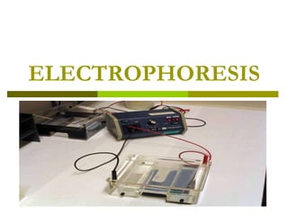
- 6. Electrophoresis Equipment Power supply Electrophoresis unit: for the separation of the required molecules. In addition a system is needed to : a- visualize separated molecules. b- quantitate separated molecules (viz: densitometer or colirometer).
- 7. Electrophoresis unit………. 1- Two electrodes : cathode(-) and anode(+) - Best type: Platinum - Stainless steel may be eroded by the buffer. 2-Buffer reservoirs 3- A support for the electrophoresis medium. 4- An insulating cover to minimize evaporation of buffer.
- 13. Buffers…… Importance: A- Transmit current. B- Adjust pH determine charge on solute C- Facilitate the migration of substances to be separated.
- 14. Supporting medium. Paper (old -fashioned) Cellulose acetate membrane (made by treating cellulose with acetic anhydride). Gels (most common). a- Agarose b- Polyacrylamide (PAG). They act also as molecular sieves and so separate according to M.W. (size & shape) in addition to charge.
- 15. The actual process of DNA electrophoresis
- 18. The instrument being used to load the samples is called a Micropipettor and can pipette very small volumes
- 19. Once the gel has been prepared and loaded, the electrical leads on the running tank are connected to a power supply like the one shown in the photo. The power supply has 3 needle gauges on it, showing what the voltage, current, and power levels
- 21. The box the gel is sitting on is called a UV Transilluminator, wear a UV-protective face shield.
- 23. A gel is prepared which will act as a support for separation of the fragments of DNA. The gel is a jello-like material, usually agarose, a substance derived from seaweed. Holes are created in the gel. These will serve as a reservoir to hold the DNA solution.
- 24. DNA solutions (mixtures of different sizes of DNA fragments) are loaded in a well in the gel.
- 25. The gel matrix acts as a sieve for DNA molecules. Large molecules have difficulty getting through the holes in the matrix. Small molecules move easily through the holes Because of this, large fragments will lag behind small fragments as DNAs migrate through the gel .
- 26. As the separation process continues, the separation between the larger and smaller fragments increases
- 27. The box the gel is sitting on is called a UV Transilluminator, wear a UV-protective face shield.
- 28. Molecular weight markers are often electrophoresed with DNAs . Molecular weight markers are usually a mixture of DNAs with known molecular weights Molecular weight markers are used to estimate the sizes of DNA fragments in your DNA sample
- 29. The actual process of DNA electrophoresis is again shown in the next few slides .The first step is to prepare a tray to hold the gel matrix (agarose).The ends of the tray are taped.
- 30. A "gel comb" is used to create holes in the gel.
- 31. The gel comb is placed in the tray.
- 32. Agarose powder is mixed with a buffer solution, usually tris borate EDTA (TBE buffer). The solution is heated until the agarose is dissolved. The hot agarose solution is poured into the tray and allowed to cool.
- 33. After the gel is cooled, tape is removed from the ends of the gel tray and the gel tray is placed in an electrophoresis chamber. The electrophoresis chamber is filled with buffer, covering the gel. This allows electrical current from poles at either end of the gel to flow through the gel. Finally, DNA samples are mixed with a "loading dye". The loading dye allows you to see the DNA as you load it and contains glycerol or sucrose to make the DNA sample heavy so that it will sink to the bottom of the well.
- 34. A safety cover is placed over the gel and electrodes are attached to a power supply. Electrical current is applied. DNA fragments will migrate through the gel at various rates, depending on their size. When the dye marker indicates that DNA fragments have moved through the gel, the current is turned off and the gel is removed from the tray.
- 35. DNAs are visualized by staining the gel with ethidium bromide which binds to DNA and will fluoresce in UV light. This photograph is of various types of DNA that have been electrophoresed on the same gel. Note that high molecular weight DNAs do not separate well on this gel. This can be corrected by altering gel density.
- 36. Visualization and Quantitation Visualization:…………………… Staining: a- Ethidium bromide for DNA, then visualize bands or zones by a transilluminator. b-Bromophenol blue or commasssie blue for proteins. c- Ninhydrin for amino acids. d- Sudan black for lipoproteins. e- Iodine for polysaccharides.
- 37. TYPES OF ELECTROPHORESIS Zone electrophoresis Iso-electric Focusing. Capillary electrophoresis. Two-dimensional PAGE. Pulsed field electrophoresis.
- 38. Zone electrophoresis It is migration of charged molecules as zones in porous supporting medium. Paper electrophoresis Cellulose acetate Agarose gel Polyacrylamide gel Membrane electrophoresis electrophoresis. (CAE) (AGE) (PAGE(
- 39. Paper electrophoresis It was the original support medium but is of limited use now due to the disadvantages of: - Time consumption.(16 →18 hrs)
- 40. Cellulose Acetate Membrane Electrophoresis (CAE) Advantages: -rapid procedure (20- 60minutes) -ability to store the results in membrane for long periods. Disadvantages: -need presoaking with buffer. - Need clearing before densitometric scanning - Not inert
- 41. Agarose gel electrophoresis(AGE) Agarose is sulphate free agar. Agar is a polysaccharide of sulfated galactose. Advantages:………………. * Inert little adsorbtion. *Amount of sample applied is very small (0.6-3 µL). *Short electrophoretic time (30- 90 minutes). *Native clarity permitting excellent densitometic scanning.
- 42. Polyacrylamide gel electrophoresis (PAGE). PAG is formed from polymerization of acrylamide by heating in presence of small amouts of bisacrylamide ( the cross-linking agent). Advantages of PAGE : 1. PAG is chemically inert no endosmosis 2. Clarity for densitometric scanning . 3. Pore size can be controlled by changing the proportions acrylamide and bis – acrylamide. 1.
- 45. II. Iso–Electric Focusing: xxxxxxxxx This method is ideal for separation of amphoteric substances such as proteins because it is based on the separation of molecules according to their isoelectric points (I.E.P). The method has high resolution being able to separate proteins that differ in their I.E.P by as little as 0.01 of a pH unit. It is especially useful in separation of isoenzymes.
- 46. 3.Capillary Electrophoresis xxxxxxx Involves electrophoresis of samples in a narrow tube. Advantage: They reduce problems resulting from heating defects. Because of the small diameter of the tubing.
- 48. In the first dimension, proteins are resolved in according to their isoelectric points(pIs)using isoelectric focusing (IEF) In the second dimension, proteins are separated according to their approximate molecular weight using sodium dodecyl sulfate poly-acrylamide-electrophoresis (SDS-PAGE( xxxxxxxxxxxxxxxxx