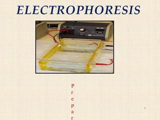
3. INTRODUCTION
In practical terms a positive electrode(anode)
and negative electrode(cathode) are placed in a
solution containing ions.
Then voltage is applied across the electrodes, so
solute ions of different charge for example,
anions (negative) and cations (positive) will
move through the solution towards the electrode
of opposite charge.
5. THEORY
Electrohoresis is the movement of ions under the
influence of electrical field. So, separation in
electrophoresis relies on differences in the speed of
migration (migration velocity) of ions or solutes.
• Migration Velocity (ν) =μe E
Where, μe=elecrophoretic mobility E=electrical field
strength
Electrophoretic mobility is a factor that determines
how fast ion or solute moves in a given medium.
• μe = q/ 6Лήr
• where, q=charge of ion ή=viscosity of solution
r=radius of ion
6. The instrumentation of capillary electrophoresis is very similar to
that of HPLC.
Power supply of E is equivalent to HPLC pump and capillary
equivalent to column.
COMPARISON OF ELECTROPHORETICAND
CHROMATOGRAPHIC TERMS
5
8. Principle of Electrophoresis
• Electromigration :
– Ions migrating in electric field
Cations cathode (-ve)
Anions anode (+ve)
7
-+
9. Structure and Properties of Protein
• PROTEINS - polymers of amino acids
• STRUCTURE of AminoAcids
– aa's have a carboxyl group (-COOH) & amino group (-NH2) and
are often ionized at physiological pH
• Proteins are amphoteric compounds and are therefore either positively
or negatively charged.
8
10. Migration of proteins in electric field
negatively charged proteins move towards the
positive pole
directly proportional to the overall charge of
proteins
inversely proportional to protein size (molecular
weight)
11. Electropherogram
An Electropherogram is the
result of an electrophoresis
which gives the movement of
charged particles over time in a
gel, paper or another medium.
10
19. Buffer solution added to the tank
This ensures that the electric current goes through the whole tank and that maintains
that ions can move in the solution
21. Electrical current applied to the chamber
Safety cover is put over the top and the current is switched on.
The dye will migrate through the gel toward the positive electrode, as will the DNA
Depending on how much voltage is applied and how warm the gel is and size and shape of
molecules will depend on how fast the ions move through the gel
Smaller fragments will move easier so they will be closer to the positive electrode
Once the dye has moved through the gel to the buffer, the electrical current is switched off
and gel is removed from the tray
22. VISUALISATION
• After the electrophoresis is complete, the molecules in the
gel can be stained to make them visible.
• Ethidium bromide, silver, or coomassie blue dye may
be used for this process.
• If the analyte molecules fluoresce under ultraviolet light,
a photograph can be taken of the gel under ultraviolet
lighting conditions.
• If the molecules to be separated contain radioactivity
added for visibility, an autoradiogram can be recorded of
the gel.
23. inversely proportional to the
logarithm
molecule.
of the size of the
22
There are molecular weight size
markers available that contain a
mixture of molecules of known
sizes. If such a marker was run on
one lane in the gel parallel to the
unknown samples, the bands
observed can be compared to
those of the unknown in order to
determine their size. The distance
a band travels is approximately
24. Quantification of separated protein band by Densitometer
Densitometer is a device that measures
the degree of darkness in photographic
or semi-transparent material.
25. Types of Electrophoresis:
Zone Electrophoresis:
a) Paper electrophoresis
b) Gel electrophoresis
c) Cellulose acetate electrophoresis
d) Thin layer electrophoresis
Moving boundary electrophoresis:
a) Capillary electrophoresis
b) Isoelectric focussing
c) Immuno electrophoresis
26. Applications of Electrophoresis
1. Forensics
DNA fingerprint of a criminal.
1. Molecular Biology To separate and organize DNA and RNA by
size
2. Genetics Provide clearer picture of DNA, it also helps prepare
DNA for cloning and genetic engineering.
3. Microbiology Information out about the organisms. Virology : to
help diagnose different strains of viruses.
4. Biochemistry Mapping of cellular components, particularly
proteins and nucleic acids.
49
27. 5. Protein and peptide determination:
50
(1) To check Purity
(2) Physical properties like MW, pI
(3) Binding studies
(4) Identification (CE-MS)
(5) Quantitation
(6) Immunoassays
1. Determination of additives in food and drug.
For example, Separation of Tartazine, Sunset yellow, amaranth,
indigo carmine
1.Separation and Quantification of Vitamins in fruits and
vegetable.
For example, Separation and quantification of Ascorbic acid in
vegetable and fruits
8. Used for separation of enatiomer from racemic mixture.
28. 9. Determination of minerals, fatty acids and carbohydrates.
1.Determination of various organic acids like Hippuric acid,
Oxalic acid, Tartaric acid, Malic acid, Lactic acid in food.
2. Pharmaceutical assay
52