Electrophoresis
•Download as PPTX, PDF•
9 likes•2,562 views

Electrophoresis is a laboratory technique used to separate DNA, RNA, or protein molecules based on their size and electrical charge. Different types of electrophoresis. Gel electrophoresis; Agarose Gel electrophoresis; polyacrylamide gel electrophoresis; pulsed-field gel electrophoresis

Recommended
More Related Content
What's hot
What's hot (20)
Similar to Electrophoresis
Similar to Electrophoresis (20)
More from Tasmina Susmi
More from Tasmina Susmi (13)
Recently uploaded
Recently uploaded (20)
Electrophoresis
- 1. ELECTROPHORESIS Tasmina Ferdous Susmi Dept. of Genetic Engineering and Biotechnology Jashore University of Science and Technology, Bangladesh
- 2. Index Electrophoresis Gel Electrophoresis Agarose Gel Electrophoresis Polyacrylamide Gel Electrophoresis Pulsed Field Gel Electrophoresis
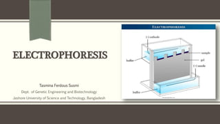
- 3. Electrophoresis A laboratory technique used to separate DNA, RNA, or protein molecules based on their size and electrical charge. Term electrophoresis describes the migration of charged particle under the influence of an electric field An electric current is used to move molecules to be separated through a medium. Pores in the gel work like a sieve, allowing smaller molecules to move faster than larger molecules. The conditions used during electrophoresis can be adjusted to separate molecules in a desired size range.
- 4. Principle of Electrophoresis The separation or purification technique of proteins, DNA, and RNA that differ in charge, size, and conformation. Charged molecules are placed at one end according to their charge and an electric field is applied. Movement of charged species in an electric field gives differential mobility to the sample molecule based on the charge and consequently resolve them On passing current, they start migrating towards opposite electrode which can be either positive or negative electrode. The size, shape, and charge of the molecule remain constant during electrophoresis and determines the mobility of ionic particles.
- 6. Factors affecting Electrophoresis Electric Field • Voltage • Current • Resistance Sample • Charge • Size • Shape Buffer • Composition • Concentratio n • pH Supporting Medium • Medium Type • Electro osmosis
- 7. Types of Electrophoresis Electrophoresis Slab Electrophoresis Zone Electrophoresis Immuno electrophoresis Isoelectro focusing Capillary Electrophoresis Gel Electrophoresis Agarose Gel Electrophoresis Polyacrylamide Gel Electrophoresis Pulse Field Gel Electrophoresis Paper Electrophoresis
- 8. Gel Electrophoresis A simple, rapid and sensitive analytical technique for the separation of charged particle. The gels, however, are porous and the size of the pores relative to that of the molecule determines whether the molecule will enter the pore and be retarded or will bypass it. The separation thus not only depends on the charge on the molecule but also on its size. Needless to say, that resolution of a sample is sharper and better in a gel than in any other type of medium. Samples are loaded into wells (indentations) at one end of a gel, and an electric current is applied to pull them through the gel. There is difference in the electrophoretic mobility of these charged molecules due to their difference in size, shape, and charge.
- 9. Gel Electrophoresis: Two Types of Materials Are Used to Make Gels Agarose Agarose is natural colloid which is isolated from the seaweed. It is linear polysaccharide. It is made up of repeating units of agarobiose, comprises alternating units of 3,6-anhydrolactose and galactose. This gel has generally larger pore size, which makes them suitable to separate larger molecules having molecular mass more than 200 kDa. It is most commonly used for the electrophoresis of both protein and nucleic acids. Agarose is used in concentration between 1% and 3%.
- 10. Gel Electrophoresis: Two Types of Materials Are Used to Make Gels Polyacrylamide Polyacrylamide gel is consisting of chains of acrylamide monomers crosslinked with N, N’- methylenebisacrylamide units, which is commonly termed as bisacrylamide. In this gel, pore size and resolving power is totally depends upon the concentration of acrylamide and bisacrylamide. The concentration of the gel normally varies from 5% to 25%. This gel is used in electrophoresis for the separation of proteins ranging from molecular weight <5000 to >200,000, and polynucleotides ranges from <5 to ~ 3000 base pairs in size.
- 11. Polyacrylamide gels Polyacrylamide gels are prepared by free radical polymerization of acrylamide and a comonomer crosslinker such as bis- acrylamide
- 12. Average Pore Size of Gel Matrix
- 13. DNA Visualization After Gel Electrophoresis Ethidium Bromide Ethidium bromide is mixed to prepare gel before electrophoresis for uniformly dispersed throughout the matrix. In the presence of ultraviolet light, ethidium bromide exhibits fluorescence. Technicians shine a specially-calibrated UV light across the gel while a machine captures the picture of the glowing fragments. Methylene Blue If a UV transilluminator is not available or practical, one can render DNA visible under normal condition by soaking the finished agarose gel, with electrophoresized DNA inside, in a solution of methylene blue overnight. The increased DNA stain density yields a deeper shade of blue, visible to the naked eye. Tracking Dyes Tracking dyes such as bromophenol blue and xylene cyanol can be used which move across the aragose gel matrices during electrophoresis at the same speed as DNA.
- 14. Agarose Gel Electrophoresis A method of gel electrophoresis used in biochemistry, molecular biology, genetics, and clinical chemistry to separate a mixed population of macromolecules such as DNA , RNA or proteins in a matrix of agarose. Agarose is a natural linear polymer extracted from seaweed that forms a gel matrix by hydrogen-bonding when heated in a buffer and allowed to cool. They are the most popular medium for the separation of moderate and large-sized nucleic acids and have a wide range of separation. Potential difference is applied across the electrodes in a horizontal electrophoretic tank containing agarose gel and biomolecules (such as nucleic acid or proteins) is loaded, then molecules migrated to their respective electrodes.
- 15. Agarose Gel Electrophoresis The rate of migration of charged particles depends on the size, shape, molecular mass etc. In this process, larger molecules have difficulty in moving through the pore size of the supporting media, whereas the smaller molecules has more mobility through it. The bands of protein or nucleic acid is visualized by using intercalating dye, i.e., ethidium bromide (Etbr), they are visualized by fluorescence when illuminated with ultraviolet lights.
- 16. Requirement Instrumentation: An electrophoretic unit, A power supply, Gel casting trays Combs Agarose gel or media Electrophoresis buffer Composition and ionic strength of electrophoresis buffer is most important factor for the separation of nucleic acids (DNA or RNA). Most routinely used buffers are: TAE- (Tris-acetate-EDTA), it has lower buffering capacity and generally used to separate larger nucleic acid fragments (>12kb).
- 17. Requirement Electrophoresis buffer Most routinely used buffers are: TBE- (Tris-borate-EDTA), it has high buffering capacity and higher ionic strength and generally used for the separation of low molecular weight compound (<1kb). Loading buffer: Nucleic acid is before loading on to a gel is first mixed with the gel loading buffer, which usually consists of:- Salts: It creates environment with favorable ionic strength and pH of the sample, e.g., Tris-HCl. Metal chelator: It prevents nucleases to degrade the nucleic acid such as EDTA. Loading dyes: It provides color for tracking and easy monitoring of sample. Such as, bromophenol blue, xylene cyanol. Transilluminator: (An ultraviolet light box), which is used to visualize bands in gels.
- 18. Steps Involved in AGE Prepare sample Prepare an agarose gel solution Gel casting Setting up the electrophoresis chamber Gel loading and sample loading Process of electrophoresis Observe the DNA Exposed under UV light
- 19. Applications of Agarose Gel Electrophoresis A routinely used method for separating proteins, DNA or RNA. Estimation of the size of DNA molecules Analysis of PCR products. Used in molecular genetic diagnosis or genetic fingerprinting. Separation of restricted genomic DNA prior to Southern analysis, or of RNA prior to Northern analysis. Widely employed to estimate the size of DNA fragments after digesting with restriction enzymes, e.g. in restriction mapping of cloned DNA.
- 20. Advantages and Disadvantages of Agarose Gel Electrophoresis Advantages For most applications, only a single-component agarose is needed and no polymerization catalysts are required. Therefore, agarose gels are simple and rapid to prepare. The gel is easily poured, does not denature the samples. The samples can also be recovered. Disadvantages Gels can melt during electrophoresis. The buffer can become exhausted. Different forms of genetic material may run in unpredictable forms.
- 21. Polyacrylamide Gel Electrophoresis (PAGE) A technique widely used in biochemistry, forensic chemistry, genetics, molecular biology and biotechnology to separate biological macromolecules, usually proteins or nucleic acids, according to their electrophoretic mobility. The most commonly used form of polyacrylamide gel electrophoresis is the Sodium dodecyl suplhate Polyacrylamide gel electrophoresis (SDS- PAGE) used mostly for the separation of proteins. Polyacrylamide gels are chemically cross-linked gels formed by the polymerization of acrylamide with a cross-linking agent, usually N,N’-methylenebisacrylamide. The goal of this technique is to separate a mixed sample of proteins to identify and quantify single proteins from the mixture. It is also possible to use PAGE to separate DNA and RNA, but proteins are the most common sample type.
- 22. Requirements for Polyacrylamide Gel Electrophoresis Acrylamide solutions (for resolving & stacking gels). Isopropanol / distilled water. Gel loading buffer. Running buffer. Staining, destaining solutions. Protein samples Molecular weight markers.
- 23. Sample preparation Preparation of polyacrylamide gel Electrophoresis Detection Steps Involved in Polyacrylamide Gel Electrophoresis Steps Involved in Polyacrylamide Gel Electrophoresis process can be described by dividing 4 steps:
- 24. Steps Involved in Polyacrylamide Gel Electrophoresis Sample preparation Samples may be any material containing proteins or nucleic acids. To analyze, the sample is optionally mixed with a chemical denaturant if so desired, usually SDS for proteins or urea for nucleic acids. Heating the samples to at least 60 °C further promotes denaturation. A tracking dye may be added to the solution. This typically has a higher electrophoretic mobility than the analytes to allow the experimenter to track the progress of the solution through the gel during the electrophoretic run.
- 25. Sample preparation Steps Involved in Polyacrylamide Gel Electrophoresis
- 26. Preparation of polyacrylamide gel + The gels typically consist of acrylamide, bisacrylamide, produces cross-linked polymerized structure. The ratio of bisacrylamide to acrylamide can be varied for special purposes, but is generally about 1 part in 35 (1:30). The acrylamide concentration of the gel can also be varied, generally in the range from 5% to 25%. Lower percentage gels are better for resolving very high molecular weight molecules, while much higher percentages of acrylamide are needed to resolve smaller proteins, Gels are usually polymerized between two glass plates in a gel caster, with a comb inserted at the top to create the sample wells. After the gel is polymerized the comb can be removed and the gel is ready for electrophoresis. Steps Involved in Polyacrylamide Gel Electrophoresis
- 27. Preparation of polyacrylamide gel Steps Involved in Polyacrylamide Gel Electrophoresis
- 28. Electrophoresis Various buffer systems are used in PAGE depending on the nature of the sample and the experimental objective. The buffers used at the anode and cathode may be the same or different. An electric field is applied across the gel, causing the negatively charged proteins or nucleic acids to migrate across the gel away from the negative and towards the positive electrode (the anode). Depending on their size, each biomolecule moves differently through the gel matrix: small molecules more easily fit through the pores in the gel, while larger ones have more difficulty. The gel is run usually for a few hours, though this depends on the voltage applied across the gel. After the set amount of time, the biomolecules will have migrated different distances based on their size. Smaller biomolecules travel farther down the gel, while larger ones remain closer to the point of origin. Steps Involved in Polyacrylamide Gel Electrophoresis
- 29. Electrophoresis Steps Involved in Polyacrylamide Gel Electrophoresis
- 30. Detection Following electrophoresis, the gel may be stained (for proteins, most commonly with Coomassie Brilliant Blue or autoradiography; for nucleic acids, ethidium bromide; or for either, silver stain), allowing visualization of the separated proteins, or processed further (e.g. Western blot). After staining, different species biomolecules appear as distinct bands within the gel. It is common to run molecular weight size markers of known molecular weight in a separate lane in the gel to calibrate the gel and determine the approximate molecular mass of unknown biomolecules by comparing the distance traveled relative to the marker. Steps Involved in Polyacrylamide Gel Electrophoresis
- 31. Applications of Polyacrylamide Gel Electrophoresis Measuring molecular weight. Peptide mapping. Estimation of protein size. Determination of protein subunits or aggregation structures. Estimation of protein purity. Protein quantitation. Monitoring protein integrity. Comparison of the polypeptide composition of different samples. Analysis of the number and size of polypeptide subunits. Post-electrophoresis applications, such as Western blotting.
- 32. Advantages and Disadvantages of Polyacrylamide Gel Electrophoresis (PAGE) Advantages Stable chemically cross-linked gel Greater resolving power (Sharp bands) Can accommodate larger quantities of DNA without significant loss in resolution The DNA recovered from polyacrylamide gels is extremely pure The pore size of the polyacrylamide gels can be altered in an easy and controllable fashion by changing the concentrations of the two monomers. Good for separation of low molecular weight fragments Disadvantages Generally more difficult to prepare and handle, involving a longer time for preparation than agarose gels. Toxic monomers Gels are tedious to prepare and often leak
- 33. Pulsed-field Gel Electrophoresis (PFGE) Pulsed-field gel electrophoresis (PFGE) is a laboratory technique used by scientists to produce a DNA fingerprint for a bacterial isolate. Pulsed Field Gel Electrophoresis (PFGE) is a technique used for the separation of large deoxyribonucleic acid (DNA) molecules by applying to a gel matrix an electric field that periodically changes direction. As DNA larger than 15-20kb migrating through a gel essentially moves together in a size- independent manner, the standard gel electrophoresis technique was unable to separate very large molecules of DNA effectively which led to the practice of pulsed field gel electrophoresis. In 1982, Schwartz introduced the concept that DNA molecules larger than 50 kb can be separated by using two alternating electric fields.
- 34. Principle of Pulsed Field Gel Electrophoresis (PFGE) While in general small fragments can find their way through the gel matrix more easily than large DNA fragments, a threshold length exists above 30–50 kb where all large fragments will run at the same rate, and appear in a gel as a single large diffuse band. However, with periodic changing of field direction, the various lengths of DNA react to the change at differing rates. That is, larger pieces of DNA will be slower to realign their charge when field direction is changed, while smaller pieces will be quicker. Over the course of time with the consistent changing of directions, each band will begin to separate more and more even at very large lengths.
- 35. Principle of Pulsed Field Gel Electrophoresis (PFGE) Thus separation of very large DNA pieces using PFGE is made possible. The procedure for this technique is relatively similar to performing a standard gel electrophoresis except that instead of constantly running the voltage in one direction, The voltage is periodically switched among three directions; one that runs through the central axis of the gel and two that run at an angle of 60 degrees either side. The pulse times are equal for each direction resulting in a net forward migration of the DNA.
- 36. Pulsed Field Gel Electrophoresis (PFGE)
- 37. Steps Involved in Pulsed-field gel electrophoresis Lysis: First, the bacterial suspension is loaded into an agarose suspension. This is done to protect the chromosomal DNA from mechanical damage by immobilizing it into agarose blocks. Then the bacterial cells are lysed to release the DNA Digestion of DNA: The bacterial DNA is treated with unusual cutting restriction enzymes so that it yields less number of larger size DNA fragments . Electrophoresis: The larger pieces of DNA are subjected to pulse field gel electrophoresis by applying electric current and altering its direction at regular intervals. Analysis: The fragments of different organisms generated by PFGE are compared to standards manually or by computer software like BioNumerics.
- 38. Steps Involved in Pulsed-field gel electrophoresis
- 39. Applications of Pulsed Field Gel Electrophoresis (PFGE) Since, field gel electrophoresis allows the separation of DNA fragments containing up to 100,000 bp (100 kilobase pairs, or kbp), characterization of such large fragments has allowed construction of a physical map for the chromosomes from several bacterial species. PFGE may be used for genotyping or genetic fingerprinting. It is commonly considered a gold standard in epidemiological studies of pathogenic organisms. Subtyping has made it easier to discriminate among strains and thus to link environmental or food isolates with clinical infections.
- 40. Advantages of Pulsed Field Gel Electrophoresis (PFGE) PFGE separates DNAs from a few kb to over 10 Mb pairs. Successfully applied to the subtyping of many pathogenic bacteria and has high concordance with epidemiological relatedness. PFGE in the same basic format can be applied as a universal generic method for subtyping of bacteria. DNA restriction patterns generated by PFGE are stable and reproducible.
- 41. Limitations of Pulsed Field Gel Electrophoresis (PFGE) Time-consuming. Requires trained and skilled technicians. Does not discriminate between all unrelated isolates. Don’t really know if bands of the same size are the same pieces of DNA. Change in one restriction site can mean more than one band change. Some strains cannot be typed by PFGE.
Editor's Notes
- NOTE: To change the image on this slide, select the picture and delete it. Then click the Pictures icon in the placeholder to insert your own image.
- The amount of resistance determines whether the circuit is a good conductor (low resistance), or a bad conductor (high resistance). The rate of migration of ions is inversely proportional to resistance. Resistance increases with the length of supporting medium but decreases with its cross-sectional area and with increase in the buffer ion concentration. An increase in temperature leads to decrease in resistance. This is due to increase mobility of ions and evaporation of the solvents from the supporting medium.