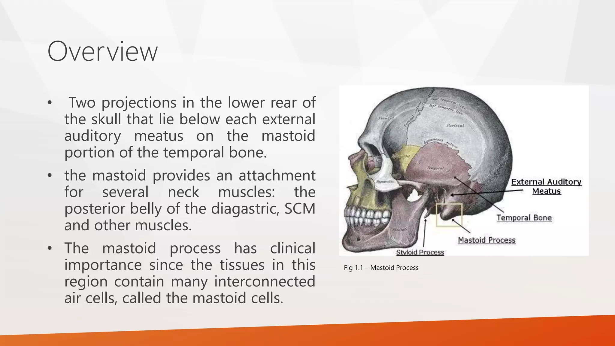
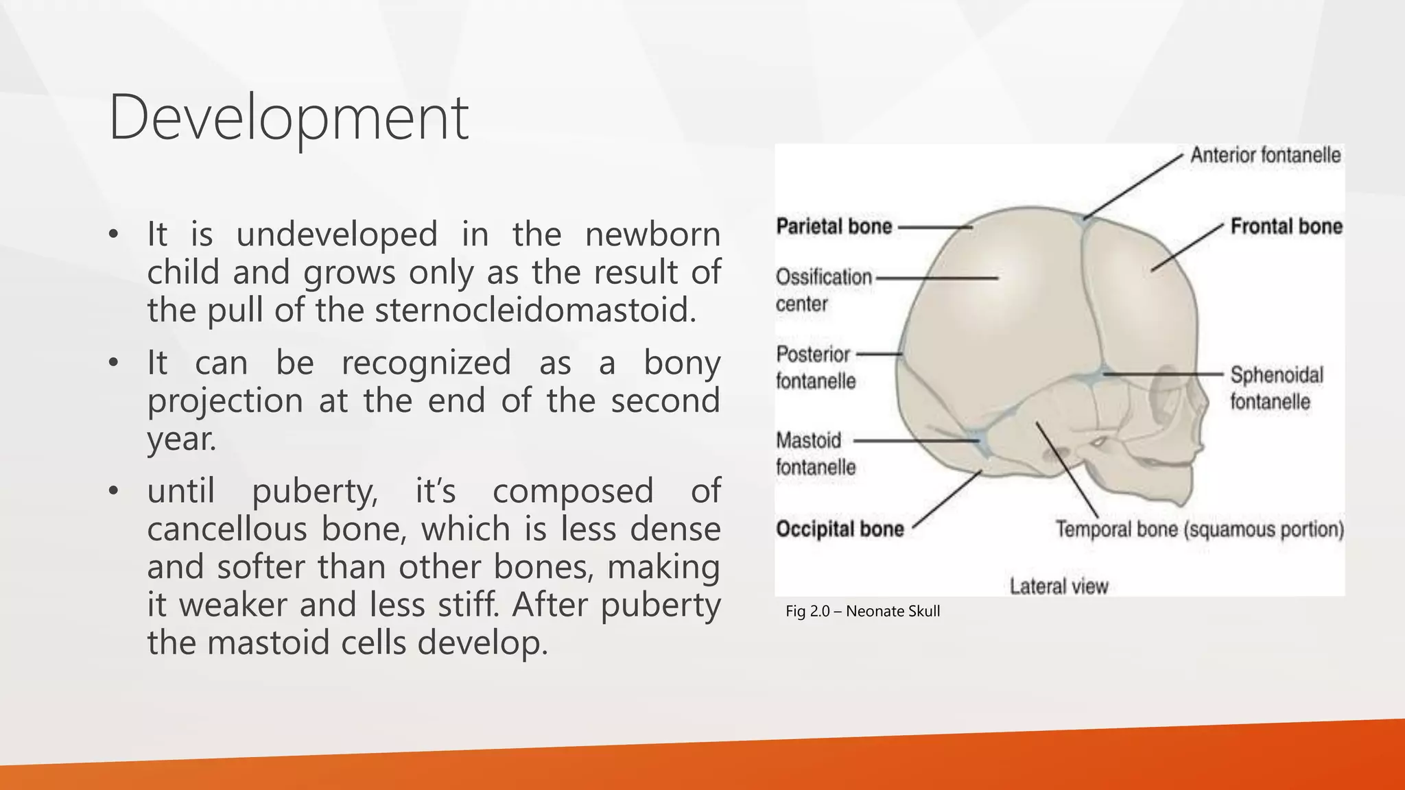
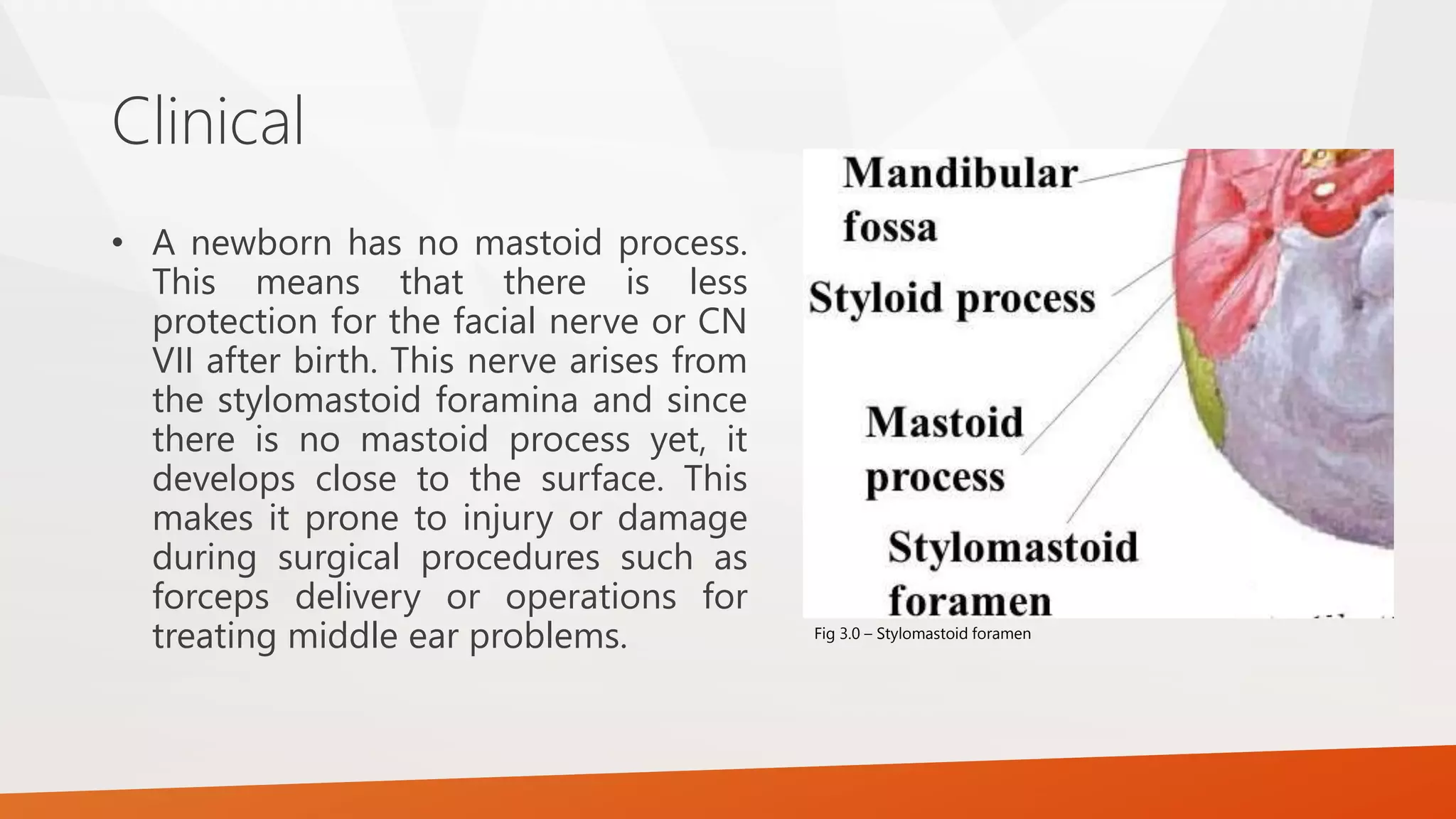
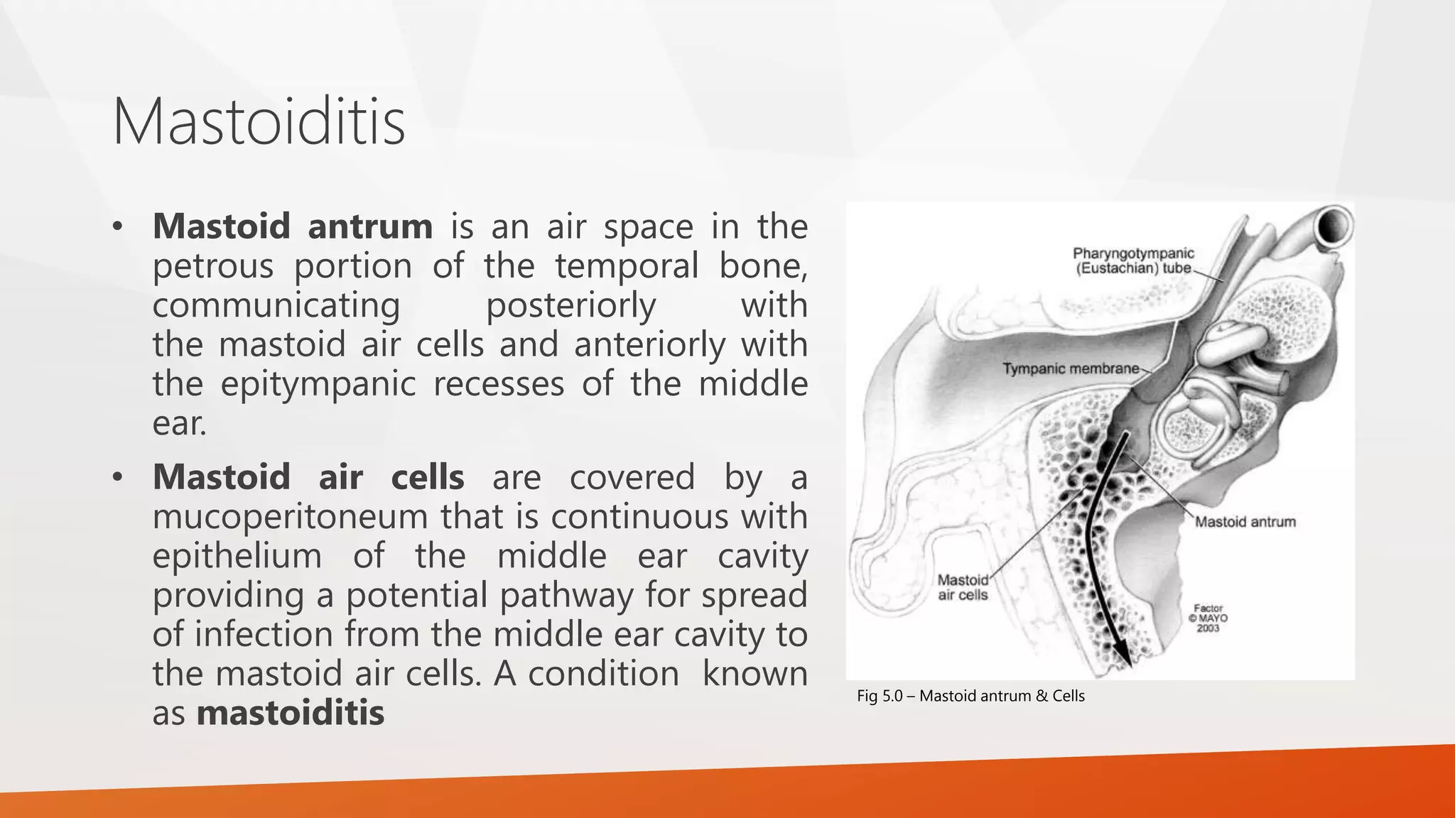
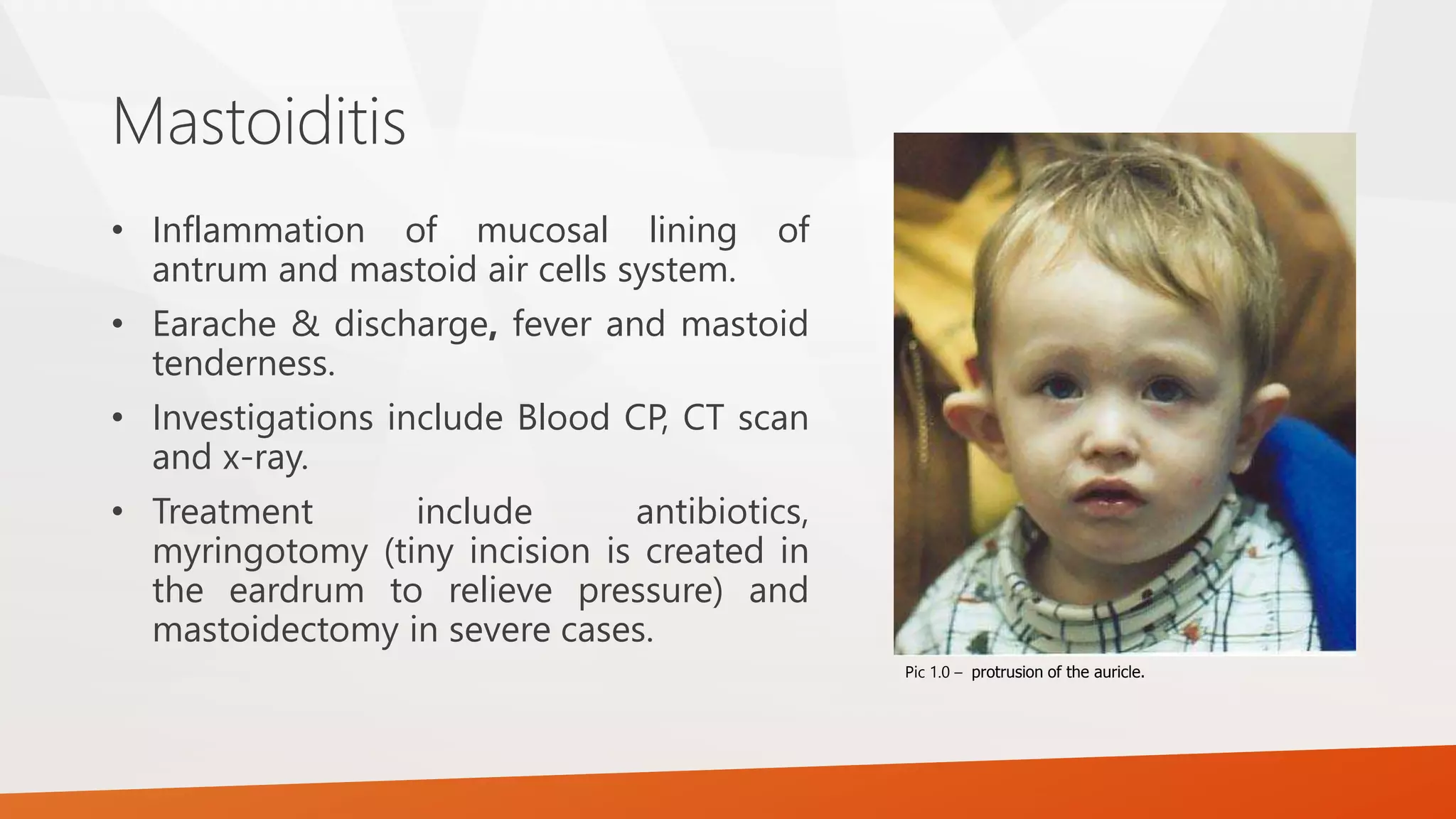
Mastoid Process
By Ali S. Mayali
Fig 1.0 – Mastoid Process
The mastoid is named for the Greek
word mastos meaning breast,
and eidos meaning resemblance.
This is reflective of the nipple-like
shape of the mastoid process.
Overview
• Two projections in the lower rear of
the skull that lie below each external
auditory meatus on the mastoid
portion of the temporal bone.
• the mastoid provides an attachment
for several neck muscles: the
posterior belly of the diagastric, SCM
and other muscles.
• The mastoid process has clinical
importance since the tissues in this
region contain many interconnected
air cells, called the mastoid cells.
Fig 1.1 – Mastoid Process
Development
• It is undeveloped in the newborn
child and grows only as the result of
the pull of the sternocleidomastoid.
• It can be recognized as a bony
projection at the end of the second
year.
• until puberty, it’s composed of
cancellous bone, which is less dense
and softer than other bones, making
it weaker and less stiff. After puberty
the mastoid cells develop.
Fig 2.0 – Neonate Skull
Clinical
• A newborn has no mastoid process.
This means that there is less
protection for the facial nerve or CN
VII after birth. This nerve arises from
the stylomastoid foramina and since
there is no mastoid process yet, it
develops close to the surface. This
makes it prone to injury or damage
during surgical procedures such as
forceps delivery or operations for
treating middle ear problems. Fig 3.0 – Stylomastoid foramen
Attachments
• Sternocleidomastoid.
• Diagastric posterior
belly.
• Splenius capitis.
• Longissimus capitis.
Fig 4.0 – Muscles attached to the mastoid bone
Mastoiditis
• Mastoid antrum is an air space in the
petrous portion of the temporal bone,
communicating posteriorly with
the mastoid air cells and anteriorly with
the epitympanic recesses of the middle
ear.
• Mastoid air cells are covered by a
mucoperitoneum that is continuous with
epithelium of the middle ear cavity
providing a potential pathway for spread
of infection from the middle ear cavity to
the mastoid air cells. A condition known
as mastoiditis
Fig 5.0 – Mastoid antrum & Cells
Mastoiditis
• Inflammation of mucosal lining of
antrum and mastoid air cells system.
• Earache & discharge, fever and mastoid
tenderness.
• Investigations include Blood CP, CT scan
and x-ray.
• Treatment include antibiotics,
myringotomy (tiny incision is created in
the eardrum to relieve pressure) and
mastoidectomy in severe cases.
Pic 1.0 – protrusion of the auricle.

Mastoid Process

  • 1.
    Mastoid Process By AliS. Mayali Fig 1.0 – Mastoid Process The mastoid is named for the Greek word mastos meaning breast, and eidos meaning resemblance. This is reflective of the nipple-like shape of the mastoid process.
  • 2.
    Overview • Two projectionsin the lower rear of the skull that lie below each external auditory meatus on the mastoid portion of the temporal bone. • the mastoid provides an attachment for several neck muscles: the posterior belly of the diagastric, SCM and other muscles. • The mastoid process has clinical importance since the tissues in this region contain many interconnected air cells, called the mastoid cells. Fig 1.1 – Mastoid Process
  • 3.
    Development • It isundeveloped in the newborn child and grows only as the result of the pull of the sternocleidomastoid. • It can be recognized as a bony projection at the end of the second year. • until puberty, it’s composed of cancellous bone, which is less dense and softer than other bones, making it weaker and less stiff. After puberty the mastoid cells develop. Fig 2.0 – Neonate Skull
  • 4.
    Clinical • A newbornhas no mastoid process. This means that there is less protection for the facial nerve or CN VII after birth. This nerve arises from the stylomastoid foramina and since there is no mastoid process yet, it develops close to the surface. This makes it prone to injury or damage during surgical procedures such as forceps delivery or operations for treating middle ear problems. Fig 3.0 – Stylomastoid foramen
  • 5.
    Attachments • Sternocleidomastoid. • Diagastricposterior belly. • Splenius capitis. • Longissimus capitis. Fig 4.0 – Muscles attached to the mastoid bone
  • 6.
    Mastoiditis • Mastoid antrumis an air space in the petrous portion of the temporal bone, communicating posteriorly with the mastoid air cells and anteriorly with the epitympanic recesses of the middle ear. • Mastoid air cells are covered by a mucoperitoneum that is continuous with epithelium of the middle ear cavity providing a potential pathway for spread of infection from the middle ear cavity to the mastoid air cells. A condition known as mastoiditis Fig 5.0 – Mastoid antrum & Cells
  • 7.
    Mastoiditis • Inflammation ofmucosal lining of antrum and mastoid air cells system. • Earache & discharge, fever and mastoid tenderness. • Investigations include Blood CP, CT scan and x-ray. • Treatment include antibiotics, myringotomy (tiny incision is created in the eardrum to relieve pressure) and mastoidectomy in severe cases. Pic 1.0 – protrusion of the auricle.