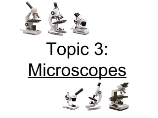
Topic 3 - Microscopes
•
100 likes•21,594 views

This topic reviews the development of the microscope, the compound microscope and other types of microscopes used in biology and science.

Recommended
More Related Content
What's hot
What's hot (20)
Viewers also liked
Viewers also liked (20)
Similar to Topic 3 - Microscopes
Similar to Topic 3 - Microscopes (20)
Recently uploaded
Recently uploaded (20)
Topic 3 - Microscopes
- 2. Definition : Microscope - a series of glass lenses in a tube that produces an enlarged, focused image of a specimen. 1. Definition
- 3. • the earliest microscope was invented around 1590 by the Janssen brothers . It had two lenses, one magnifying the other (a) The Earliest Microscopes 2. Early Microscopes
- 4. • in 1609 , Galileo (father of physics and astronomy) worked out the principles of lenses and made a much better instrument with a focusing device (a) The Earliest Microscopes 2. Early Microscopes
- 5. 2. Early Microscopes • known as the “ Father of Microscopy ” • an apprentice in a dry goods store where magnifying glasses were used to count the threads in cloth • he was able to produce lenses with magnifications of 270 diameters- the best at the time, and could see objects not visible to the naked eye. (b) Anton Van Leeuwenhoek
- 6. 2. Early Microscopes • an English physicist • improved on Leeuwenhoek's microscope design • Hooke wrote the book Micrographia , the first book to describe observations made through a microscope (c) Robert Hooke
- 7. 3. Light/Compound Microscopes The Light Microscope • uses ordinary light to magnify objects • contain two types of lenses: 1 ocular/eyepiece (10x magnification) 3 objectives (4x, 10x and 40x magnification) • the objects seen are extremely tiny and are measured in micrometers ( μ ) where 1 μ = 0.001 mm (or 1000 μ = 1 mm)
- 8. 3. Light/Compound Microscopes The ability to see an object under the microscope depends on two things: i. Magnification: how much the microscope enlarges an object . • If an object is magnified 50x, the image you see is 50 times longer and 50 times wider than if viewed 25 cm away. • total magnification is calculated by multiplying the objective lens by the ocular lens Objective Ocular (Eyepiece) Total Magnification 4x (low power) 10x = 40x 10x (medium) 10x = 100x 40x (high) 10x = 400x
- 9. 3. Light/Compound Microscopes ii. Resolution - the ability to distinguish two points as separate points. (how clear and visible an object is) • the better the resolution , the sharper the image . (but higher magnification causes a decrease in resolution) High Resolution Low Resolution
- 10. Microscope Diagram 3. Light/Compound Microscopes
- 11. 4. Other Microscopes A . Phase-Contrast Microscope • a special type of light microscope that increases differences in light and dark areas • enhances contrast of transparent and colorless specimens without the use of stains) • primarily used in biological and medical research Stages of Cell Division
- 12. 4. Other Microscopes B . Stereoscopes • has two sets of lenses for each eye that enables you to see the object in its true three dimensional form • has two eye pieces with matching objectives for each eye • magnification ranges between 3x and 50x (like a high powered magnifying glass)
- 13. C. Electron Microscopes • powerful microscopes that use electrons to create an image, instead of light waves • resolution created is thousands of times better than a compound microscope • two types: scanning electron microscope and transmission electron microscope 4. Other Microscopes
- 14. 4. Other Microscopes i. Scanning Electron Microscopes • produce a three dimensional image of the surface by bouncing electrons off of the object. • magnifies 5x – 200 000x Gun emits a beam of electrons toward the image. Beam moves across the specimen. Electrons bounce off toward the amplifying machine. An image is produced.
- 15. Scanning Electron Microscope Images Staple Through Paper (35x)- staple is seen where it ripped through the fibers of a yellow sticky note paper 4. Other Microscopes
- 16. Scanning Electron Microscope Images Mascara Brush (35x)- The bristles of this mascara brush are covered with dried mascara and flakes of skin. 4. Other Microscopes
- 17. Scanning Electron Microscope Images Black Widow Spider Claw (500x)- The claw has three hooks, the middle one is used to work the silk. 4. Other Microscopes
- 18. Scanning Electron Microscope Images Toilet Paper (500x)- The long, thin fibers you see here are really elongated cells of the pine tree. The trees are cut down, chipped up and pounded to separate the fibers from the rest of the tree material. 4. Other Microscopes
- 19. Scanning Electron Microscope Images Dentist Drill (50x)- The dentist drill is covered with tiny diamond chips. Diamonds are the hardest substance known and will easily wear away tooth particles as the drill spins at high speeds. 4. Other Microscopes
- 20. Scanning Electron Microscope Images Deer Tick (500x)- The deer tick, responsible for transmitting Lyme Disease, is about the size of a freckle. Once attached to a person, the barbed mouth, along with a special glue, allows the tick to hold fast until it has finished feeding. 4. Other Microscopes
- 21. Scanning Electron Microscope Images Porcupine Quill (50x)- Porcupine quills are sharp as needles. Unlike needles, quills have backwards facing barbs that catch on the skin making them difficult to extract. 4. Other Microscopes
- 22. Scanning Electron Microscope Images Scratch and Sniff (1000x)- These tiny glass capsules contain a liquid scent and are glued onto paper. When the paper is scratched, some of the capsules are ruptured and the scent is released. 4. Other Microscopes
- 23. Scanning Electron Microscope Images Mosquito Head (200x)- The mosquito's head is mostly eye. The eyes of most insects are compound eyes, made up of many tiny lenses. Each lens sees a slightly different picture, making up a mosaic of the object it is looking at. This type of vision is very efficient at noticing very slight motions. 4. Other Microscopes
- 24. ii. Transmission Electron Microscopes • shows the internal structure of specimens by passing beams of electrons through the object to be magnified • objects must be sliced extremely thin • magnifies 5x to 1000000 x. 4. Other Microscopes
- 25. Transmission Electron Microscope Images Ecoli Bacteria Dividing (400,000x) 4. Other Microscopes
- 26. Transmission Electron Microscope Images 4. Other Microscopes Chloroplast (40,000x) - responsible for photosynthesis in plants
- 27. A. Care of the Microscope 1. Always carry the microscope with two hands- one beneath the base and one on arm 2. Keep microscope away from the edge 3. Avoid tilting the microscope when looking in 4. Do not touch glass surfaces of the lenses with fingers (grease, dirt & perspiration) 5. Always return objective lens to low power when finished 5. Using the Microscope
- 29. Topic 3 Assignment : 1. Color the Microscope Diagram (next page) 2. Read page 19-25 and complete questions #1-3 (page 27). 3. Microscope Lab #1 – The Basics 4. Microscope Lab #2 – Live Specimens 5. Microscope Lab #3 – Abstract Slide Art