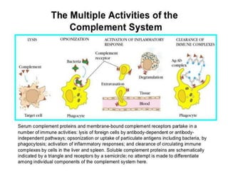
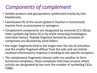
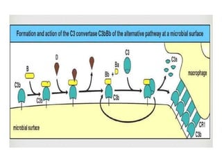
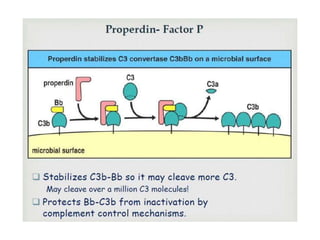
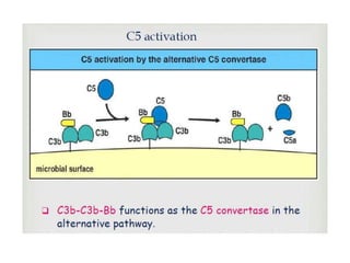
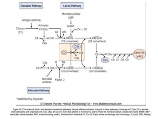
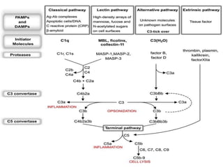
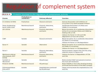
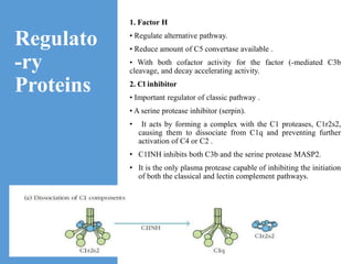
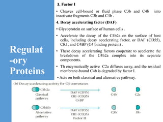
This document provides an overview of the complement system submitted by five MSc students to their professor. It describes that complement was first discovered in 1890 and plays a major role in innate immunity through lysis of cells, opsonization, and inflammation. It summarizes the three pathways of complement activation (classical, lectin, and alternative), components and regulation of the complement system, and consequences of complement activation including cell lysis, inflammation, opsonization, viral neutralization, and solubilization of immune complexes.
1. • SUBMITTED TO:
• Dr. BINDU BATTAN
• ASSISTANT PROFESSOR
• (DEPARTMMENT OF
BIOTECHNOLOGY)
• IMMUNOLOGY
• SUBMITTED BY:
• SHASHI (1)
• TEENA(5)
• MEHEK(9)
• CHITRANGNA(15)
• KIRTI(16)
• (MSc. FINAL YEAR STUDENTS)
2. COMPLEMENT SYSTEM
• Major effector of the humoral branch of immune
system .
•Research on complement began in 1890 when Jules
Border at Institute Pasteur in Paris showed that sheep
antiserum to the bacterium Vibrio cholerae caused lysis of
the bacteria and that heating the antiserum destroyed it's
activity.
•Paul Ehrlich coined term 'complement' defining it
as"activity of blood serum that completes the action of
antibody".
Activation of complement cascade may be initiated by
bye several proteins that circulate in normal
serum.These molecules, termed accused face protein
acute phase protein, dresses pattern recognition
capacity and undergo changes in concentration during
inflammation.
3. Functions of the complement
• Lysis of cells ,bacteria and viruses
• Opsonization, which promotes phagocytosis of
particulate antigen
• binding to specific complement receptors on cells of
the immune system, triggering specific cell functions,
inflammation, and secretion of immunoregulatory
molecules
• Immune clearance, which removes immune
complexes from the circulation and deposits them in
the spleen and liver
4.
5. Components of complement
• Soluble proteins and glycoproteins synthesized mainly by liver
hepatocytes
• Constituted 5% of the serum global in fraction in functionally
inactive form as proenzyme or zymogens.
• Complement components are designated by numerals (C1-C8),by
letter symbols (eg factor D) or by trivial names(eg homologous
restriction factor). Peptide fragment formed by activation of
component are denoted by small letters.
• the larger fragments bind to the target near the site of activation
and the smaller fragment diffuse from the side and can initiate
localised inflammatory response is binding to the specific receptors.
• The complement fragments interact with one another to form
functional complexes, those complexes that have enzyme attack
activity are designated by bar over the number of symbol(eg C42a,
C3Bb)
6. Complement activation
• The early steps culminating in the formation of C5b
can occur by classical pathway, the alternative
pathway or the lectin pathway.
• the final steps that lead to the formation of the
membrane attack complex or MAC are identical in
all three pathways.
7. Classical Pathway
• The classical pathway of complement activation is
considered part of adaptive immune response since it
begins with the formation of antigen-antibody complexes.
• These complexes may be soluble, or they may be formed
when an antibody binds to antigenic determinants, or
epitopes, situated on viral, fungal, parasitic, or bacterial
cell membranes.
• Only complexes formed by IgM or certain subclasses of
IgG antibodies are capable of activating the classical
complement pathway.
• The initial stage of activation involves the complement
components C1, C2, C3 and C4, which are present in
plasma as zymogens.
8. • The formation of an antigen-anribody complex induces
conformational changes in the nonantigen-binding (Fc)
portion of the antibody molecule. This conformational
change exposes a binding site for the C1 component of
complement.
• In serum, C1 exists as a macromolecular complex complex
consisting of one molecule of C1q and two molecules each of
serine proteases, C1r and C1s, held together in a Ca+²
stabilized complex.
• The C1q molecule itself is composed of 18 polypeptide chains
that associate to form six collagen like triple helical arms, the
tips of which bind the CH² domain of antigen bound antibody
molecule.
• Binding of C1q to the CH²domains of Fc regions of the antigen
complexed antibody molecule induces a conformational
change in one of the C1r molecules that converts it to an
avtive serine protease enzyme.This C1r molecule then cleaves
and activate its partner C1r molecule.
9.
10. • The two C1r proteases then cleave and activate the two
C1s molecules.C1s has two substrates, C4 and C2.C4 is
activated when C1s hydrolyzes a small fragment (C4a) from
the amino terminus of one of its chains.
• The C4b fragment attaches covalently to the target
membrane surface in the vicinity of C1, & then binds
C2.On binding C4b, C2 becomes susceptible to cleavage by
the neighboring C1s enzyme, and the smaller C2b
fragment diffuses away, leaving behind an enzymatically
active C4b2a complex. C4b2a complex is called C3
convertase.
• The membrane bound C3 convertase enzyme, C4b2a,now
hydrolyzes C3, generating two unequal fragments; the
small anaphylatoxin C3a and the pivotal fragment C3b.
11.
12. • C3b acts in three important and different way to protect
the host.
① Similar to that of C4b, C3b binds covalently to microbial surfaces,
providing a molecular "tag" & thereby allowing phagocytic cells with
C3b receptors to engulf the tagged microbes.This process is called
Opsonization.
② C3b molecules can attach to the Fc portion of antibodies
participating in soluble antigen-antibody complexes. These C3b -
tagged immune complexes are bound by C3b receptors on
phagocytes or red blood cells, & are either phagocytosed, or
conveyed to the liver where they are destroyed.
③ Some molecules of C3b bind the membrane - localized C4b2a
enzyme to form the trimolecular, membrane bound, C5 convertase
complex C4b2a3b.The C3b component of this complex binds C5 , &
the complex then cleaves C5 into two fragments: C5b and C5a.
• This trio of tasks accomplished by C3b molecule places it
right at the centre of complement attack pathways.
13.
14.
15. LECTIN PATHWAY
• Also called as MBL pathway.
• The pathway is independent of antibodies for
its activation like classical pathway
• For initiation of complement activation
process this pathway uses lectin proteins that
recognise specific carbohydrate components
primarily found on microbial surface as it’s
receptor molecules.
16. MBL-mannose binding lectin, the first lectin
demonstrated to be capable of Initiating complement
activation.
MBL are found in several microorganisms such as
Salmonella, Listeria and Niesser of bacteria.
Cryptococcus neoformans and Candida albicans strains.
MBL also recognise structure in addition to mannose,
including N- acetylglucosamine, D-glucose and L-fucose.
17.
18.
19. LECTINPATHWAY
• After MBL binds to the surface of a cell or a pathogen, MBL
associated Serine protease, MASP- 1 and MASP-2, binds to
MBL.
• The active complex formed by this association causes
cleavage and activation of C4 and C2 .
• The MASP-1 and MASP-2 proteins have structural similarity to
C1r and C1s and mimic their activities .
• This means of activating the C2-C4 components to form a C3
andC5 convertase without need for specific antibody binding
represents an important innate defence mechanism
comparable to the alternative pathway , but utilising the
elements of the classical pathway except for C1 protein.
21. TheAlternativePathway
• The alternative pathway generates bound C5b, the same product
that the classical pathway generates, but it does so without the
need for antigen-antibody complexes for initiation.
• Because no antibody is required, the alternative pathway is a
component of the innate immunesystem.
• This major pathway of complement activation involves four serum
proteins: C3,factor B,factor D,andproperdin.
• The alternative pathway is initiated in most cases by cell-surface
constituents that are foreign to the host . For example, both gram-
negative and gram-positive bacteria have cell-wall constituents that
canactivate the alternativepathway.
22. • In the classicalpathway,C3is rapidly cleaved to C3aand C3bby
the enzymatic activity of the C3convertase. In the alternative
pathway, serum C3,which contains an unstable thioester bond,
is subject to slow spontaneous hydrolysisto yield C3aand C3b.
• The C3b component can bind to foreign surface antigens (such
as those on bacterial cells or viral particles) or even to the host’s
own cells.
• The membranes of most mammalian cells have high levels of
sialic acid, which contributes to the rapid inactivation of bound
C3b molecules on host cells; consequently this binding rarely
leads to further reactions on the host cellmembrane.
• Because many foreign antigenic surfaces (e.g., bacterial cell
walls, yeast cell walls, and certain viral envelopes) have only low
levels of sialic acid, C3b bound to these surfaces remains active
for alonger time.
31. Regulato
-ry
Proteins
1. Factor H
• Regulate alternative pathway.
• Reduce amount of C5 convertase available .
• With both cofactor activity for the factor (-mediated C3b
cleavage, and decay accelerating activity.
2. Cl inhibitor
• Important regulator of classic pathway .
• A serine protease inhibitor (serpin).
• It acts by forming a complex with the C1 proteases, C1r2s2,
causing them to dissociate from C1q and preventing further
activation of C4 or C2 .
• C1INH inhibits both C3b and the serine protease MASP2.
• It is the only plasma protease capable of inhibiting the initiation
of both the classical and lectin complement pathways.
32. Regulat
-ory
Proteins
3. Factor I
• Cleaves cell-bound or fluid phase C3b and C4b into
inactivate fragments C3b and C4b .
4. Decay accelerating factor (DAF)
• Glycoprotein on surface of human cells .
• Accelerate the decay of the C4b2a on the surface of host
cells, including decay accelerating factor, or DAF (CD55),
CR1, and C4BP (C4 binding protein) .
• These decay accelerating factors cooperate to accelerate the
breakdown of the C4b2a complex into its separate
components.
• Th enzymatically active C2a diffuses away, and the residual
membrane-bound C4b is degraded by factor I.
• Acts on both classical and alternative pathway.
33. Regulat
ory
Proteins
6. C4b-binding protein (C4BP)
• Inhibits the action of C4b in classical pathway
• Splits C4 convertase and is a cofactor for
factor I
7. Complement Receptor 1 (CR-1)
• Co-factor for factor I, together with CD46
8. Protectin (CD59) and Vitronectin (S
protein)
• Inhibits formation of MAC by binding C5b678
• Present on "self" cells to prevent complement
from damaging them
34. Regulat
ory
Protein
10.Carboxypeptidases Can Inactivate the Anaphylatoxins C3a
and C5a
• Anaphylatoxin activity is regulated by cleavage of the
C-terminal arginine residues from both C3a and C5a by
serum carboxypeptidases, resulting in rapid inactivation
of
the anaphylatoxin activity .
• The specific enzymes that mediate the control of
anaphylatoxin concentrations are carboxypeptidases N,
B, and R.
• These enzymes remove arginine residues from the
carboxyl termini of C3a and C5a to form the so-called
des-Arg (“without Arginine”), inactive forms of the
molecules.
35.
36. Consequences
Of
Complement
System
1. Cell lysis by MAC
• MAC damages cell membrane by
making pores or channels in it and
allowing the free passage of various
ions and water in to the cell. This
ultimately leads to cell death.
Bacteria, enveloped viruses,
irreversibly damaged cells,
cancerous cells etc. are killed by
this mechanism commonly referred
to as complement mediated cell
lysis.
• Gram positive bacteria, which are
protected by their thick
peptidoglycan layer, bacteria with a
capsule or slime layer around their
cell wall, or non-enveloped viruses
are less susceptible to lysis.
37. 2. Inflammatory Response
• C3a, C4a and C5a are called anaphylatoxins. They bind to
receptors on mast cells and induce its degranulation leading to
release of histamine and other pharmacologically active
mediators of inflammation.
• They cause vasoconstriction and increase in vascular
permeability. Along with the C5b67, they also induce the
migration of neutrophils and monocytes to the site of
complement activation.
• This leads to an inflammatory response which is a local
protective response.
38. 3. Opsonization
• C3b and C4b act as major opsonin that coat the immune
complexes and particulate antigen.
• Phagocytic cells express receptors (CR1, CR3 and CR4) for
complement components (C3b, C4b etc.) and are able to bind
the complement coated antigen and enhance its phagocytosis.
• C5a augments this process by enhancing the CR1 expression
on phagocytes by 10 folds.
39. 4. Viral Neutralization
• Complement plays a very important role in viral neutralization
in many ways:
• Most viral particles bind to their serum antibody and form
particulate immune complex which stimulates the classical
pathway. Many viruses are also capable of activating the
alternative and lectin pathway in the absence of antibody. So,
the MAC is able to destroy the viruses.
• C3b helps in the formation of viral aggregates by acting as
opsonin and thus decrease the net number of infective viral
particles. This effect is enhanced in the presence of
serum antibody.
• Complement products also coat the viral particles. This coating
neutralizes the viral infectivity by blocking its attachment to
target cell and enhancing its phagocytosis by macrophages
through complement.
• Complement cytolyze most enveloped viruses causing
fragmentation and disintegration.
40. 5. Solubilization of Immune Complex
• C3b plays an important role in removing immune complex from
the blood. its binding to complexes facilitates their binding to
CR1 on RBCs.
• CR1 are present in higher number is granulocytes than the
RBCs, but because of much larger number of RBCs than
granulocytes in blood, RBCs account for the 90% of the total
CR1 in blood.
• Immune complexes bound to the RBCs are taken to liver and
spleen where they are phagocyted after separation from RBCs.