This document discusses programmed cell death (PCD) and apoptosis. It contains the following key points:
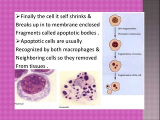
PCD is responsible for balancing cell proliferation and maintaining constant cell numbers in tissues. During apoptosis, chromosomal DNA fragments, the nucleus condenses and breaks into apoptotic bodies which are then cleared from tissues. Studies in C. elegans identified three genes, ced-3, ced-4, and ced-9, that regulate and execute apoptosis. Recent research includes developing assays to analyze apoptosis in tumor samples and investigating drugs that can fight cancer's evasion of apoptosis.
iPSCs are pluripotent; unlike ESC, iPSCs are not derived from the embryo, but instead created from differentiated cells in the lab through a process – cellular reprogramming.
Apoptosis is an orderly process in which the cell's contents are packaged into small packets of membrane for “garbage collection” by immune cells. Apoptosis removes cells during development, eliminates potentially cancerous and virus-infected cells, and maintains balance in the body.
Apoptosis(from the Greek words apo=from and ptosis=falling,)is a highly regulated biochemical mechanism of programmed cell death.
Between 50 -70 billion cells die each day due to apoptosis in the average human adult. For an average child between the age of 8-15 approximately 20-30 billion cells die a day.
German scientist Carl Vogt was first to describe the principle of apoptosis in 1842.
In 1972 Ker first introduced the term apoptosis in a publication.
The 2002 Nobel prize in medicine was awarded to Syndeney Brenner,Horvitz and John E.Sulstone for their work identifiying genes that control apoptosis.The gene were identified by studies in the Nematode C. elegans(CED4) and these same genes function in humans(APAF) for apoptosis.
There is a alignment between the nucleotide sequence of C.elegans and APAF.
Apoptosis can be normally noticed during embryogenesis,metamorphosis and aging.For example,the differentiation of human fingers in a developing embryo requires the cells between the fingers to initiate apoptosis so that the fingers could separate.
Apoptosis is needed to destroy cells infected with viruses, cells with damage DNA.
Caspase dependent apoptosis is inititate either by exrtinsc or intrinsic factors.
There are two main caspase dependent apoptotic pathway
The extrinsic or death receptor pathway
The intrinsic or mitochondrial pathway
Extra cellular signal molecules bind with cell surface receptors(termed death receptors).One typical example is a Fas receptor(FasR),a member of and FasRTNF receptor family.Its ligand is Fasl(Fatty acid synthetase ligand) expressed in cytotoxic t-lymphocytes and natural killer cells.
FasL binds FasR on the surface of a cell recruits the adptor protein, FADD(Fas-associated death domain).This occurs through interaction between the death domain of FADD and FasR.
The death effector domain(DED) of FADD active the caspase-8 from procaspase-8.
Caspase 8 activates execuitor caspases which cleave substrates within the cell.Nucleases are activated, chromosomal DNA is degraded and cell dies by apoptosis.
Apoptosis is a process of programmed cell death that occurs in multicellular organisms. Biochemical events lead to characteristic cell changes (morphology) and death.
Apoptosis is the programmed cell death. Aim of cancer therapy is to destroy the invading cells. Cancerous cells can be destroyed by increasing apoptosis.
it can occur in both physiological and pathological conditions. It is different from necrosis. In necrosis, the cell contents leak out and lead to inflammation. But in apoptosis there is no cellular leakage, only apoptotic bodies are formed. They are then engulfed by macrophages.
Pluripotent Stem Cells and their applications in disease modelling, drug disc...tara singh rawat
This ppt gives an insight of the potential and possibilities of pluripotent stem cells research in disease modelling, drug discovery and regenerative medicine
The Indian Dental Academy is the Leader in continuing dental education , training dentists in all aspects of dentistry and
offering a wide range of dental certified courses in different formats.for more details please visit
www.indiandentalacademy.com
Content-
1. Background
2. Introduction
3. Difference between apoptosis and necrosis
4. Apoptosis in biologic processes
5. Apoptosis in pathologic processes
6. Morphologic features
7. Techniques to identify and count apoptotic cells
8. Biochemical changes
9. Molecular mechanism of apoptosis
10. Recent advancement and emerging trends in apoptosis
11. References
Introduction
Definition
History
Evolution and origin of apoptosis
Significance
Purpose of apoptosis
Steps /process
Morphological and biochemical changes
Mechanism of apoptosis
Caspases
Regulation of apoptosis
Disorders of apoptosis
Application
Conclusion
Referances
Apoptosis is a
-pathway of cell death that is
-induced by an internally regulated program
-in which cells destined to die activate intrinsic enzymes that --degrade the cells’ own nuclear DNA and also nuclear and cytoplasmic proteins
-With minimal host reaction.
Nutraceutical market, scope and growth: Herbal drug technologyLokesh Patil
As consumer awareness of health and wellness rises, the nutraceutical market—which includes goods like functional meals, drinks, and dietary supplements that provide health advantages beyond basic nutrition—is growing significantly. As healthcare expenses rise, the population ages, and people want natural and preventative health solutions more and more, this industry is increasing quickly. Further driving market expansion are product formulation innovations and the use of cutting-edge technology for customized nutrition. With its worldwide reach, the nutraceutical industry is expected to keep growing and provide significant chances for research and investment in a number of categories, including vitamins, minerals, probiotics, and herbal supplements.
Cancer cell metabolism: special Reference to Lactate PathwayAADYARAJPANDEY1
Normal Cell Metabolism:
Cellular respiration describes the series of steps that cells use to break down sugar and other chemicals to get the energy we need to function.
Energy is stored in the bonds of glucose and when glucose is broken down, much of that energy is released.
Cell utilize energy in the form of ATP.
The first step of respiration is called glycolysis. In a series of steps, glycolysis breaks glucose into two smaller molecules - a chemical called pyruvate. A small amount of ATP is formed during this process.
Most healthy cells continue the breakdown in a second process, called the Kreb's cycle. The Kreb's cycle allows cells to “burn” the pyruvates made in glycolysis to get more ATP.
The last step in the breakdown of glucose is called oxidative phosphorylation (Ox-Phos).
It takes place in specialized cell structures called mitochondria. This process produces a large amount of ATP. Importantly, cells need oxygen to complete oxidative phosphorylation.
If a cell completes only glycolysis, only 2 molecules of ATP are made per glucose. However, if the cell completes the entire respiration process (glycolysis - Kreb's - oxidative phosphorylation), about 36 molecules of ATP are created, giving it much more energy to use.
IN CANCER CELL:
Unlike healthy cells that "burn" the entire molecule of sugar to capture a large amount of energy as ATP, cancer cells are wasteful.
Cancer cells only partially break down sugar molecules. They overuse the first step of respiration, glycolysis. They frequently do not complete the second step, oxidative phosphorylation.
This results in only 2 molecules of ATP per each glucose molecule instead of the 36 or so ATPs healthy cells gain. As a result, cancer cells need to use a lot more sugar molecules to get enough energy to survive.
introduction to WARBERG PHENOMENA:
WARBURG EFFECT Usually, cancer cells are highly glycolytic (glucose addiction) and take up more glucose than do normal cells from outside.
Otto Heinrich Warburg (; 8 October 1883 – 1 August 1970) In 1931 was awarded the Nobel Prize in Physiology for his "discovery of the nature and mode of action of the respiratory enzyme.
WARNBURG EFFECT : cancer cells under aerobic (well-oxygenated) conditions to metabolize glucose to lactate (aerobic glycolysis) is known as the Warburg effect. Warburg made the observation that tumor slices consume glucose and secrete lactate at a higher rate than normal tissues.
Richard's aventures in two entangled wonderlandsRichard Gill
Since the loophole-free Bell experiments of 2020 and the Nobel prizes in physics of 2022, critics of Bell's work have retreated to the fortress of super-determinism. Now, super-determinism is a derogatory word - it just means "determinism". Palmer, Hance and Hossenfelder argue that quantum mechanics and determinism are not incompatible, using a sophisticated mathematical construction based on a subtle thinning of allowed states and measurements in quantum mechanics, such that what is left appears to make Bell's argument fail, without altering the empirical predictions of quantum mechanics. I think however that it is a smoke screen, and the slogan "lost in math" comes to my mind. I will discuss some other recent disproofs of Bell's theorem using the language of causality based on causal graphs. Causal thinking is also central to law and justice. I will mention surprising connections to my work on serial killer nurse cases, in particular the Dutch case of Lucia de Berk and the current UK case of Lucy Letby.
This pdf is about the Schizophrenia.
For more details visit on YouTube; @SELF-EXPLANATORY;
https://www.youtube.com/channel/UCAiarMZDNhe1A3Rnpr_WkzA/videos
Thanks...!
Seminar of U.V. Spectroscopy by SAMIR PANDASAMIR PANDA
Spectroscopy is a branch of science dealing the study of interaction of electromagnetic radiation with matter.
Ultraviolet-visible spectroscopy refers to absorption spectroscopy or reflect spectroscopy in the UV-VIS spectral region.
Ultraviolet-visible spectroscopy is an analytical method that can measure the amount of light received by the analyte.
A brief information about the SCOP protein database used in bioinformatics.
The Structural Classification of Proteins (SCOP) database is a comprehensive and authoritative resource for the structural and evolutionary relationships of proteins. It provides a detailed and curated classification of protein structures, grouping them into families, superfamilies, and folds based on their structural and sequence similarities.
Slide 1: Title Slide
Extrachromosomal Inheritance
Slide 2: Introduction to Extrachromosomal Inheritance
Definition: Extrachromosomal inheritance refers to the transmission of genetic material that is not found within the nucleus.
Key Components: Involves genes located in mitochondria, chloroplasts, and plasmids.
Slide 3: Mitochondrial Inheritance
Mitochondria: Organelles responsible for energy production.
Mitochondrial DNA (mtDNA): Circular DNA molecule found in mitochondria.
Inheritance Pattern: Maternally inherited, meaning it is passed from mothers to all their offspring.
Diseases: Examples include Leber’s hereditary optic neuropathy (LHON) and mitochondrial myopathy.
Slide 4: Chloroplast Inheritance
Chloroplasts: Organelles responsible for photosynthesis in plants.
Chloroplast DNA (cpDNA): Circular DNA molecule found in chloroplasts.
Inheritance Pattern: Often maternally inherited in most plants, but can vary in some species.
Examples: Variegation in plants, where leaf color patterns are determined by chloroplast DNA.
Slide 5: Plasmid Inheritance
Plasmids: Small, circular DNA molecules found in bacteria and some eukaryotes.
Features: Can carry antibiotic resistance genes and can be transferred between cells through processes like conjugation.
Significance: Important in biotechnology for gene cloning and genetic engineering.
Slide 6: Mechanisms of Extrachromosomal Inheritance
Non-Mendelian Patterns: Do not follow Mendel’s laws of inheritance.
Cytoplasmic Segregation: During cell division, organelles like mitochondria and chloroplasts are randomly distributed to daughter cells.
Heteroplasmy: Presence of more than one type of organellar genome within a cell, leading to variation in expression.
Slide 7: Examples of Extrachromosomal Inheritance
Four O’clock Plant (Mirabilis jalapa): Shows variegated leaves due to different cpDNA in leaf cells.
Petite Mutants in Yeast: Result from mutations in mitochondrial DNA affecting respiration.
Slide 8: Importance of Extrachromosomal Inheritance
Evolution: Provides insight into the evolution of eukaryotic cells.
Medicine: Understanding mitochondrial inheritance helps in diagnosing and treating mitochondrial diseases.
Agriculture: Chloroplast inheritance can be used in plant breeding and genetic modification.
Slide 9: Recent Research and Advances
Gene Editing: Techniques like CRISPR-Cas9 are being used to edit mitochondrial and chloroplast DNA.
Therapies: Development of mitochondrial replacement therapy (MRT) for preventing mitochondrial diseases.
Slide 10: Conclusion
Summary: Extrachromosomal inheritance involves the transmission of genetic material outside the nucleus and plays a crucial role in genetics, medicine, and biotechnology.
Future Directions: Continued research and technological advancements hold promise for new treatments and applications.
Slide 11: Questions and Discussion
Invite Audience: Open the floor for any questions or further discussion on the topic.
1. Made by-Teena
Roll no -5
Submitted to-Dr.sulekha Chahal
BIOTECHNOLOGY DEPARTMENT
KURUKSHETRA UNIVERSITY.
2. PCD responsible for balancing cell proliferation and
maintaining constant cell no. in tissue.
•About 1011 blood cells eliminated daily
•Provides defense mechanism by which damaged and
dangerous cells can be eliminated.eg virus infected cells
•DNA damage also induce cell death
•It eliminates unwanted cell from tissue during development
eg,in amphibians there is elimination of tissue between digits
and elimination of larval tissue
•In mammalian cell about 50% neurons are eliminated
3.
4.
5.
6.
7.
8. 1. Chromosomal DNA
2. Fragmentation
3. Nucleosome cleavage
4. Chromatin condensation
5. Nucleus breaks into small pieces
6. Cell shrinks and break into membrane
enclosed fragments called apoptotic
bodies.
9.
10.
11.
12. STUDY OF C.ELEGANS
Robert identified 3 genes that regulate and execute
apoptosis
In 1986 mutagenesis of C.Elegans identified 2 genes
ced3 and ced4
3rd gene ced9 is negative regulator of apoptosis
13.
14.
15.
16.
17.
18.
19.
20.
21.
22.
23.
24.
25.
26.
27.
28.
29.
30. TAG 1 induced apoptosis-related gene
expression without tiggering apoptosis in
glioma cells
Micro RNA 29 molecule makes brain cells
resistant to programmed cell death
Researchers develop highly specific apoptosis
assay for pharmacodyanamic analysis of
tumour specimens
Role of protein in initiation of apoptosis in
melanoma
Drugs fighting aginst apoptosis evasion in
cancer