
Recommended
More Related Content
What's hot
What's hot (20)
Similar to Burn
Similar to Burn (20)
More from suchismita sethi
More from suchismita sethi (20)
Recently uploaded
Recently uploaded (20)
Burn
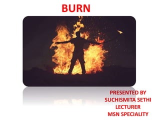
- 1. BURN PRESENTED BY SUCHISMITA SETHI LECTURER MSN SPECIALITY
- 2. DEFINITION • A burn is the injury to the tissue of the body caused by heat, chemicals, electrical current or radiation. (Lewies, 2015)
- 3. INCIDENCE:- • In India the incidence is quite high in young females due to various social factors. • The mortality rate due to burn injury is 3.5 per 100000 population. • The highest fatality rate occur in children age 4 year & younger & adult above age 65 • Nearly 11 million people need medical attention annually for burn injury & about 300000 die.
- 4. TYPES OF BURN INJURY:- 1. THERMAL BURN 2. CHEMICAL BURN 3. SMOKE & INHALATION INJURY 4. ELECTRICAL BURN 5. COLD THERMAL INJURY
- 5. 1. THERMAL BURN • Thermal burn is caused by flame, flash, scald or contact with hot object. • The severity of injury depends on the temperature of the burning agent & duration of contact time. • EX:-Scald injury are occur in during cooking.
- 6. 2. CHEMICAL BURNS:- • Chemical burn are result of acids, alkalis & organic compounds • Acids like hydrochloric acid, oxalic acid which are found in home for various purposes. • Alkalis are found in oven & drain cleaner, fertilizers. • Alkali burn difficult to manage because it adhere to tissue, causing protein hydrolysis.
- 7. 3. SMOKING & INHALATION INJURY:- • This injury caused by breathing hot air or noxious chemical which damage to the respiratory tract. • There are three type of smoke & inhalation injuries, those are:- • Metabolic asphyxiation • Upper airway injury • Lower airway injury
- 8. • Metabolic asphyxiation:- It is the inhalation of smoke elements such as carbon monoxide or hydrogen cyanide which impaired the oxygen carrying capacity of blood & caused hypoxia. Here carboxyhemoglobin causes hypoxia. • Upper airway injury:- It is the injury to the mouth, orophrynx & larynx which is caused by hot air, steam or smoke. • Upper air way injury is manifested by redness, blistering & edema. Flame burn in the neck and chest causes breathing difficulties. • Lower airway injury:- It is the injury to the trachea, bronchioles alveoli which is caused by breathing of toxic chemicals or smoke. • Clinical manifestation includes dyspnea, wheezing, altered mental status & ARDS
- 9. 4. ELECTRICAL BURN:- • Electrical burn caused by electrical current. The electrical current damage the nerve & blood vessels which cause tissue anoxia & death. • The severity of the electrical injury depend upon the amount of voltage, surface area in contact with current. 5. COLD THERMAL INJURY:- • It is the tissue injury caused by intense cold to the tissue.
- 10. CLASSIFICATION OF BURN INJURY ACCORDING TO DEAPTH • According to depth of tissue involvement the burn injury classified in to 2 categorise 1. Partial thickness burn 2. Full thickness burn
- 11. 1. PARTIAL THICKNESS BURN:- Partial thickness burn again categorised in to 2 types a. Superficial partial thickness burn( First degree burn):- • Here there is damage of epidermis, which causes hyperaemia, pain sensation present hear. • Ex:- sun burn or quick heat flash. • C/F:- Erythema, pain, mild swelling, no vesicles or blisters ( after 24 hour may blister found) • Healing time:- About 3-6 days, the superficial skin layer over the burn may be peel off in 1-2 days.
- 12. b. Deep partial thickness burn( Second degree burn):- • Here there is involvement of epidermis & dermis layer. • This type of burn may be caused by flame, flashes, scaled, chemicals or electrical current. • C/F:- Fluid field vesicles that are red, shiny, wet( if vesicle rupture). Severe pain caused by nerve injury, mild to moderate edema. • Healing time:- It depends upon the severity of burn. It may take 1-3 weeks to heal.
- 13. 3. FULL THICKNESS BURN ( THIRD & FOURTH DEGREE BURN):- • Here all the skin elements & local nerve endings destroyed, coagulation necrosis present which need surgical intervention for healing. • It s caused by flame, scald, chemical, tar & electrical current. • C/F:- Dry, waxy white hard skin, visible thrombosed vessels. Insensitive to pain due to loss of nerve. Possible involvement of muscle, tendon & bones. • Healing time:- Depth second & third degree burn need to be treated with skin graft in which healthy skin is taken from another part of body & grafted.
- 14. ACCORDING TO BURN SEVERITY:- • According to burn severity burn is classified in to 3 three categories:- 1. Minor 2. Moderate 3. Severe
- 15. 1. Minor:- All the first degree burn as well as second degree burn that involves less then 10% of body surface area. 2. Moderate burn:- Burn involving the hands, feet, face or genitals, second degree burns involving more than 10% body surface area. 3. Severe burn:- It involves more than 25% of TBSA. The third degree burn are classified as moderate or more then often as severe. Deep burn of head, hands, feet, perineum, inhalation injury & chemicals or high voltage electrical burn.
- 16. EXTENT OF BURN:- Various methods are use to estimate the extent of burn/ Total body surface area(TBSA). Those are 1. Rule of nine 2. Lund & Browder method 3. Palmer method. 4. Jackson’s Burn model.
- 17. RULE OF NINE:- • The rule of nine was devised by Pulaski & Tennison in 1947 & published by Alexander Burn Wallace in 1951. • It is the most commonly used method to estimate extent of burn in adult. • This system is based on dividing anatomic region, each represents approximately 9% of TBSA. • BODY SURFACE AREA PERCENTAGE (%) • Head & neck 9% • Arms(each) 18% ( anterior 4.5%, Posterior 4.5%) • Ant. Trunk 18% • Post. Trunk 18% • Legs(each anterior posterior) 36%( anterior 9%, posterior 9%) • Perineum 1% • ________ • 100%
- 19. 2. LUND & BROWDER METHOD: • It is the most accurate form of assessment form • BODY SURFACE AREA PERCENTAGE • Head 7% • Neck 2% • Anterior trunk 13% • Posterior trunk 13% • Right Buttock 2½ • Left Buttock 2½ • Genitalia 1 • Right upper arm 4 • Left upper arm 4 • Right lower arm 3 • Left lower arm 3 • R. Hand 2½ • L. Hand 2½ • R. Thigh 9 ½ • L. Thigh 9 ½ • R. Leg 7% • L. Leg 7% • R. foot 3 ½ • L. Foot 3 ½ • ___ 100%
- 21. 3. PALMER METHOD
- 22. SYSTEMIC EFFECT DUE TO BURN/ PATHOPHYSIOLOGY:- • FLUID & ELECTROLYTE SHIFT:- • • BURN • INCREASE VASCULAR PERMIABILITY • EDEMA INCREASE INTRAVASCULAR VOLUME • DECREASE BLOOD VOLUME INCREASE HEMATOCRITE • INCREASE VISCOCITY • INCREASE PEREPHERAL RESISTANCE • BURN SHOCK
- 23. CARDIOVASCULAR SYSTEM:- • BURN • TISSUE INJURY • ACTIVATE SYSTEMIC INFLAMATORY MEDIATORS • IT RELEASE OXYGEN REDICALS • THESE CAUSES INCREASE VASCULAR PERMIABLILITY • FLUID SHIFT FROM INTRACELLULAR SPACE TO EXTRACELLULAR SPACE • INTRAVASCULAR FLUID LOSS • HYPOVOLEMIA • SHOCK
- 24. CARDIOVASCULAR • CLINICAL MANIFESTATION:- • Angina • Jugular venous distension • Tachycardia • Dysrhythmia • Irreversible shock • Venous thromboembolism
- 25. RESPIRATORY SYSTEM:- • BURN INJURY • ADDHERANCE OF IRRITANT TO THE UPPER RESPIRATORY TRACT • RELEASE INFLAMATORY MEDIATORS • INCREASE AVASCULAR PERMIABLITY • EDIMA FORMATION • AIR WAY OBSTRUCTION & BRONCHOSPASM • EXCESSIVE SECRETION • ARDS • DEATH
- 26. EDEMA IN LUNGS
- 27. CLINICAL MANIFESTATION:- • Dyspnea • Orthopnea • Persistent hacking cough • Crackle • Restlessness
- 28. NURVOUS SYSTEM:- • BURN INJURY • INCREASE VASCULAR PERMIABILITY • CEREBRAL EDEMA • DECREASE BLOOD SUPPLY TO BRAIN • CELLULAR HYPOXIA • LOSS OF CONSCIOUSNESS/SHOCK
- 29. • CLINICAL MANIFESTATION:- • Altered mental status • Lethargy
- 30. MANAGEMENT OF BURN:- 1. Emergent 2. Acute 3. Rehabilitation
- 31. 1. EMERGENT PHASE:- • This period starts from time of burn up to 42 hour. • This phase required to resolve immediately, life threatening problems resulting from burn injury. • The emergent phase end when the fluid mobilization& Diuresis begin. • The prime goal is to prevent hypovolemic shock & prevent vital organ dysfunction, It includes I. Pre Hospital care II. Airway management. III. Fluid management IV. Wound management V. Analgesia & sedation VI. Feeding Protocol VII. Tetanus immunization VIII. Antimicrobial agent IX. Venous thromboemboilism prophylaxis
- 32. i. PRE HOSPITAL CARE:- • Remove the person from source of burn • For small thermal burn TBSA less then 10% cover the burn area with clean, cool, tap water dampened towel to minimise the injury • Do not emerge the burn wound which more then 10% because it may cause excessive heat loss. • If burn is larger then 10% or electrical burn or inhalation injury then check ABC • In case of chemical burn quickly remove the chemical particle or powder from the skin • If burn is due to CO poisoning treat the patient with 100% humidified O2.
- 33. ii. Airway management:- • Assess the breath sound, respiratory rate, rhythm, symmetry of chest excursion. • Monitor sign of hypoxia . • Early endotracheal intubation needed in case of burn with more then 10% TBSA or burn in neck & chest area. • Check ABG value frequently to know the level of hypoxia & also know metabolic acidosis & alkalosis. • Extubation is indicated when edema is resolved & usually 3-6 day after burn injury.
- 34. • Escharotomies of the chest wall may be needed in case of burn in neck & trunk which may cause respiratory distress. • Fibro optic Bronchoscopy is needed in case of smoke & inhalation injury after 6 to 12 hour to know lower airway condition. • Reposition the patient every 1-2 hours & provide suctioning & chest physiotherapy. • Bronchodilator provide to treat severe bronchospasm. • For CO poisoning administer 100% oxygen until carboxy haemoglobin level return to normal. • Fluid management
- 35. iii. Fluid management:- • Assess the fluid need of the patient. • Begin IV fluid replacement. • Insert urinary catheter to monitor the urinary out put every hour. • If more then 15% TBSA burn insert 2 large bore IV cannula. • If more then 30% TBSA burn maintain central line , arterial line for fluid medication & blood assess. • Fluid administration during the first 45 hours help to maintain circulating fluid volume. • There are three type fluid used to maintain body need, those are:- • Electrolyte such as RL • Colloids which includes plasma & plasma expanders • 5% dextrose.
- 36. Formula used for fluid calculation are:- a) Parkland formula b) Brooke army formula c) Evans formula d) Consensus formula
- 37. a. PARKLAND FORMULA:- • Parkland formula for fluid giving is:- 4ml*kg body weight*%TBSA • Colloid formula is:- 0.3 to 0.5 ml*kg body weight*% TBSA • Application of fluid:- • ½ of total fluid in first 8 hour • ¼ of total fluid in 2nd 8 hour. • ¼ of total fluid in 3rd 8 hour • After 24 hour colloid should be given when capillary permeability return to normal. • Ex- For a 70 kg patient with 50% TBSA BURN
- 38. • Ex- For a 70 kg patient with 50% TBSA BURN • Ans:- • Fluid need:- 4ml* kg body weight*%TBSA • 4ML*70KG*50= 14000Ml in 24 hour • Application of Fluid:- • ½ in first 8 hour means =7000ml in 8 hour • ¼ in 2nd 8 hour means=3500 ml in next 8 hour • ¼ of total in 3rd 8 hour means= 3500 ml in next 8 hour • Colloids Administration:- After 24 hour colloids should be given • Formula:- 0.3 to 0.5 ml * kg in body weight*% TBSA • 0.3*70*50=1050 ml= 1 lit 50 ml in 24 hour.
- 39. b. BROOKE ARMY FORMULA • Electrolyte:- 1.5ml*kg body weight*% TBSA • Colloid:- 0.5ml*kg body weight*%TBSA • Glucose:- 2000 ml for insensible loss. • APPLICATION:- • DAY-1:- Half to be given in first 8 hour, remaining half is given next 16 hour • DAY-2:- Half of colloids half of electrolyte, all sensible fluid replacement
- 40. CONT... • EX:- For 70 kg patient with 50 % burn • FLUID:- 1.5ml*70kg*50=5250, 5 lit 250ml • 1st 8 hour=2,625ml • 2nd 8 hour:-1312 ml • 3rd 8 hour:-1312ml • COLLOIDS:- 0.5ml*kg body weight*% TBSA • 0.5ml*70*50= 1750ml • In day 2nd :- half of the colloids, half of the electrolyte, all sensible fluid replace • 875 colloids+2625+2000ml= 5500ml
- 41. EVENS FORMULA:- • Electrolyte:- 1ml* body weight*% TBSA • Colloids:- 1ml*kg body weight*% TBSA • Glucose:- 2000ml for insensible loss • DAY 1:- First 24 hour:- crystalloid 1ml*kg*% TBSA+Colloid at 1ml/kg/% TBSA+2000ML glucose in water • Next 24 hour:- crystalloid at 0.5ml/kg/%TBSA+ Colloids at 0.5 ml * kg * % TBSA+2000ml of glucose in water • Ex- 70 kg patient with 50% TBSA • Day 1 :- Fluid:- 0.5ml*kg*%TBSA+0.5 ml*kg*%TBSA+2000ml=3500+3500+2000=9000ml total fluid. • Day 2:- crystalloid- 0.5ml*kg*%TBSA+Colloids 0.5ml*kg*%TBSA+2000=1750+1750+200=5500ML
- 42. • CONSENSUS FORMULA:- • RL solution 2-4 ml*kg body weight*% TBSA • Half of the fluid is given in the first 8 hour • next half of the fluid is given in next 8 hour
- 43. Wound care:- • Once the patient airway, circulation & fluid replacement is established next priority should given towards burn wound care. • Wound cleaning should perform by using gentile debridement by using scissor & forceps. • Surgical debridement should be done in Operation room, wound cleansing can be done on pt bed. • A daily shower or dressing changing done in morning & in evening dressing change should be done in patient room. • In case of antimicrobial dressing, dressing left for 3-14 day, no need to repeated dressing to prevent infection.
- 44. CONT... Patient wound treatment in done by two method • Open method:- Here patient wound covered with topical antimicrobial with no dressing over wound. • Multiple dressing change:- Sterile dressing with tropical agent, dressing done every 12-24 hour.
- 45. Feeding Protocol:- • Feeding Protocol:- • Early & aggressive nutritional nutritional support within several hour of burn injury help to decrease mortality risk, optimizing healing of burn wound. • Patient less then 20% TBSA are able to eat enough to meet their nutritional need. • For patient with ventilator provide enteral feeding (gastric or intestinal) which help to preserve GI function increase intestinal blood flow & promote optimal condition for wound healing. • Start 20-40 ml/hr & increase to the goal rate with in 24-48 hour. • Check bowel sound every 8 hourly
- 46. CONT... • High calorie, high protein , iron, multivitamin is required. • Dietary protein started at 1.2gm/kg/day & increase with subsequent increase in protein marker. • Caloric need to be meet according to Harris Benedict formula(adult), Galveston formula (children) • Fat 30% of calories to be provided as fat. • Supplemental vitamin A,C,E promote wound healing • Mineral iron, zinc promote cell integrity & haemoglobin formation.
- 47. Analgesic & sedation:- • Analgesic need to be provided for comfort. Ex- Morphin, Fentanyl, Methadone. • Sedative, hypnotics & antidepressant given to control anxiety, insomenia. Ex- Lorazepam, Midazolam.
- 48. Tetanus immunization • TT should be provide to prevent anaerobic wound contamination. If the patient not receive TT within 10 year before burn injury TT immunoglobin should be considered.
- 49. Antimicrobial Agent:- • Systemic antibiotic are not routinely used to control burn wound flora because the burn eschar has little or no blood supply & may cause multidrug resistance organism develop. • Topical antiviral agent used. Ex- Silver sulfadiazine & Mafenide acetate. • Oral infection should treated with nystatin mouth wash.
- 50. Venous thromboemboilism prophylaxis:- • Burn patient are more prone to develop DVT. • Low molecular heparine & compression stokins, sequencial compression device needed to prevent DVT.
- 51. NURSING MANAGEMENT:- • Impaired gas exchange related to carbon monoxide poisoning, smoke inhalation & upper airway obstruction. • Goal:- To maintain the adequate tissue oxygenation • Intervention:- • Provide adequate 100% humidified oxygen. • Assess the breath sound, respiratory rate, rhythm, symmetry of chest excursion. • Monitor sign of hypoxia . • Observe erythma or blister in the buccal mucosa. • Monitor ABG value • Prepare the patient for escharotomies or intubation.
- 52. Ineffective airway clearance related to edema & effects of smoke & inhalation. • Goal:- Maintain patient airway & adequate airway clearance. • Intervention:- • Maintain patient airway through proper patient positioning, removal of secretion & artificial airway if needed. • Provide humidified oxygen • Encourage patient to turn, cough & breathing. • Encourage the patient to use incentive spirometry.
- 53. Deficit fluid volume related to increase capillary permeability & evaporative loss from the burn wound. • Goal:- Restore the fluid & electrical balance. • Intervention:- • Monitor vital sign, hemodynamic monitoring, urine output as well as strict intake output. • Maintain IV line & regulate IV flui1d at appropriate rate. • Elevate head of the patient bed & elevate extremities. • Notify the physician immediately of decrease urine output.
- 55. ACUTE PHASE:- • It is the phase begin with mobilization of extracellular fluid & subsequent dieresis. • Pathophysiology:- • Diuresis from fluid mobilization & patient is less edematous. • Bowel sound return • Wound healing begin as WBC surround the burn wound & phagocytosis occur. • Necrotic tissue begin to slough. • Fibroblast lay down matrices of the collagen precursors that eventually from granulation tissue.
- 56. Clinical manifestation:- • Eschar begin separating. • Re-epithelialisation begin separating . • Hyponatremia CF:- Weakness, dizziness. Muscle cramps, fatigue, headache, tachycardia. • Hypernatremia:- Hypernatremia due to copious amount of hypertonic solution administration. • CF:- Lethargy, confusion, thirst. • Hyperkalemia:- If the patient have renal failure, adrenocortical insufficiency, with large amount of potassium release from damaged cell. • CF:- Muscle weakness, cramping, paralysis. • Hypokalemia:- Due to vomiting, diarrhea, prolonged GI suction, IV without potassium supplementation. • CF:- Fatigue, muscle weakness, leg cramp.
- 57. Laboratory Value:- • Hyponatremia develop from excessive GI suction, diarrhea, & water intake. • Hypernatremia • Hypokalemia • Hyperkalemia
- 58. Complication:- • Infection:- Due to skin destruction, decrease immunity & malpractice in wound dressing. • Cardiovascular Respiratory complication • Neurological:- Stress, edema, sepsis, sleep disturbance. • Musculoskeletal System:- • Restricted ROM • GI Syatem:- • Diarrhea, paralytic ileus, curling ulcer • Endocrine system:- increase glucose level due to stress mediated cortisol, chatacholamine release.
- 59. NURSING CARE:- • Wound care & Excision & grafting • Pain management • Physical & occupation therapy • Nutritional Therapy • Wound care & excision & grafting:-
- 60. Goal:- • Prevent infection by cleaning & debriding the area of necrotic tissue that would promote bacterial growth, promote wound re- epithelialisation & successful skin grafting.
- 61. Intervention:- • Wound cleaning should be done with soap & water or normal saline moisture gauze to gently remove the old antimicrobial agent, necrotic tissue & dried blood. • During debridement phase cover the wound with antimicrobial agent. • When wound is fully debride petroleum or paraffin gauze dressing is applied to protect the re- epithellializing keratinocytes • In case of grafting protect the graft by applying petroleum gauze dressing following by applying petroleum gauze dressing followed by saline moist middle& dry gauze outer dressing. • In case there is formation of sero sanguineous exudates aspiration needed by syringe or vacume to promote graft reepithelialisation.
- 62. Excision & Grafting:- • Management of full thickness burn wound involve removal of necrotic tissue & application of split thickness autograft skin. • To decrease the blood loss topical application of epinephrine, injection of saline. • The graft is then is placed on clean, viable tissue to achive good adherence. • The wound is covered with autograft & stappled to attached the wound. • The average healing time for a donor site is 10-14 days. • Sometimes artificial skin must replaced all function of skin & consist of both dermal & epidermal elements.
- 63. Pain management:- • Burn patient experience two kind of pain. One is continuous back ground pain that exist through out the day & night, Another type is treatment induced pain which associated with dressing change, ambulation& rehabilitation activity. • IV infusion of morphine or hydromorphone provide. • Provide lorazipam or midazolam to reduce anxiety. • Patient control analgesia used some time if patient not able to tolerate. • Pain also managed with by using non pharmacologic strategies such as relaxation, hypnosis, guided imagery, bio feed back & medication.
- 64. Physical occupational therapy:- • Physical therapy should be given in all stage of burn to regain the muscle strength & joint function. • The good timing for exercise during & after wound dressing/cleaning when skin is soft. • Passive & active ROM exercise should be done to promote healing. • For patient with neck burn encourage to sleep without pillow with hand hanging slightly over the top of the metres to encourage hyper extension • Use custom feting splint to keep the joint in functional position. More splint pressure cause skin damage or nerve damage.
- 65. Nutritional therapy:- • Adequate calories & protein is needed for wound healing. • Burn patient is in highly catabolic & highly catabolic state as a result of burn injury. • Alert patient should be encouraged to eat high protein , high carbohydrate food to met increased caloric needs • Record the patient caloric intake daily using calorie count sheet.
- 67. REHABILITATION:- • The rehabilitation begins when patient burn wound have healed & patient able to resume a level of self care activity. • It may happen as early as 2 week or as long as 7-8 month after burn injury. • Goal is to Work towards resuming the functional role towards society. Rehabilitate from any functional & cosmetic post burn reconstructive surgery that may be necessary. • When patient is able to go home teach patient & family member about wound care & dressing changing. • Pain management & nutritional need based on the individual status • Water based cream used to heal the wound area. • Low dose of histamine used at bed time if itching persist. • Continue encourage the patient for physical & occupational therapy.
- 69. Plastic surgery:- • • The plastic derived from the Greek ward “ plastikos” which means to module or to shape. • Plastic surgery is the medical speciality concerned with the correction or restoration of form & function of body structure damaged by trauma, transformed by ageing process, changed by disease process & malformation as a result of congenital defect. • Goal:- • Correction of perceived disfigurement • Restoration of impaired function • Improvement of physical appearance.
- 70. TECHNIQUE USE FOR PLASTIC SURGERY:- • Incision • Excision • Microsurgery • Chemosurgery • Electro surgery • Laser surgery • Dermabrasion • Liposuction
- 71. • Plastic surgery divided in to two major areas • RECONSTRUCTIVE SURGERY • COSMETIC SURGERY • RECONSTRUCTIVE SURGERY:- • Reconstructive surgery perform to correct functional impairment caused by burn, traumatic injury, congenital abnormality such as cleft lip or palate, removal of cancer or trauma such as mastectomy. • Common reconstructive surgeries:- • Breast reconstruction • Burn contracture surgery • Cleft lip & palate. • Injury to the limb. • Amputation • Pressure sore
- 72. RECONSTRUCTIVE MODALITIES:- • Skin Grafting:- Here a portion of skin is detached from its own blood supply & transferred as free tissue to a recipient site. It provide protection underlying tissue. • Indication:- Extensive wound, burn, surgery which require skin graft for healing. • Classification:- • Autograft:- Tissue obtained from patient own skin. • Allograft:- Obtain from donor of same species. • Xenograft:- Obtained from donor of different species.
- 73. Graft application:- • The wound is prepare for surgery . The wound is cleaned & measured. Administration of anaesthesia. • Donor skin harvested & prepaired with skin grafting knive. • The graft meshed ( multiple incision on graft ) to removed the excessive fluid leakout from underlying tissue. • Then the graft hold in place in several stitches. • Apressure bandage is applied in over the graft recipient site. • Then a VAC( vacuum apparatus called) placed over the area for 3-5 days to drain fluid & easy wound healing. • New blood vessel begin to grow within 36 hours of grafting followed by new skin.
- 74. Skin Flap:- • It is a segment of tissue that remain attached at one end while other end is moved to recipient area. • Its survival depends upon functioning of arterial & venous blood supply & lymphatic drainage. • Flap may consist of skin, mucous, muscle, adipose tissue, omentum & bone.
- 75. POST OPERATIVE MANAGEMENT :- • Initial pressure dressing will be left in place for 24- 48 hour. • If wound begin to ooze apply firm pressure for 10-15 min. • Do not give aspirin or aspirin containing medication. • Most skin graft are hold in place by a bolster dressing( cotton ball or foam) • Clean site & apply ointment to the surrounding area of the blister dressing. • Keep the graft edge moist with antibiotic ointment. • Protect the graft from sun because it may cause pigmentation changes. • Free flap:- • It is harvested from one area of body & reconstruct a defect area. •
- 76. COSMETIC SURGERY:- • Cosmetic surgery refers to the surgery that design to improve appearance. • It is perform for changes that result from ageing, to altered inherited features or because of client personal desire.
- 77. TYPES OF RECONSTRUCTIVE & COSMETIC SURGERIES:- • Liposuction:- Here there is removal of fat from different site of human body. • Tummy tuck:- It is the removal of excessive skin & excessive fat from middle & lower abdomen in order to tighten the muscle & facia of the abdominal wall. • Brachioplasty:- Here the excessive fat & skin is removed from the upper arm area in order to create a youthful look. • Thigh lift:- It is done to remove excess skin from the thigh & buttock area. • Buttock augmentation surgery:- Fat grafting done to increase the size of buttock. • Blepharoplasty:- It is the eye lid surgery which is done in case of droopy or saggy skin as well as bag around the eye.
- 78. • Endoscopic forehead & browlift :- Brow & forhead lift are perform to raise eyebrows & reduce ridges on forehead, it gives youthful look. • Rhytidectomy surgery:- It involves tightening of the facial & neck muscle as well as the removal of excessive wrinkled skin. • Rhinoplasty:- It is the reshaping of nose. • Cleft lip nose deformity:- • Cleft lip • Cleft palate • Breast reconstruction • Breast reduction • Breast lift:- Reposition of breast for youth look. • Hair transplant
- 79. DRUG USED IN TREATMENT OF BURN, RECONSTRUCTIVE & COSMETIC SURGERY • THERMAL BURN MEDICATIONS:- • Topical antibiotics:- Ex- Noosporin • Analgesic:- Morphine sulphate, • NSAID:- Ex- Advil or mortin • DRUG USED IN RECONSTRUCTIVE & COSMETIC SURGERY • Altabax:- Bacterial protein synthesis inhibitors. • Amevive:- Immunosuppresive • Avita Gel:- Treatment of acne • Bactroban cream:- Topical antibiotics • Benzomycin:- Reduce acne infection • Botox Cometics:- It blocking the nerve impulse to relax the contraction of the forhead muscle which cause the wrinkle between eyebrows.