SlideShare a Scribd company logo
1 of 8
Download to read offline
Figure:
Immunohistochemical
analysis of paraffin
embedded Human
uterus tissue. 1: α-
tubulin (Acetyl Lys40)
Monoclonal
Antibody(4A8) was
diluted at 1:200 (4
degree
Celsius,overnight). 2:
Sodium citrate pH 6.0
was used for antibody
retrieval (>98 degree
Celsius,20min). 3:
Secondary antibody was
diluted at 1:200 (room
temperature, 30min).
Negative control was
used by secondary
antibody only.
Report Number 97537-a Host Mouse
Application IHC-P Clonality Monoclonal
Model Number STJ97537 Clone ID 4A8
Antibody Name Anti-α-tubulin (Acetyl Lys40) antibody
Testing Species HUMAN Testing Tissue UTERUS
ANTIBODY VALIDATION REPORT
a. (A small amount of distilled water was added into the incubation
box to prevent evaporation of antibody).
51. Secondary antibody incubation
a. Slides were washed 3 times, with PBS on a shaker for 5min.
Shortly after the slides were dried the corresponding secondary
antibody solution was added (HRP labelled), covering the
tissues, and incubated at room temperature for 30min.
b.
52. DAB staining
a. Slides were washed 3 times, with PBS on a shaker for 5min.
b. Shortly after, the slides were dried and fresh DAB staining buffer
was added inside the circles. The staining time was adjusted
under a microscope. Yellow-brown colour represented a positive
result. Slides were washed with water to stop the staining.
c.
53. Haematoxylin staining
a. Haematoxylin was used to counter-staining for 1min, and then
the slides were washed with water. 1% Hydrochloric acid and
alcohol was added for several seconds and then washed with
water. Ammonia was used to reveal blue colour, and then
flushed with water.
b.
54. Desolation and Clearing
i. Slides were incubated sequentially into: 75% alcohol 5min, 85%
alcohol 5min, Anhydrous ethanol - 5min, Anhydrous ethanol -
5min & Xylene - 5min. Shortly after slides were dried and neutral
gum was used to seal the slides.
ii.
55. Visualization
a. Results were validated with microscope, and the slides were
scanned.
Paraffin-Embedded
Immunohistochemistry Protocol
45.
46. Tissue processing
a. Slides were incubated sequentially into Xylene; 15min –
Xylene, 15min - Anhydrous ethanol, 5min - Anhydrous
ethanol, 5min - 85% alcohol, 5min - 75% alcohol & 5min –
wash in distilled water.
b.
47. Antigen retrieval
a. Tissue slides were incubated with citric acid (PH6.0) antigen
retrieval buffer and microwaved for antigen retrieval (heated
until boiled and then stopped heating) for 8min. Slides were
then heated with medium power for 7min. During this
process slides were kept from drying out. After cooling down
at room temperature, slides were washed with PBS on
shaker for 5min, repeated for 3 times.
b.
48. Inhibition of endogenous peroxidase
a. Slides were placed in 3% Hydrogen peroxide solution, and
incubated for 10 min at room temperature without light
exposure. Slides were then washed 3 times with PBS on a
shaker for 5mins.
b.
49. BSA Blocking
a. Shortly after slides were dried, a PAP pen was used to draw
circles around the tissue sections (and to prevent draining of
the antibody solution). Inside the circles, BSA was used to
cover the tissue evenly, blocking for 30min.
b.
50. Primary antibody incubation
After blocking solution was removed a 1:200 solution of
primary antibody/PBS was added on the slide, and incubated
overnight at 4°C.
St John's Laboratory Ltd.
www.stjohnslabs.com
Figure:
Immunohistochemical
analysis of paraffin
embedded Human liver
tissue. 1: α-tubulin
(Acetyl Lys40)
Monoclonal
Antibody(4A8) was
diluted at 1:200 (4
degree
Celsius,overnight). 2:
Sodium citrate pH 6.0
was used for antibody
retrieval (>98 degree
Celsius,20min). 3:
Secondary antibody was
diluted at 1:200 (room
temperature, 30min).
Negative control was
used by secondary
antibody only.
Report Number 97537-b Host Mouse
Application IHC-P Clonality Monoclonal
Model Number STJ97537 Clone ID 4A8
Antibody Name Anti-α-tubulin (Acetyl Lys40) antibody
Testing Species HUMAN Testing Tissue LIVER
ANTIBODY VALIDATION REPORT
a. (A small amount of distilled water was added into the incubation
box to prevent evaporation of antibody).
40. Secondary antibody incubation
a. Slides were washed 3 times, with PBS on a shaker for 5min.
Shortly after the slides were dried the corresponding secondary
antibody solution was added (HRP labelled), covering the
tissues, and incubated at room temperature for 30min.
b.
41. DAB staining
a. Slides were washed 3 times, with PBS on a shaker for 5min.
b. Shortly after, the slides were dried and fresh DAB staining buffer
was added inside the circles. The staining time was adjusted
under a microscope. Yellow-brown colour represented a positive
result. Slides were washed with water to stop the staining.
c.
42. Haematoxylin staining
a. Haematoxylin was used to counter-staining for 1min, and then
the slides were washed with water. 1% Hydrochloric acid and
alcohol was added for several seconds and then washed with
water. Ammonia was used to reveal blue colour, and then
flushed with water.
b.
43. Desolation and Clearing
i. Slides were incubated sequentially into: 75% alcohol 5min, 85%
alcohol 5min, Anhydrous ethanol - 5min, Anhydrous ethanol -
5min & Xylene - 5min. Shortly after slides were dried and neutral
gum was used to seal the slides.
ii.
44. Visualization
a. Results were validated with microscope, and the slides were
scanned.
Paraffin-Embedded
Immunohistochemistry Protocol
34.
35. Tissue processing
a. Slides were incubated sequentially into Xylene; 15min –
Xylene, 15min - Anhydrous ethanol, 5min - Anhydrous
ethanol, 5min - 85% alcohol, 5min - 75% alcohol & 5min –
wash in distilled water.
b.
36. Antigen retrieval
a. Tissue slides were incubated with citric acid (PH6.0) antigen
retrieval buffer and microwaved for antigen retrieval (heated
until boiled and then stopped heating) for 8min. Slides were
then heated with medium power for 7min. During this
process slides were kept from drying out. After cooling down
at room temperature, slides were washed with PBS on
shaker for 5min, repeated for 3 times.
b.
37. Inhibition of endogenous peroxidase
a. Slides were placed in 3% Hydrogen peroxide solution, and
incubated for 10 min at room temperature without light
exposure. Slides were then washed 3 times with PBS on a
shaker for 5mins.
b.
38. BSA Blocking
a. Shortly after slides were dried, a PAP pen was used to draw
circles around the tissue sections (and to prevent draining of
the antibody solution). Inside the circles, BSA was used to
cover the tissue evenly, blocking for 30min.
b.
39. Primary antibody incubation
After blocking solution was removed a 1:200 solution of
primary antibody/PBS was added on the slide, and incubated
overnight at 4°C.
St John's Laboratory Ltd.
www.stjohnslabs.com
Figure:
Immunohistochemical
analysis of paraffin
embedded Human liver
cancer tissue. 1: α-
tubulin (Acetyl Lys40)
Monoclonal
Antibody(4A8) was
diluted at 1:200 (4
degree
Celsius,overnight). 2:
Sodium citrate pH 6.0
was used for antibody
retrieval (>98 degree
Celsius,20min). 3:
Secondary antibody was
diluted at 1:200 (room
temperature, 30min).
Negative control was
used by secondary
antibody only.
Report Number 97537-c Host Mouse
Application IHC-P Clonality Monoclonal
Model Number STJ97537 Clone ID 4A8
Antibody Name Anti-α-tubulin (Acetyl Lys40) antibody
Testing Species HUMAN Testing Tissue LIVER CANCER
ANTIBODY VALIDATION REPORT
a. (A small amount of distilled water was added into the incubation
box to prevent evaporation of antibody).
29. Secondary antibody incubation
a. Slides were washed 3 times, with PBS on a shaker for 5min.
Shortly after the slides were dried the corresponding secondary
antibody solution was added (HRP labelled), covering the
tissues, and incubated at room temperature for 30min.
b.
30. DAB staining
a. Slides were washed 3 times, with PBS on a shaker for 5min.
b. Shortly after, the slides were dried and fresh DAB staining buffer
was added inside the circles. The staining time was adjusted
under a microscope. Yellow-brown colour represented a positive
result. Slides were washed with water to stop the staining.
c.
31. Haematoxylin staining
a. Haematoxylin was used to counter-staining for 1min, and then
the slides were washed with water. 1% Hydrochloric acid and
alcohol was added for several seconds and then washed with
water. Ammonia was used to reveal blue colour, and then
flushed with water.
b.
32. Desolation and Clearing
i. Slides were incubated sequentially into: 75% alcohol 5min, 85%
alcohol 5min, Anhydrous ethanol - 5min, Anhydrous ethanol -
5min & Xylene - 5min. Shortly after slides were dried and neutral
gum was used to seal the slides.
ii.
33. Visualization
a. Results were validated with microscope, and the slides were
scanned.
Paraffin-Embedded
Immunohistochemistry Protocol
23.
24. Tissue processing
a. Slides were incubated sequentially into Xylene; 15min –
Xylene, 15min - Anhydrous ethanol, 5min - Anhydrous
ethanol, 5min - 85% alcohol, 5min - 75% alcohol & 5min –
wash in distilled water.
b.
25. Antigen retrieval
a. Tissue slides were incubated with citric acid (PH6.0) antigen
retrieval buffer and microwaved for antigen retrieval (heated
until boiled and then stopped heating) for 8min. Slides were
then heated with medium power for 7min. During this
process slides were kept from drying out. After cooling down
at room temperature, slides were washed with PBS on
shaker for 5min, repeated for 3 times.
b.
26. Inhibition of endogenous peroxidase
a. Slides were placed in 3% Hydrogen peroxide solution, and
incubated for 10 min at room temperature without light
exposure. Slides were then washed 3 times with PBS on a
shaker for 5mins.
b.
27. BSA Blocking
a. Shortly after slides were dried, a PAP pen was used to draw
circles around the tissue sections (and to prevent draining of
the antibody solution). Inside the circles, BSA was used to
cover the tissue evenly, blocking for 30min.
b.
28. Primary antibody incubation
After blocking solution was removed a 1:200 solution of
primary antibody/PBS was added on the slide, and incubated
overnight at 4°C.
St John's Laboratory Ltd.
www.stjohnslabs.com
Figure:
Immunohistochemical
analysis of paraffin
embedded Human
stomach cancer tissue.
1: α-tubulin (Acetyl
Lys40) Monoclonal
Antibody(4A8) was
diluted at 1:200 (4
degree
Celsius,overnight). 2:
Sodium citrate pH 6.0
was used for antibody
retrieval (>98 degree
Celsius,20min). 3:
Secondary antibody was
diluted at 1:200 (room
temperature, 30min).
Negative control was
used by secondary
antibody only.
Report Number 97537-d Host Mouse
Application IHC-P Clonality Monoclonal
Model Number STJ97537 Clone ID 4A8
Antibody Name Anti-α-tubulin (Acetyl Lys40) antibody
Testing Species HUMAN Testing Tissue STOMACH CANCER
ANTIBODY VALIDATION REPORT
a. (A small amount of distilled water was added into the incubation
box to prevent evaporation of antibody).
18. Secondary antibody incubation
a. Slides were washed 3 times, with PBS on a shaker for 5min.
Shortly after the slides were dried the corresponding secondary
antibody solution was added (HRP labelled), covering the
tissues, and incubated at room temperature for 30min.
b.
19. DAB staining
a. Slides were washed 3 times, with PBS on a shaker for 5min.
b. Shortly after, the slides were dried and fresh DAB staining buffer
was added inside the circles. The staining time was adjusted
under a microscope. Yellow-brown colour represented a positive
result. Slides were washed with water to stop the staining.
c.
20. Haematoxylin staining
a. Haematoxylin was used to counter-staining for 1min, and then
the slides were washed with water. 1% Hydrochloric acid and
alcohol was added for several seconds and then washed with
water. Ammonia was used to reveal blue colour, and then
flushed with water.
b.
21. Desolation and Clearing
i. Slides were incubated sequentially into: 75% alcohol 5min, 85%
alcohol 5min, Anhydrous ethanol - 5min, Anhydrous ethanol -
5min & Xylene - 5min. Shortly after slides were dried and neutral
gum was used to seal the slides.
ii.
22. Visualization
a. Results were validated with microscope, and the slides were
scanned.
Paraffin-Embedded
Immunohistochemistry Protocol
12.
13. Tissue processing
a. Slides were incubated sequentially into Xylene; 15min –
Xylene, 15min - Anhydrous ethanol, 5min - Anhydrous
ethanol, 5min - 85% alcohol, 5min - 75% alcohol & 5min –
wash in distilled water.
b.
14. Antigen retrieval
a. Tissue slides were incubated with citric acid (PH6.0) antigen
retrieval buffer and microwaved for antigen retrieval (heated
until boiled and then stopped heating) for 8min. Slides were
then heated with medium power for 7min. During this
process slides were kept from drying out. After cooling down
at room temperature, slides were washed with PBS on
shaker for 5min, repeated for 3 times.
b.
15. Inhibition of endogenous peroxidase
a. Slides were placed in 3% Hydrogen peroxide solution, and
incubated for 10 min at room temperature without light
exposure. Slides were then washed 3 times with PBS on a
shaker for 5mins.
b.
16. BSA Blocking
a. Shortly after slides were dried, a PAP pen was used to draw
circles around the tissue sections (and to prevent draining of
the antibody solution). Inside the circles, BSA was used to
cover the tissue evenly, blocking for 30min.
b.
17. Primary antibody incubation
After blocking solution was removed a 1:200 solution of
primary antibody/PBS was added on the slide, and incubated
overnight at 4°C.
St John's Laboratory Ltd.
www.stjohnslabs.com
Figure:
Immunohistochemical
analysis of paraffin
embedded Rat heart
tissue. 1: α-tubulin
(Acetyl Lys40)
Monoclonal
Antibody(4A8) was
diluted at 1:200 (4
degree
Celsius,overnight). 2:
Sodium citrate pH 6.0
was used for antibody
retrieval (>98 degree
Celsius,20min). 3:
Secondary antibody was
diluted at 1:200 (room
temperature, 30min).
Negative control was
used by secondary
antibody only.
Report Number 97537-e Host Mouse
Application IHC-P Clonality Monoclonal
Model Number STJ97537 Clone ID 4A8
Antibody Name Anti-α-tubulin (Acetyl Lys40) antibody
Testing Species RAT Testing Tissue HEART
ANTIBODY VALIDATION REPORT
a. (A small amount of distilled water was added into the incubation
box to prevent evaporation of antibody).
7. Secondary antibody incubation
a. Slides were washed 3 times, with PBS on a shaker for 5min.
Shortly after the slides were dried the corresponding secondary
antibody solution was added (HRP labelled), covering the
tissues, and incubated at room temperature for 30min.
b.
8. DAB staining
a. Slides were washed 3 times, with PBS on a shaker for 5min.
b. Shortly after, the slides were dried and fresh DAB staining buffer
was added inside the circles. The staining time was adjusted
under a microscope. Yellow-brown colour represented a positive
result. Slides were washed with water to stop the staining.
c.
9. Haematoxylin staining
a. Haematoxylin was used to counter-staining for 1min, and then
the slides were washed with water. 1% Hydrochloric acid and
alcohol was added for several seconds and then washed with
water. Ammonia was used to reveal blue colour, and then
flushed with water.
b.
10. Desolation and Clearing
i. Slides were incubated sequentially into: 75% alcohol 5min, 85%
alcohol 5min, Anhydrous ethanol - 5min, Anhydrous ethanol -
5min & Xylene - 5min. Shortly after slides were dried and neutral
gum was used to seal the slides.
ii.
11. Visualization
a. Results were validated with microscope, and the slides were
scanned.
Paraffin-Embedded
Immunohistochemistry Protocol
1.
2. Tissue processing
a. Slides were incubated sequentially into Xylene; 15min –
Xylene, 15min - Anhydrous ethanol, 5min - Anhydrous
ethanol, 5min - 85% alcohol, 5min - 75% alcohol & 5min –
wash in distilled water.
b.
3. Antigen retrieval
a. Tissue slides were incubated with citric acid (PH6.0) antigen
retrieval buffer and microwaved for antigen retrieval (heated
until boiled and then stopped heating) for 8min. Slides were
then heated with medium power for 7min. During this
process slides were kept from drying out. After cooling down
at room temperature, slides were washed with PBS on
shaker for 5min, repeated for 3 times.
b.
4. Inhibition of endogenous peroxidase
a. Slides were placed in 3% Hydrogen peroxide solution, and
incubated for 10 min at room temperature without light
exposure. Slides were then washed 3 times with PBS on a
shaker for 5mins.
b.
5. BSA Blocking
a. Shortly after slides were dried, a PAP pen was used to draw
circles around the tissue sections (and to prevent draining of
the antibody solution). Inside the circles, BSA was used to
cover the tissue evenly, blocking for 30min.
b.
6. Primary antibody incubation
After blocking solution was removed a 1:200 solution of
primary antibody/PBS was added on the slide, and incubated
overnight at 4°C.
St John's Laboratory Ltd.
www.stjohnslabs.com
Figure:
Immunohistochemical
analysis of paraffin
embedded Rat kidney
tissue. 1: α-tubulin
(Acetyl Lys40)
Monoclonal
Antibody(4A8) was
diluted at 1:200 (4
degree
Celsius,overnight). 2:
Sodium citrate pH 6.0
was used for antibody
retrieval (>98 degree
Celsius,20min). 3:
Secondary antibody was
diluted at 1:200 (room
temperature, 30min).
Negative control was
used by secondary
antibody only.
Report Number 97537-f Host Mouse
Application IHC-P Clonality Monoclonal
Model Number STJ97537 Clone ID 4A8
Antibody Name Anti-α-tubulin (Acetyl Lys40) antibody
Testing Species RAT Testing Tissue KIDNEY
ANTIBODY VALIDATION REPORT
c. (A small amount of distilled water was added into the incubation
box to prevent evaporation of antibody).
56. Secondary antibody incubation
a. Slides were washed 3 times, with PBS on a shaker for 5min.
Shortly after the slides were dried the corresponding secondary
antibody solution was added (HRP labelled), covering the
tissues, and incubated at room temperature for 30min.
b.
57. DAB staining
a. Slides were washed 3 times, with PBS on a shaker for 5min.
b. Shortly after, the slides were dried and fresh DAB staining buffer
was added inside the circles. The staining time was adjusted
under a microscope. Yellow-brown colour represented a positive
result. Slides were washed with water to stop the staining.
c.
58. Haematoxylin staining
a. Haematoxylin was used to counter-staining for 1min, and then
the slides were washed with water. 1% Hydrochloric acid and
alcohol was added for several seconds and then washed with
water. Ammonia was used to reveal blue colour, and then
flushed with water.
b.
59. Desolation and Clearing
i. Slides were incubated sequentially into: 75% alcohol 5min, 85%
alcohol 5min, Anhydrous ethanol - 5min, Anhydrous ethanol -
5min & Xylene - 5min. Shortly after slides were dried and neutral
gum was used to seal the slides.
ii.
60. Visualization
a. Results were validated with microscope, and the slides were
scanned.
Paraffin-Embedded
Immunohistochemistry Protocol
61.
62. Tissue processing
a. Slides were incubated sequentially into Xylene; 15min –
Xylene, 15min - Anhydrous ethanol, 5min - Anhydrous
ethanol, 5min - 85% alcohol, 5min - 75% alcohol & 5min –
wash in distilled water.
b.
63. Antigen retrieval
a. Tissue slides were incubated with citric acid (PH6.0) antigen
retrieval buffer and microwaved for antigen retrieval (heated
until boiled and then stopped heating) for 8min. Slides were
then heated with medium power for 7min. During this
process slides were kept from drying out. After cooling down
at room temperature, slides were washed with PBS on
shaker for 5min, repeated for 3 times.
b.
64. Inhibition of endogenous peroxidase
a. Slides were placed in 3% Hydrogen peroxide solution, and
incubated for 10 min at room temperature without light
exposure. Slides were then washed 3 times with PBS on a
shaker for 5mins.
b.
65. BSA Blocking
a. Shortly after slides were dried, a PAP pen was used to draw
circles around the tissue sections (and to prevent draining of
the antibody solution). Inside the circles, BSA was used to
cover the tissue evenly, blocking for 30min.
b.
66. Primary antibody incubation
After blocking solution was removed a 1:200 solution of
primary antibody/PBS was added on the slide, and incubated
overnight at 4°C.
St John's Laboratory Ltd.
www.stjohnslabs.com
Figure:
Immunohistochemical
analysis of paraffin
embedded Mouse heart
tissue. 1: α-tubulin
(Acetyl Lys40)
Monoclonal
Antibody(4A8) was
diluted at 1:200 (4
degree
Celsius,overnight). 2:
Sodium citrate pH 6.0
was used for antibody
retrieval (>98 degree
Celsius,20min). 3:
Secondary antibody was
diluted at 1:200 (room
temperature, 30min).
Negative control was
used by secondary
antibody only.
Report Number 97537-g Host Mouse
Application IHC-P Clonality Monoclonal
Model Number STJ97537 Clone ID 4A8
Antibody Name Anti-α-tubulin (Acetyl Lys40) antibody
Testing Species MOUSE Testing Tissue HEART
ANTIBODY VALIDATION REPORT
b. (A small amount of distilled water was added into the incubation
box to prevent evaporation of antibody).
67. Secondary antibody incubation
a. Slides were washed 3 times, with PBS on a shaker for 5min.
Shortly after the slides were dried the corresponding secondary
antibody solution was added (HRP labelled), covering the
tissues, and incubated at room temperature for 30min.
b.
68. DAB staining
a. Slides were washed 3 times, with PBS on a shaker for 5min.
b. Shortly after, the slides were dried and fresh DAB staining buffer
was added inside the circles. The staining time was adjusted
under a microscope. Yellow-brown colour represented a positive
result. Slides were washed with water to stop the staining.
c.
69. Haematoxylin staining
a. Haematoxylin was used to counter-staining for 1min, and then
the slides were washed with water. 1% Hydrochloric acid and
alcohol was added for several seconds and then washed with
water. Ammonia was used to reveal blue colour, and then
flushed with water.
b.
70. Desolation and Clearing
i. Slides were incubated sequentially into: 75% alcohol 5min, 85%
alcohol 5min, Anhydrous ethanol - 5min, Anhydrous ethanol -
5min & Xylene - 5min. Shortly after slides were dried and neutral
gum was used to seal the slides.
ii.
71. Visualization
a. Results were validated with microscope, and the slides were
scanned.
Paraffin-Embedded
Immunohistochemistry Protocol
72.
73. Tissue processing
a. Slides were incubated sequentially into Xylene; 15min –
Xylene, 15min - Anhydrous ethanol, 5min - Anhydrous
ethanol, 5min - 85% alcohol, 5min - 75% alcohol & 5min –
wash in distilled water.
b.
74. Antigen retrieval
a. Tissue slides were incubated with citric acid (PH6.0) antigen
retrieval buffer and microwaved for antigen retrieval (heated
until boiled and then stopped heating) for 8min. Slides were
then heated with medium power for 7min. During this
process slides were kept from drying out. After cooling down
at room temperature, slides were washed with PBS on
shaker for 5min, repeated for 3 times.
b.
75. Inhibition of endogenous peroxidase
a. Slides were placed in 3% Hydrogen peroxide solution, and
incubated for 10 min at room temperature without light
exposure. Slides were then washed 3 times with PBS on a
shaker for 5mins.
b.
76. BSA Blocking
a. Shortly after slides were dried, a PAP pen was used to draw
circles around the tissue sections (and to prevent draining of
the antibody solution). Inside the circles, BSA was used to
cover the tissue evenly, blocking for 30min.
b.
77. Primary antibody incubation
After blocking solution was removed a 1:200 solution of
primary antibody/PBS was added on the slide, and incubated
overnight at 4°C.
St John's Laboratory Ltd.
www.stjohnslabs.com
Figure:
Immunohistochemical
analysis of paraffin
embedded Mouse
kidney tissue. 1: α-
tubulin (Acetyl Lys40)
Monoclonal
Antibody(4A8) was
diluted at 1:200 (4
degree
Celsius,overnight). 2:
Sodium citrate pH 6.0
was used for antibody
retrieval (>98 degree
Celsius,20min). 3:
Secondary antibody was
diluted at 1:200 (room
temperature, 30min).
Negative control was
used by secondary
antibody only.
Report Number 97537-h Host Mouse
Application IHC-P Clonality Monoclonal
Model Number STJ97537 Clone ID 4A8
Antibody Name Anti-α-tubulin (Acetyl Lys40) antibody
Testing Species MOUSE Testing Tissue KIDNEY
ANTIBODY VALIDATION REPORT
b. (A small amount of distilled water was added into the incubation
box to prevent evaporation of antibody).
78. Secondary antibody incubation
a. Slides were washed 3 times, with PBS on a shaker for 5min.
Shortly after the slides were dried the corresponding secondary
antibody solution was added (HRP labelled), covering the
tissues, and incubated at room temperature for 30min.
b.
79. DAB staining
a. Slides were washed 3 times, with PBS on a shaker for 5min.
b. Shortly after, the slides were dried and fresh DAB staining buffer
was added inside the circles. The staining time was adjusted
under a microscope. Yellow-brown colour represented a positive
result. Slides were washed with water to stop the staining.
c.
80. Haematoxylin staining
a. Haematoxylin was used to counter-staining for 1min, and then
the slides were washed with water. 1% Hydrochloric acid and
alcohol was added for several seconds and then washed with
water. Ammonia was used to reveal blue colour, and then
flushed with water.
b.
81. Desolation and Clearing
i. Slides were incubated sequentially into: 75% alcohol 5min, 85%
alcohol 5min, Anhydrous ethanol - 5min, Anhydrous ethanol -
5min & Xylene - 5min. Shortly after slides were dried and neutral
gum was used to seal the slides.
ii.
82. Visualization
a. Results were validated with microscope, and the slides were
scanned.
Paraffin-Embedded
Immunohistochemistry Protocol
83.
84. Tissue processing
a. Slides were incubated sequentially into Xylene; 15min –
Xylene, 15min - Anhydrous ethanol, 5min - Anhydrous
ethanol, 5min - 85% alcohol, 5min - 75% alcohol & 5min –
wash in distilled water.
b.
85. Antigen retrieval
a. Tissue slides were incubated with citric acid (PH6.0) antigen
retrieval buffer and microwaved for antigen retrieval (heated
until boiled and then stopped heating) for 8min. Slides were
then heated with medium power for 7min. During this
process slides were kept from drying out. After cooling down
at room temperature, slides were washed with PBS on
shaker for 5min, repeated for 3 times.
b.
86. Inhibition of endogenous peroxidase
a. Slides were placed in 3% Hydrogen peroxide solution, and
incubated for 10 min at room temperature without light
exposure. Slides were then washed 3 times with PBS on a
shaker for 5mins.
b.
87. BSA Blocking
a. Shortly after slides were dried, a PAP pen was used to draw
circles around the tissue sections (and to prevent draining of
the antibody solution). Inside the circles, BSA was used to
cover the tissue evenly, blocking for 30min.
b.
88. Primary antibody incubation
After blocking solution was removed a 1:200 solution of
primary antibody/PBS was added on the slide, and incubated
overnight at 4°C.
St John's Laboratory Ltd.
www.stjohnslabs.com

More Related Content

What's hot

Immunohistochemistry Antibody Validation Report for Anti-Histone H4 (Acetyl L...
Immunohistochemistry Antibody Validation Report for Anti-Histone H4 (Acetyl L...Immunohistochemistry Antibody Validation Report for Anti-Histone H4 (Acetyl L...
Immunohistochemistry Antibody Validation Report for Anti-Histone H4 (Acetyl L...St John's Laboratory Ltd
 
Immunohistochemistry Antibody Validation Report for Anti-α skeletal muscle ac...
Immunohistochemistry Antibody Validation Report for Anti-α skeletal muscle ac...Immunohistochemistry Antibody Validation Report for Anti-α skeletal muscle ac...
Immunohistochemistry Antibody Validation Report for Anti-α skeletal muscle ac...St John's Laboratory Ltd
 
Immunohistochemistry Antibody Validation Report for Anti-Phospho-PI 3-kinase ...
Immunohistochemistry Antibody Validation Report for Anti-Phospho-PI 3-kinase ...Immunohistochemistry Antibody Validation Report for Anti-Phospho-PI 3-kinase ...
Immunohistochemistry Antibody Validation Report for Anti-Phospho-PI 3-kinase ...St John's Laboratory Ltd
 
Immunohistochemistry Antibody Validation Report for Anti-Phospho-PTEN (S380/T...
Immunohistochemistry Antibody Validation Report for Anti-Phospho-PTEN (S380/T...Immunohistochemistry Antibody Validation Report for Anti-Phospho-PTEN (S380/T...
Immunohistochemistry Antibody Validation Report for Anti-Phospho-PTEN (S380/T...St John's Laboratory Ltd
 
Immunohistochemistry Antibody Validation Report for Anti-Phospho-mTOR (S2448)...
Immunohistochemistry Antibody Validation Report for Anti-Phospho-mTOR (S2448)...Immunohistochemistry Antibody Validation Report for Anti-Phospho-mTOR (S2448)...
Immunohistochemistry Antibody Validation Report for Anti-Phospho-mTOR (S2448)...St John's Laboratory Ltd
 
Immunohistochemistry Antibody Validation Report for Anti-Histone H3 (Acetyl K...
Immunohistochemistry Antibody Validation Report for Anti-Histone H3 (Acetyl K...Immunohistochemistry Antibody Validation Report for Anti-Histone H3 (Acetyl K...
Immunohistochemistry Antibody Validation Report for Anti-Histone H3 (Acetyl K...St John's Laboratory Ltd
 
Immunohistochemistry Antibody Validation Report for Anti-Phospho-Catenin-β (S...
Immunohistochemistry Antibody Validation Report for Anti-Phospho-Catenin-β (S...Immunohistochemistry Antibody Validation Report for Anti-Phospho-Catenin-β (S...
Immunohistochemistry Antibody Validation Report for Anti-Phospho-Catenin-β (S...St John's Laboratory Ltd
 
Immunohistochemistry Antibody Validation Report for Anti-COX IV Antibody (STJ...
Immunohistochemistry Antibody Validation Report for Anti-COX IV Antibody (STJ...Immunohistochemistry Antibody Validation Report for Anti-COX IV Antibody (STJ...
Immunohistochemistry Antibody Validation Report for Anti-COX IV Antibody (STJ...St John's Laboratory Ltd
 
Immunohistochemistry Antibody Validation Report for Anti-Phospho-IκB-α (S32/S...
Immunohistochemistry Antibody Validation Report for Anti-Phospho-IκB-α (S32/S...Immunohistochemistry Antibody Validation Report for Anti-Phospho-IκB-α (S32/S...
Immunohistochemistry Antibody Validation Report for Anti-Phospho-IκB-α (S32/S...St John's Laboratory Ltd
 
Immunohistochemistry Antibody Validation Report for Anti-Lactoferrin Antibody...
Immunohistochemistry Antibody Validation Report for Anti-Lactoferrin Antibody...Immunohistochemistry Antibody Validation Report for Anti-Lactoferrin Antibody...
Immunohistochemistry Antibody Validation Report for Anti-Lactoferrin Antibody...St John's Laboratory Ltd
 
Immunohistochemistry Antibody Validation Report for Anti-Cytokeratin 19 Antib...
Immunohistochemistry Antibody Validation Report for Anti-Cytokeratin 19 Antib...Immunohistochemistry Antibody Validation Report for Anti-Cytokeratin 19 Antib...
Immunohistochemistry Antibody Validation Report for Anti-Cytokeratin 19 Antib...St John's Laboratory Ltd
 
Immunohistochemistry Antibody Validation Report for Anti-Carcinoembryonic Ant...
Immunohistochemistry Antibody Validation Report for Anti-Carcinoembryonic Ant...Immunohistochemistry Antibody Validation Report for Anti-Carcinoembryonic Ant...
Immunohistochemistry Antibody Validation Report for Anti-Carcinoembryonic Ant...St John's Laboratory Ltd
 
Immunohistochemistry Antibody Validation Report for Anti-Survivin Antibody (S...
Immunohistochemistry Antibody Validation Report for Anti-Survivin Antibody (S...Immunohistochemistry Antibody Validation Report for Anti-Survivin Antibody (S...
Immunohistochemistry Antibody Validation Report for Anti-Survivin Antibody (S...St John's Laboratory Ltd
 
Immunohistochemistry Antibody Validation Report for Anti-ABCB5 Antibody (STJ9...
Immunohistochemistry Antibody Validation Report for Anti-ABCB5 Antibody (STJ9...Immunohistochemistry Antibody Validation Report for Anti-ABCB5 Antibody (STJ9...
Immunohistochemistry Antibody Validation Report for Anti-ABCB5 Antibody (STJ9...St John's Laboratory Ltd
 
Immunohistochemistry Antibody Validation Report for Anti-Phospho-JNK1/2/3 (Y1...
Immunohistochemistry Antibody Validation Report for Anti-Phospho-JNK1/2/3 (Y1...Immunohistochemistry Antibody Validation Report for Anti-Phospho-JNK1/2/3 (Y1...
Immunohistochemistry Antibody Validation Report for Anti-Phospho-JNK1/2/3 (Y1...St John's Laboratory Ltd
 
Immunohistochemistry Antibody Validation Report for Anti-Phospho-MEK-1/2 (S21...
Immunohistochemistry Antibody Validation Report for Anti-Phospho-MEK-1/2 (S21...Immunohistochemistry Antibody Validation Report for Anti-Phospho-MEK-1/2 (S21...
Immunohistochemistry Antibody Validation Report for Anti-Phospho-MEK-1/2 (S21...St John's Laboratory Ltd
 
Immunohistochemistry Antibody Validation Report for Anti-phospho-MLKL (S358) ...
Immunohistochemistry Antibody Validation Report for Anti-phospho-MLKL (S358) ...Immunohistochemistry Antibody Validation Report for Anti-phospho-MLKL (S358) ...
Immunohistochemistry Antibody Validation Report for Anti-phospho-MLKL (S358) ...St John's Laboratory Ltd
 
Immunohistochemistry Antibody Validation Report for Anti-IL-1β Antibody (STJ9...
Immunohistochemistry Antibody Validation Report for Anti-IL-1β Antibody (STJ9...Immunohistochemistry Antibody Validation Report for Anti-IL-1β Antibody (STJ9...
Immunohistochemistry Antibody Validation Report for Anti-IL-1β Antibody (STJ9...St John's Laboratory Ltd
 
Immunohistochemistry Antibody Validation Report for Anti-Cleaved PARP Antibod...
Immunohistochemistry Antibody Validation Report for Anti-Cleaved PARP Antibod...Immunohistochemistry Antibody Validation Report for Anti-Cleaved PARP Antibod...
Immunohistochemistry Antibody Validation Report for Anti-Cleaved PARP Antibod...St John's Laboratory Ltd
 
Immunohistochemistry Antibody Validation Report for Anti-Akt1 Antibody (STJ91...
Immunohistochemistry Antibody Validation Report for Anti-Akt1 Antibody (STJ91...Immunohistochemistry Antibody Validation Report for Anti-Akt1 Antibody (STJ91...
Immunohistochemistry Antibody Validation Report for Anti-Akt1 Antibody (STJ91...St John's Laboratory Ltd
 

What's hot (20)

Immunohistochemistry Antibody Validation Report for Anti-Histone H4 (Acetyl L...
Immunohistochemistry Antibody Validation Report for Anti-Histone H4 (Acetyl L...Immunohistochemistry Antibody Validation Report for Anti-Histone H4 (Acetyl L...
Immunohistochemistry Antibody Validation Report for Anti-Histone H4 (Acetyl L...
 
Immunohistochemistry Antibody Validation Report for Anti-α skeletal muscle ac...
Immunohistochemistry Antibody Validation Report for Anti-α skeletal muscle ac...Immunohistochemistry Antibody Validation Report for Anti-α skeletal muscle ac...
Immunohistochemistry Antibody Validation Report for Anti-α skeletal muscle ac...
 
Immunohistochemistry Antibody Validation Report for Anti-Phospho-PI 3-kinase ...
Immunohistochemistry Antibody Validation Report for Anti-Phospho-PI 3-kinase ...Immunohistochemistry Antibody Validation Report for Anti-Phospho-PI 3-kinase ...
Immunohistochemistry Antibody Validation Report for Anti-Phospho-PI 3-kinase ...
 
Immunohistochemistry Antibody Validation Report for Anti-Phospho-PTEN (S380/T...
Immunohistochemistry Antibody Validation Report for Anti-Phospho-PTEN (S380/T...Immunohistochemistry Antibody Validation Report for Anti-Phospho-PTEN (S380/T...
Immunohistochemistry Antibody Validation Report for Anti-Phospho-PTEN (S380/T...
 
Immunohistochemistry Antibody Validation Report for Anti-Phospho-mTOR (S2448)...
Immunohistochemistry Antibody Validation Report for Anti-Phospho-mTOR (S2448)...Immunohistochemistry Antibody Validation Report for Anti-Phospho-mTOR (S2448)...
Immunohistochemistry Antibody Validation Report for Anti-Phospho-mTOR (S2448)...
 
Immunohistochemistry Antibody Validation Report for Anti-Histone H3 (Acetyl K...
Immunohistochemistry Antibody Validation Report for Anti-Histone H3 (Acetyl K...Immunohistochemistry Antibody Validation Report for Anti-Histone H3 (Acetyl K...
Immunohistochemistry Antibody Validation Report for Anti-Histone H3 (Acetyl K...
 
Immunohistochemistry Antibody Validation Report for Anti-Phospho-Catenin-β (S...
Immunohistochemistry Antibody Validation Report for Anti-Phospho-Catenin-β (S...Immunohistochemistry Antibody Validation Report for Anti-Phospho-Catenin-β (S...
Immunohistochemistry Antibody Validation Report for Anti-Phospho-Catenin-β (S...
 
Immunohistochemistry Antibody Validation Report for Anti-COX IV Antibody (STJ...
Immunohistochemistry Antibody Validation Report for Anti-COX IV Antibody (STJ...Immunohistochemistry Antibody Validation Report for Anti-COX IV Antibody (STJ...
Immunohistochemistry Antibody Validation Report for Anti-COX IV Antibody (STJ...
 
Immunohistochemistry Antibody Validation Report for Anti-Phospho-IκB-α (S32/S...
Immunohistochemistry Antibody Validation Report for Anti-Phospho-IκB-α (S32/S...Immunohistochemistry Antibody Validation Report for Anti-Phospho-IκB-α (S32/S...
Immunohistochemistry Antibody Validation Report for Anti-Phospho-IκB-α (S32/S...
 
Immunohistochemistry Antibody Validation Report for Anti-Lactoferrin Antibody...
Immunohistochemistry Antibody Validation Report for Anti-Lactoferrin Antibody...Immunohistochemistry Antibody Validation Report for Anti-Lactoferrin Antibody...
Immunohistochemistry Antibody Validation Report for Anti-Lactoferrin Antibody...
 
Immunohistochemistry Antibody Validation Report for Anti-Cytokeratin 19 Antib...
Immunohistochemistry Antibody Validation Report for Anti-Cytokeratin 19 Antib...Immunohistochemistry Antibody Validation Report for Anti-Cytokeratin 19 Antib...
Immunohistochemistry Antibody Validation Report for Anti-Cytokeratin 19 Antib...
 
Immunohistochemistry Antibody Validation Report for Anti-Carcinoembryonic Ant...
Immunohistochemistry Antibody Validation Report for Anti-Carcinoembryonic Ant...Immunohistochemistry Antibody Validation Report for Anti-Carcinoembryonic Ant...
Immunohistochemistry Antibody Validation Report for Anti-Carcinoembryonic Ant...
 
Immunohistochemistry Antibody Validation Report for Anti-Survivin Antibody (S...
Immunohistochemistry Antibody Validation Report for Anti-Survivin Antibody (S...Immunohistochemistry Antibody Validation Report for Anti-Survivin Antibody (S...
Immunohistochemistry Antibody Validation Report for Anti-Survivin Antibody (S...
 
Immunohistochemistry Antibody Validation Report for Anti-ABCB5 Antibody (STJ9...
Immunohistochemistry Antibody Validation Report for Anti-ABCB5 Antibody (STJ9...Immunohistochemistry Antibody Validation Report for Anti-ABCB5 Antibody (STJ9...
Immunohistochemistry Antibody Validation Report for Anti-ABCB5 Antibody (STJ9...
 
Immunohistochemistry Antibody Validation Report for Anti-Phospho-JNK1/2/3 (Y1...
Immunohistochemistry Antibody Validation Report for Anti-Phospho-JNK1/2/3 (Y1...Immunohistochemistry Antibody Validation Report for Anti-Phospho-JNK1/2/3 (Y1...
Immunohistochemistry Antibody Validation Report for Anti-Phospho-JNK1/2/3 (Y1...
 
Immunohistochemistry Antibody Validation Report for Anti-Phospho-MEK-1/2 (S21...
Immunohistochemistry Antibody Validation Report for Anti-Phospho-MEK-1/2 (S21...Immunohistochemistry Antibody Validation Report for Anti-Phospho-MEK-1/2 (S21...
Immunohistochemistry Antibody Validation Report for Anti-Phospho-MEK-1/2 (S21...
 
Immunohistochemistry Antibody Validation Report for Anti-phospho-MLKL (S358) ...
Immunohistochemistry Antibody Validation Report for Anti-phospho-MLKL (S358) ...Immunohistochemistry Antibody Validation Report for Anti-phospho-MLKL (S358) ...
Immunohistochemistry Antibody Validation Report for Anti-phospho-MLKL (S358) ...
 
Immunohistochemistry Antibody Validation Report for Anti-IL-1β Antibody (STJ9...
Immunohistochemistry Antibody Validation Report for Anti-IL-1β Antibody (STJ9...Immunohistochemistry Antibody Validation Report for Anti-IL-1β Antibody (STJ9...
Immunohistochemistry Antibody Validation Report for Anti-IL-1β Antibody (STJ9...
 
Immunohistochemistry Antibody Validation Report for Anti-Cleaved PARP Antibod...
Immunohistochemistry Antibody Validation Report for Anti-Cleaved PARP Antibod...Immunohistochemistry Antibody Validation Report for Anti-Cleaved PARP Antibod...
Immunohistochemistry Antibody Validation Report for Anti-Cleaved PARP Antibod...
 
Immunohistochemistry Antibody Validation Report for Anti-Akt1 Antibody (STJ91...
Immunohistochemistry Antibody Validation Report for Anti-Akt1 Antibody (STJ91...Immunohistochemistry Antibody Validation Report for Anti-Akt1 Antibody (STJ91...
Immunohistochemistry Antibody Validation Report for Anti-Akt1 Antibody (STJ91...
 

Viewers also liked

Immunofluorescence Antibody Validation Report for Anti-Cleaved PARP Antibody ...
Immunofluorescence Antibody Validation Report for Anti-Cleaved PARP Antibody ...Immunofluorescence Antibody Validation Report for Anti-Cleaved PARP Antibody ...
Immunofluorescence Antibody Validation Report for Anti-Cleaved PARP Antibody ...St John's Laboratory Ltd
 
Immunohistochemistry Antibody Validation Report for Anti-α-tubulin Antibody (...
Immunohistochemistry Antibody Validation Report for Anti-α-tubulin Antibody (...Immunohistochemistry Antibody Validation Report for Anti-α-tubulin Antibody (...
Immunohistochemistry Antibody Validation Report for Anti-α-tubulin Antibody (...St John's Laboratory Ltd
 
Immunohistochemistry Antibody Validation Report for Anti-Cleaved-PARP-1 (D214...
Immunohistochemistry Antibody Validation Report for Anti-Cleaved-PARP-1 (D214...Immunohistochemistry Antibody Validation Report for Anti-Cleaved-PARP-1 (D214...
Immunohistochemistry Antibody Validation Report for Anti-Cleaved-PARP-1 (D214...St John's Laboratory Ltd
 
Immunofluorescence Antibody Validation Report for Anti-Cleaved-PARP-1 (D214) ...
Immunofluorescence Antibody Validation Report for Anti-Cleaved-PARP-1 (D214) ...Immunofluorescence Antibody Validation Report for Anti-Cleaved-PARP-1 (D214) ...
Immunofluorescence Antibody Validation Report for Anti-Cleaved-PARP-1 (D214) ...St John's Laboratory Ltd
 
Immunofluorescence Antibody Validation Report for Anti-HSP90β Antibody (STJ97...
Immunofluorescence Antibody Validation Report for Anti-HSP90β Antibody (STJ97...Immunofluorescence Antibody Validation Report for Anti-HSP90β Antibody (STJ97...
Immunofluorescence Antibody Validation Report for Anti-HSP90β Antibody (STJ97...St John's Laboratory Ltd
 
Immunofluorescence Antibody Validation Report for Anti-CYCS Antibody (STJ97419)
Immunofluorescence Antibody Validation Report for Anti-CYCS Antibody (STJ97419)Immunofluorescence Antibody Validation Report for Anti-CYCS Antibody (STJ97419)
Immunofluorescence Antibody Validation Report for Anti-CYCS Antibody (STJ97419)St John's Laboratory Ltd
 
Immunofluorescence Antibody Validation Report for Anti-Ub Antibody (STJ97236)
Immunofluorescence Antibody Validation Report for Anti-Ub Antibody (STJ97236)Immunofluorescence Antibody Validation Report for Anti-Ub Antibody (STJ97236)
Immunofluorescence Antibody Validation Report for Anti-Ub Antibody (STJ97236)St John's Laboratory Ltd
 
Immunofluorescence Antibody Validation Report for Anti-XRCC4 Antibody (STJ96988)
Immunofluorescence Antibody Validation Report for Anti-XRCC4 Antibody (STJ96988)Immunofluorescence Antibody Validation Report for Anti-XRCC4 Antibody (STJ96988)
Immunofluorescence Antibody Validation Report for Anti-XRCC4 Antibody (STJ96988)St John's Laboratory Ltd
 
Immunofluorescence Antibody Validation Report for Anti-OCT1 Antibody (STJ96991)
Immunofluorescence Antibody Validation Report for Anti-OCT1 Antibody (STJ96991)Immunofluorescence Antibody Validation Report for Anti-OCT1 Antibody (STJ96991)
Immunofluorescence Antibody Validation Report for Anti-OCT1 Antibody (STJ96991)St John's Laboratory Ltd
 
Immunofluorescence Antibody Validation Report for Anti-α skeletal muscle acti...
Immunofluorescence Antibody Validation Report for Anti-α skeletal muscle acti...Immunofluorescence Antibody Validation Report for Anti-α skeletal muscle acti...
Immunofluorescence Antibody Validation Report for Anti-α skeletal muscle acti...St John's Laboratory Ltd
 
Immunofluorescence Antibody Validation Report for Anti-CD15 Antibody (STJ97007)
Immunofluorescence Antibody Validation Report for Anti-CD15 Antibody (STJ97007)Immunofluorescence Antibody Validation Report for Anti-CD15 Antibody (STJ97007)
Immunofluorescence Antibody Validation Report for Anti-CD15 Antibody (STJ97007)St John's Laboratory Ltd
 
Immunofluorescence Antibody Validation Report for Anti-IκB β Antibody (STJ97539)
Immunofluorescence Antibody Validation Report for Anti-IκB β Antibody (STJ97539)Immunofluorescence Antibody Validation Report for Anti-IκB β Antibody (STJ97539)
Immunofluorescence Antibody Validation Report for Anti-IκB β Antibody (STJ97539)St John's Laboratory Ltd
 
Immunofluorescence Antibody Validation Report for Anti-PR Antibody (STJ97365)
Immunofluorescence Antibody Validation Report for Anti-PR Antibody (STJ97365)Immunofluorescence Antibody Validation Report for Anti-PR Antibody (STJ97365)
Immunofluorescence Antibody Validation Report for Anti-PR Antibody (STJ97365)St John's Laboratory Ltd
 
Immunofluorescence Antibody Validation Report for Anti-Cystatin C Antibody (S...
Immunofluorescence Antibody Validation Report for Anti-Cystatin C Antibody (S...Immunofluorescence Antibody Validation Report for Anti-Cystatin C Antibody (S...
Immunofluorescence Antibody Validation Report for Anti-Cystatin C Antibody (S...St John's Laboratory Ltd
 
Immunofluorescence Antibody Validation Report for Anti-β-tubulin (HRP)Antibo...
Immunofluorescence Antibody Validation Report for Anti-β-tubulin  (HRP)Antibo...Immunofluorescence Antibody Validation Report for Anti-β-tubulin  (HRP)Antibo...
Immunofluorescence Antibody Validation Report for Anti-β-tubulin (HRP)Antibo...St John's Laboratory Ltd
 
Immunofluorescence Antibody Validation Report for Anti-Catenin-β Antibody (ST...
Immunofluorescence Antibody Validation Report for Anti-Catenin-β Antibody (ST...Immunofluorescence Antibody Validation Report for Anti-Catenin-β Antibody (ST...
Immunofluorescence Antibody Validation Report for Anti-Catenin-β Antibody (ST...St John's Laboratory Ltd
 
Immunofluorescence Antibody Validation Report for Anti-PPAR Delta Antibody (S...
Immunofluorescence Antibody Validation Report for Anti-PPAR Delta Antibody (S...Immunofluorescence Antibody Validation Report for Anti-PPAR Delta Antibody (S...
Immunofluorescence Antibody Validation Report for Anti-PPAR Delta Antibody (S...St John's Laboratory Ltd
 
Immunofluorescence Antibody Validation Report for Anti-HDAC1 Antibody (STJ97722)
Immunofluorescence Antibody Validation Report for Anti-HDAC1 Antibody (STJ97722)Immunofluorescence Antibody Validation Report for Anti-HDAC1 Antibody (STJ97722)
Immunofluorescence Antibody Validation Report for Anti-HDAC1 Antibody (STJ97722)St John's Laboratory Ltd
 
Immunofluorescence Antibody Validation Report for Anti-AR Antibody (STJ97233)
Immunofluorescence Antibody Validation Report for Anti-AR Antibody (STJ97233)Immunofluorescence Antibody Validation Report for Anti-AR Antibody (STJ97233)
Immunofluorescence Antibody Validation Report for Anti-AR Antibody (STJ97233)St John's Laboratory Ltd
 
Immunofluorescence Antibody Validation Report for Anti-BMP-2 Antibody (STJ97346)
Immunofluorescence Antibody Validation Report for Anti-BMP-2 Antibody (STJ97346)Immunofluorescence Antibody Validation Report for Anti-BMP-2 Antibody (STJ97346)
Immunofluorescence Antibody Validation Report for Anti-BMP-2 Antibody (STJ97346)St John's Laboratory Ltd
 

Viewers also liked (20)

Immunofluorescence Antibody Validation Report for Anti-Cleaved PARP Antibody ...
Immunofluorescence Antibody Validation Report for Anti-Cleaved PARP Antibody ...Immunofluorescence Antibody Validation Report for Anti-Cleaved PARP Antibody ...
Immunofluorescence Antibody Validation Report for Anti-Cleaved PARP Antibody ...
 
Immunohistochemistry Antibody Validation Report for Anti-α-tubulin Antibody (...
Immunohistochemistry Antibody Validation Report for Anti-α-tubulin Antibody (...Immunohistochemistry Antibody Validation Report for Anti-α-tubulin Antibody (...
Immunohistochemistry Antibody Validation Report for Anti-α-tubulin Antibody (...
 
Immunohistochemistry Antibody Validation Report for Anti-Cleaved-PARP-1 (D214...
Immunohistochemistry Antibody Validation Report for Anti-Cleaved-PARP-1 (D214...Immunohistochemistry Antibody Validation Report for Anti-Cleaved-PARP-1 (D214...
Immunohistochemistry Antibody Validation Report for Anti-Cleaved-PARP-1 (D214...
 
Immunofluorescence Antibody Validation Report for Anti-Cleaved-PARP-1 (D214) ...
Immunofluorescence Antibody Validation Report for Anti-Cleaved-PARP-1 (D214) ...Immunofluorescence Antibody Validation Report for Anti-Cleaved-PARP-1 (D214) ...
Immunofluorescence Antibody Validation Report for Anti-Cleaved-PARP-1 (D214) ...
 
Immunofluorescence Antibody Validation Report for Anti-HSP90β Antibody (STJ97...
Immunofluorescence Antibody Validation Report for Anti-HSP90β Antibody (STJ97...Immunofluorescence Antibody Validation Report for Anti-HSP90β Antibody (STJ97...
Immunofluorescence Antibody Validation Report for Anti-HSP90β Antibody (STJ97...
 
Immunofluorescence Antibody Validation Report for Anti-CYCS Antibody (STJ97419)
Immunofluorescence Antibody Validation Report for Anti-CYCS Antibody (STJ97419)Immunofluorescence Antibody Validation Report for Anti-CYCS Antibody (STJ97419)
Immunofluorescence Antibody Validation Report for Anti-CYCS Antibody (STJ97419)
 
Immunofluorescence Antibody Validation Report for Anti-Ub Antibody (STJ97236)
Immunofluorescence Antibody Validation Report for Anti-Ub Antibody (STJ97236)Immunofluorescence Antibody Validation Report for Anti-Ub Antibody (STJ97236)
Immunofluorescence Antibody Validation Report for Anti-Ub Antibody (STJ97236)
 
Immunofluorescence Antibody Validation Report for Anti-XRCC4 Antibody (STJ96988)
Immunofluorescence Antibody Validation Report for Anti-XRCC4 Antibody (STJ96988)Immunofluorescence Antibody Validation Report for Anti-XRCC4 Antibody (STJ96988)
Immunofluorescence Antibody Validation Report for Anti-XRCC4 Antibody (STJ96988)
 
Immunofluorescence Antibody Validation Report for Anti-OCT1 Antibody (STJ96991)
Immunofluorescence Antibody Validation Report for Anti-OCT1 Antibody (STJ96991)Immunofluorescence Antibody Validation Report for Anti-OCT1 Antibody (STJ96991)
Immunofluorescence Antibody Validation Report for Anti-OCT1 Antibody (STJ96991)
 
Immunofluorescence Antibody Validation Report for Anti-α skeletal muscle acti...
Immunofluorescence Antibody Validation Report for Anti-α skeletal muscle acti...Immunofluorescence Antibody Validation Report for Anti-α skeletal muscle acti...
Immunofluorescence Antibody Validation Report for Anti-α skeletal muscle acti...
 
Immunofluorescence Antibody Validation Report for Anti-CD15 Antibody (STJ97007)
Immunofluorescence Antibody Validation Report for Anti-CD15 Antibody (STJ97007)Immunofluorescence Antibody Validation Report for Anti-CD15 Antibody (STJ97007)
Immunofluorescence Antibody Validation Report for Anti-CD15 Antibody (STJ97007)
 
Immunofluorescence Antibody Validation Report for Anti-IκB β Antibody (STJ97539)
Immunofluorescence Antibody Validation Report for Anti-IκB β Antibody (STJ97539)Immunofluorescence Antibody Validation Report for Anti-IκB β Antibody (STJ97539)
Immunofluorescence Antibody Validation Report for Anti-IκB β Antibody (STJ97539)
 
Immunofluorescence Antibody Validation Report for Anti-PR Antibody (STJ97365)
Immunofluorescence Antibody Validation Report for Anti-PR Antibody (STJ97365)Immunofluorescence Antibody Validation Report for Anti-PR Antibody (STJ97365)
Immunofluorescence Antibody Validation Report for Anti-PR Antibody (STJ97365)
 
Immunofluorescence Antibody Validation Report for Anti-Cystatin C Antibody (S...
Immunofluorescence Antibody Validation Report for Anti-Cystatin C Antibody (S...Immunofluorescence Antibody Validation Report for Anti-Cystatin C Antibody (S...
Immunofluorescence Antibody Validation Report for Anti-Cystatin C Antibody (S...
 
Immunofluorescence Antibody Validation Report for Anti-β-tubulin (HRP)Antibo...
Immunofluorescence Antibody Validation Report for Anti-β-tubulin  (HRP)Antibo...Immunofluorescence Antibody Validation Report for Anti-β-tubulin  (HRP)Antibo...
Immunofluorescence Antibody Validation Report for Anti-β-tubulin (HRP)Antibo...
 
Immunofluorescence Antibody Validation Report for Anti-Catenin-β Antibody (ST...
Immunofluorescence Antibody Validation Report for Anti-Catenin-β Antibody (ST...Immunofluorescence Antibody Validation Report for Anti-Catenin-β Antibody (ST...
Immunofluorescence Antibody Validation Report for Anti-Catenin-β Antibody (ST...
 
Immunofluorescence Antibody Validation Report for Anti-PPAR Delta Antibody (S...
Immunofluorescence Antibody Validation Report for Anti-PPAR Delta Antibody (S...Immunofluorescence Antibody Validation Report for Anti-PPAR Delta Antibody (S...
Immunofluorescence Antibody Validation Report for Anti-PPAR Delta Antibody (S...
 
Immunofluorescence Antibody Validation Report for Anti-HDAC1 Antibody (STJ97722)
Immunofluorescence Antibody Validation Report for Anti-HDAC1 Antibody (STJ97722)Immunofluorescence Antibody Validation Report for Anti-HDAC1 Antibody (STJ97722)
Immunofluorescence Antibody Validation Report for Anti-HDAC1 Antibody (STJ97722)
 
Immunofluorescence Antibody Validation Report for Anti-AR Antibody (STJ97233)
Immunofluorescence Antibody Validation Report for Anti-AR Antibody (STJ97233)Immunofluorescence Antibody Validation Report for Anti-AR Antibody (STJ97233)
Immunofluorescence Antibody Validation Report for Anti-AR Antibody (STJ97233)
 
Immunofluorescence Antibody Validation Report for Anti-BMP-2 Antibody (STJ97346)
Immunofluorescence Antibody Validation Report for Anti-BMP-2 Antibody (STJ97346)Immunofluorescence Antibody Validation Report for Anti-BMP-2 Antibody (STJ97346)
Immunofluorescence Antibody Validation Report for Anti-BMP-2 Antibody (STJ97346)
 

Similar to Immunohistochemistry Antibody Validation Report for Anti-α-tubulin (Acetyl Lys40) Antibody (STJ97537)

Immunohistochemistry Antibody Validation Report for Anti-Cleaved-Caspase-8 (D...
Immunohistochemistry Antibody Validation Report for Anti-Cleaved-Caspase-8 (D...Immunohistochemistry Antibody Validation Report for Anti-Cleaved-Caspase-8 (D...
Immunohistochemistry Antibody Validation Report for Anti-Cleaved-Caspase-8 (D...St John's Laboratory Ltd
 
Immunohistochemistry Antibody Validation Report for Anti-Cleaved-Caspase-3 p1...
Immunohistochemistry Antibody Validation Report for Anti-Cleaved-Caspase-3 p1...Immunohistochemistry Antibody Validation Report for Anti-Cleaved-Caspase-3 p1...
Immunohistochemistry Antibody Validation Report for Anti-Cleaved-Caspase-3 p1...St John's Laboratory Ltd
 
Immunohistochemistry Antibody Validation Report for Anti-Synapsin I Antibody ...
Immunohistochemistry Antibody Validation Report for Anti-Synapsin I Antibody ...Immunohistochemistry Antibody Validation Report for Anti-Synapsin I Antibody ...
Immunohistochemistry Antibody Validation Report for Anti-Synapsin I Antibody ...St John's Laboratory Ltd
 
Immunohistochemistry Antibody Validation Report for Anti-NFκB-p65 (Acetyl K31...
Immunohistochemistry Antibody Validation Report for Anti-NFκB-p65 (Acetyl K31...Immunohistochemistry Antibody Validation Report for Anti-NFκB-p65 (Acetyl K31...
Immunohistochemistry Antibody Validation Report for Anti-NFκB-p65 (Acetyl K31...St John's Laboratory Ltd
 
Immunohistochemistry Antibody Validation Report for Anti-Cleaved-Caspase-9 (D...
Immunohistochemistry Antibody Validation Report for Anti-Cleaved-Caspase-9 (D...Immunohistochemistry Antibody Validation Report for Anti-Cleaved-Caspase-9 (D...
Immunohistochemistry Antibody Validation Report for Anti-Cleaved-Caspase-9 (D...St John's Laboratory Ltd
 
Immunohistochemistry Antibody Validation Report for Anti-Cleaved-Caspase-3 p1...
Immunohistochemistry Antibody Validation Report for Anti-Cleaved-Caspase-3 p1...Immunohistochemistry Antibody Validation Report for Anti-Cleaved-Caspase-3 p1...
Immunohistochemistry Antibody Validation Report for Anti-Cleaved-Caspase-3 p1...St John's Laboratory Ltd
 
Immunohistochemistry Antibody Validation Report for Anti-Gamma Tubulin Antibo...
Immunohistochemistry Antibody Validation Report for Anti-Gamma Tubulin Antibo...Immunohistochemistry Antibody Validation Report for Anti-Gamma Tubulin Antibo...
Immunohistochemistry Antibody Validation Report for Anti-Gamma Tubulin Antibo...St John's Laboratory Ltd
 
Immunohistochemistry Antibody Validation Report for Anti-Cleaved-Caspase-9 p3...
Immunohistochemistry Antibody Validation Report for Anti-Cleaved-Caspase-9 p3...Immunohistochemistry Antibody Validation Report for Anti-Cleaved-Caspase-9 p3...
Immunohistochemistry Antibody Validation Report for Anti-Cleaved-Caspase-9 p3...St John's Laboratory Ltd
 
Immunohistochemistry Antibody Validation Report for Anti-Phospho-Tau (S396) A...
Immunohistochemistry Antibody Validation Report for Anti-Phospho-Tau (S396) A...Immunohistochemistry Antibody Validation Report for Anti-Phospho-Tau (S396) A...
Immunohistochemistry Antibody Validation Report for Anti-Phospho-Tau (S396) A...St John's Laboratory Ltd
 
Immunohistochemistry Antibody Validation Report for Anti-Cleaved-Caspase-1 (D...
Immunohistochemistry Antibody Validation Report for Anti-Cleaved-Caspase-1 (D...Immunohistochemistry Antibody Validation Report for Anti-Cleaved-Caspase-1 (D...
Immunohistochemistry Antibody Validation Report for Anti-Cleaved-Caspase-1 (D...St John's Laboratory Ltd
 
Immunohistochemistry Antibody Validation Report for Anti-Phospho-NFκB-p65 (S5...
Immunohistochemistry Antibody Validation Report for Anti-Phospho-NFκB-p65 (S5...Immunohistochemistry Antibody Validation Report for Anti-Phospho-NFκB-p65 (S5...
Immunohistochemistry Antibody Validation Report for Anti-Phospho-NFκB-p65 (S5...St John's Laboratory Ltd
 
Immunohistochemistry Antibody Validation Report for Anti-Phospho-NFκB-p65 (S2...
Immunohistochemistry Antibody Validation Report for Anti-Phospho-NFκB-p65 (S2...Immunohistochemistry Antibody Validation Report for Anti-Phospho-NFκB-p65 (S2...
Immunohistochemistry Antibody Validation Report for Anti-Phospho-NFκB-p65 (S2...St John's Laboratory Ltd
 
Immunohistochemistry Antibody Validation Report for Anti-Collagen IV Antibody...
Immunohistochemistry Antibody Validation Report for Anti-Collagen IV Antibody...Immunohistochemistry Antibody Validation Report for Anti-Collagen IV Antibody...
Immunohistochemistry Antibody Validation Report for Anti-Collagen IV Antibody...St John's Laboratory Ltd
 
Immunohistochemistry Antibody Validation Report for Anti-Cystatin C Antibody ...
Immunohistochemistry Antibody Validation Report for Anti-Cystatin C Antibody ...Immunohistochemistry Antibody Validation Report for Anti-Cystatin C Antibody ...
Immunohistochemistry Antibody Validation Report for Anti-Cystatin C Antibody ...St John's Laboratory Ltd
 
Immunohistochemistry Antibody Validation Report for Anti-Ki 67 Antibody (STJ9...
Immunohistochemistry Antibody Validation Report for Anti-Ki 67 Antibody (STJ9...Immunohistochemistry Antibody Validation Report for Anti-Ki 67 Antibody (STJ9...
Immunohistochemistry Antibody Validation Report for Anti-Ki 67 Antibody (STJ9...St John's Laboratory Ltd
 
Immunohistochemistry Antibody Validation Report for Anti-Phospho-Stat3 (Y705)...
Immunohistochemistry Antibody Validation Report for Anti-Phospho-Stat3 (Y705)...Immunohistochemistry Antibody Validation Report for Anti-Phospho-Stat3 (Y705)...
Immunohistochemistry Antibody Validation Report for Anti-Phospho-Stat3 (Y705)...St John's Laboratory Ltd
 
Immunohistochemistry Antibody Validation Report for Anti-Phospho-JAK1 (Y1022)...
Immunohistochemistry Antibody Validation Report for Anti-Phospho-JAK1 (Y1022)...Immunohistochemistry Antibody Validation Report for Anti-Phospho-JAK1 (Y1022)...
Immunohistochemistry Antibody Validation Report for Anti-Phospho-JAK1 (Y1022)...St John's Laboratory Ltd
 
Immunohistochemistry Antibody Validation Report for Anti-HSP A5 Antibody (STJ...
Immunohistochemistry Antibody Validation Report for Anti-HSP A5 Antibody (STJ...Immunohistochemistry Antibody Validation Report for Anti-HSP A5 Antibody (STJ...
Immunohistochemistry Antibody Validation Report for Anti-HSP A5 Antibody (STJ...St John's Laboratory Ltd
 
Immunohistochemistry Antibody Validation Report for Anti-Epsilon Tubulin Anti...
Immunohistochemistry Antibody Validation Report for Anti-Epsilon Tubulin Anti...Immunohistochemistry Antibody Validation Report for Anti-Epsilon Tubulin Anti...
Immunohistochemistry Antibody Validation Report for Anti-Epsilon Tubulin Anti...St John's Laboratory Ltd
 

Similar to Immunohistochemistry Antibody Validation Report for Anti-α-tubulin (Acetyl Lys40) Antibody (STJ97537) (19)

Immunohistochemistry Antibody Validation Report for Anti-Cleaved-Caspase-8 (D...
Immunohistochemistry Antibody Validation Report for Anti-Cleaved-Caspase-8 (D...Immunohistochemistry Antibody Validation Report for Anti-Cleaved-Caspase-8 (D...
Immunohistochemistry Antibody Validation Report for Anti-Cleaved-Caspase-8 (D...
 
Immunohistochemistry Antibody Validation Report for Anti-Cleaved-Caspase-3 p1...
Immunohistochemistry Antibody Validation Report for Anti-Cleaved-Caspase-3 p1...Immunohistochemistry Antibody Validation Report for Anti-Cleaved-Caspase-3 p1...
Immunohistochemistry Antibody Validation Report for Anti-Cleaved-Caspase-3 p1...
 
Immunohistochemistry Antibody Validation Report for Anti-Synapsin I Antibody ...
Immunohistochemistry Antibody Validation Report for Anti-Synapsin I Antibody ...Immunohistochemistry Antibody Validation Report for Anti-Synapsin I Antibody ...
Immunohistochemistry Antibody Validation Report for Anti-Synapsin I Antibody ...
 
Immunohistochemistry Antibody Validation Report for Anti-NFκB-p65 (Acetyl K31...
Immunohistochemistry Antibody Validation Report for Anti-NFκB-p65 (Acetyl K31...Immunohistochemistry Antibody Validation Report for Anti-NFκB-p65 (Acetyl K31...
Immunohistochemistry Antibody Validation Report for Anti-NFκB-p65 (Acetyl K31...
 
Immunohistochemistry Antibody Validation Report for Anti-Cleaved-Caspase-9 (D...
Immunohistochemistry Antibody Validation Report for Anti-Cleaved-Caspase-9 (D...Immunohistochemistry Antibody Validation Report for Anti-Cleaved-Caspase-9 (D...
Immunohistochemistry Antibody Validation Report for Anti-Cleaved-Caspase-9 (D...
 
Immunohistochemistry Antibody Validation Report for Anti-Cleaved-Caspase-3 p1...
Immunohistochemistry Antibody Validation Report for Anti-Cleaved-Caspase-3 p1...Immunohistochemistry Antibody Validation Report for Anti-Cleaved-Caspase-3 p1...
Immunohistochemistry Antibody Validation Report for Anti-Cleaved-Caspase-3 p1...
 
Immunohistochemistry Antibody Validation Report for Anti-Gamma Tubulin Antibo...
Immunohistochemistry Antibody Validation Report for Anti-Gamma Tubulin Antibo...Immunohistochemistry Antibody Validation Report for Anti-Gamma Tubulin Antibo...
Immunohistochemistry Antibody Validation Report for Anti-Gamma Tubulin Antibo...
 
Immunohistochemistry Antibody Validation Report for Anti-Cleaved-Caspase-9 p3...
Immunohistochemistry Antibody Validation Report for Anti-Cleaved-Caspase-9 p3...Immunohistochemistry Antibody Validation Report for Anti-Cleaved-Caspase-9 p3...
Immunohistochemistry Antibody Validation Report for Anti-Cleaved-Caspase-9 p3...
 
Immunohistochemistry Antibody Validation Report for Anti-Phospho-Tau (S396) A...
Immunohistochemistry Antibody Validation Report for Anti-Phospho-Tau (S396) A...Immunohistochemistry Antibody Validation Report for Anti-Phospho-Tau (S396) A...
Immunohistochemistry Antibody Validation Report for Anti-Phospho-Tau (S396) A...
 
Immunohistochemistry Antibody Validation Report for Anti-Cleaved-Caspase-1 (D...
Immunohistochemistry Antibody Validation Report for Anti-Cleaved-Caspase-1 (D...Immunohistochemistry Antibody Validation Report for Anti-Cleaved-Caspase-1 (D...
Immunohistochemistry Antibody Validation Report for Anti-Cleaved-Caspase-1 (D...
 
Immunohistochemistry Antibody Validation Report for Anti-Phospho-NFκB-p65 (S5...
Immunohistochemistry Antibody Validation Report for Anti-Phospho-NFκB-p65 (S5...Immunohistochemistry Antibody Validation Report for Anti-Phospho-NFκB-p65 (S5...
Immunohistochemistry Antibody Validation Report for Anti-Phospho-NFκB-p65 (S5...
 
Immunohistochemistry Antibody Validation Report for Anti-Phospho-NFκB-p65 (S2...
Immunohistochemistry Antibody Validation Report for Anti-Phospho-NFκB-p65 (S2...Immunohistochemistry Antibody Validation Report for Anti-Phospho-NFκB-p65 (S2...
Immunohistochemistry Antibody Validation Report for Anti-Phospho-NFκB-p65 (S2...
 
Immunohistochemistry Antibody Validation Report for Anti-Collagen IV Antibody...
Immunohistochemistry Antibody Validation Report for Anti-Collagen IV Antibody...Immunohistochemistry Antibody Validation Report for Anti-Collagen IV Antibody...
Immunohistochemistry Antibody Validation Report for Anti-Collagen IV Antibody...
 
Immunohistochemistry Antibody Validation Report for Anti-Cystatin C Antibody ...
Immunohistochemistry Antibody Validation Report for Anti-Cystatin C Antibody ...Immunohistochemistry Antibody Validation Report for Anti-Cystatin C Antibody ...
Immunohistochemistry Antibody Validation Report for Anti-Cystatin C Antibody ...
 
Immunohistochemistry Antibody Validation Report for Anti-Ki 67 Antibody (STJ9...
Immunohistochemistry Antibody Validation Report for Anti-Ki 67 Antibody (STJ9...Immunohistochemistry Antibody Validation Report for Anti-Ki 67 Antibody (STJ9...
Immunohistochemistry Antibody Validation Report for Anti-Ki 67 Antibody (STJ9...
 
Immunohistochemistry Antibody Validation Report for Anti-Phospho-Stat3 (Y705)...
Immunohistochemistry Antibody Validation Report for Anti-Phospho-Stat3 (Y705)...Immunohistochemistry Antibody Validation Report for Anti-Phospho-Stat3 (Y705)...
Immunohistochemistry Antibody Validation Report for Anti-Phospho-Stat3 (Y705)...
 
Immunohistochemistry Antibody Validation Report for Anti-Phospho-JAK1 (Y1022)...
Immunohistochemistry Antibody Validation Report for Anti-Phospho-JAK1 (Y1022)...Immunohistochemistry Antibody Validation Report for Anti-Phospho-JAK1 (Y1022)...
Immunohistochemistry Antibody Validation Report for Anti-Phospho-JAK1 (Y1022)...
 
Immunohistochemistry Antibody Validation Report for Anti-HSP A5 Antibody (STJ...
Immunohistochemistry Antibody Validation Report for Anti-HSP A5 Antibody (STJ...Immunohistochemistry Antibody Validation Report for Anti-HSP A5 Antibody (STJ...
Immunohistochemistry Antibody Validation Report for Anti-HSP A5 Antibody (STJ...
 
Immunohistochemistry Antibody Validation Report for Anti-Epsilon Tubulin Anti...
Immunohistochemistry Antibody Validation Report for Anti-Epsilon Tubulin Anti...Immunohistochemistry Antibody Validation Report for Anti-Epsilon Tubulin Anti...
Immunohistochemistry Antibody Validation Report for Anti-Epsilon Tubulin Anti...
 

More from St John's Laboratory Ltd

Immunohistochemistry Customer Review Anti-Smo antibody (STJ95710)
Immunohistochemistry Customer Review Anti-Smo antibody (STJ95710)Immunohistochemistry Customer Review Anti-Smo antibody (STJ95710)
Immunohistochemistry Customer Review Anti-Smo antibody (STJ95710)St John's Laboratory Ltd
 
Western Blot Customer Review Anti Glucocorticoid Receptor antibody (STJ97101)
Western Blot Customer Review Anti Glucocorticoid Receptor antibody (STJ97101)Western Blot Customer Review Anti Glucocorticoid Receptor antibody (STJ97101)
Western Blot Customer Review Anti Glucocorticoid Receptor antibody (STJ97101)St John's Laboratory Ltd
 
Western Blot Customer Review Anti-Phospho-Cofilin (S3) Antibody (STJ90230)
Western Blot Customer Review Anti-Phospho-Cofilin (S3) Antibody (STJ90230)Western Blot Customer Review Anti-Phospho-Cofilin (S3) Antibody (STJ90230)
Western Blot Customer Review Anti-Phospho-Cofilin (S3) Antibody (STJ90230)St John's Laboratory Ltd
 
Western Blot Customer Review Anti-WAVE2 Antibody (STJ96263)
Western Blot Customer Review Anti-WAVE2 Antibody (STJ96263)Western Blot Customer Review Anti-WAVE2 Antibody (STJ96263)
Western Blot Customer Review Anti-WAVE2 Antibody (STJ96263)St John's Laboratory Ltd
 
Western Blot Customer Review Anti-Endophilin I Antibody (STJ92924)
Western Blot Customer Review Anti-Endophilin I Antibody (STJ92924)Western Blot Customer Review Anti-Endophilin I Antibody (STJ92924)
Western Blot Customer Review Anti-Endophilin I Antibody (STJ92924)St John's Laboratory Ltd
 
Western Blot Customer Review Anti-β-tubulin Antibody (STJ97037)
Western Blot Customer Review Anti-β-tubulin Antibody (STJ97037)Western Blot Customer Review Anti-β-tubulin Antibody (STJ97037)
Western Blot Customer Review Anti-β-tubulin Antibody (STJ97037)St John's Laboratory Ltd
 
Immunohistochemistry Antibody Validation Report for Anti-Caspase-8 Antibody (...
Immunohistochemistry Antibody Validation Report for Anti-Caspase-8 Antibody (...Immunohistochemistry Antibody Validation Report for Anti-Caspase-8 Antibody (...
Immunohistochemistry Antibody Validation Report for Anti-Caspase-8 Antibody (...St John's Laboratory Ltd
 
Immunohistochemistry Antibody Validation Report for Anti-Luciferase Antibody ...
Immunohistochemistry Antibody Validation Report for Anti-Luciferase Antibody ...Immunohistochemistry Antibody Validation Report for Anti-Luciferase Antibody ...
Immunohistochemistry Antibody Validation Report for Anti-Luciferase Antibody ...St John's Laboratory Ltd
 
Immunohistochemistry Antibody Validation Report for Anti-LC3A Antibody (STJ97...
Immunohistochemistry Antibody Validation Report for Anti-LC3A Antibody (STJ97...Immunohistochemistry Antibody Validation Report for Anti-LC3A Antibody (STJ97...
Immunohistochemistry Antibody Validation Report for Anti-LC3A Antibody (STJ97...St John's Laboratory Ltd
 
Immunohistochemistry Antibody Validation Report for Anti-CHOP Antibody (STJ97...
Immunohistochemistry Antibody Validation Report for Anti-CHOP Antibody (STJ97...Immunohistochemistry Antibody Validation Report for Anti-CHOP Antibody (STJ97...
Immunohistochemistry Antibody Validation Report for Anti-CHOP Antibody (STJ97...St John's Laboratory Ltd
 
Immunohistochemistry Antibody Validation Report for Anti-ERK1 Antibody (STJ97...
Immunohistochemistry Antibody Validation Report for Anti-ERK1 Antibody (STJ97...Immunohistochemistry Antibody Validation Report for Anti-ERK1 Antibody (STJ97...
Immunohistochemistry Antibody Validation Report for Anti-ERK1 Antibody (STJ97...St John's Laboratory Ltd
 
Immunohistochemistry Antibody Validation Report for Anti-PDGFRα Antibody (STJ...
Immunohistochemistry Antibody Validation Report for Anti-PDGFRα Antibody (STJ...Immunohistochemistry Antibody Validation Report for Anti-PDGFRα Antibody (STJ...
Immunohistochemistry Antibody Validation Report for Anti-PDGFRα Antibody (STJ...St John's Laboratory Ltd
 
Immunohistochemistry Antibody Validation Report for Anti-Caspase-1 Antibody (...
Immunohistochemistry Antibody Validation Report for Anti-Caspase-1 Antibody (...Immunohistochemistry Antibody Validation Report for Anti-Caspase-1 Antibody (...
Immunohistochemistry Antibody Validation Report for Anti-Caspase-1 Antibody (...St John's Laboratory Ltd
 
Immunohistochemistry Antibody Validation Report for Anti-Amyloid-β Antibody (...
Immunohistochemistry Antibody Validation Report for Anti-Amyloid-β Antibody (...Immunohistochemistry Antibody Validation Report for Anti-Amyloid-β Antibody (...
Immunohistochemistry Antibody Validation Report for Anti-Amyloid-β Antibody (...St John's Laboratory Ltd
 
Immunohistochemistry Antibody Validation Report for Anti-Bcl-2 Antibody (STJ9...
Immunohistochemistry Antibody Validation Report for Anti-Bcl-2 Antibody (STJ9...Immunohistochemistry Antibody Validation Report for Anti-Bcl-2 Antibody (STJ9...
Immunohistochemistry Antibody Validation Report for Anti-Bcl-2 Antibody (STJ9...St John's Laboratory Ltd
 
Immunohistochemistry Antibody Validation Report for Anti-α-SMA Antibody (STJ9...
Immunohistochemistry Antibody Validation Report for Anti-α-SMA Antibody (STJ9...Immunohistochemistry Antibody Validation Report for Anti-α-SMA Antibody (STJ9...
Immunohistochemistry Antibody Validation Report for Anti-α-SMA Antibody (STJ9...St John's Laboratory Ltd
 
Immunohistochemistry Antibody Validation Report for Anti-E2F-1 Antibody (STJ9...
Immunohistochemistry Antibody Validation Report for Anti-E2F-1 Antibody (STJ9...Immunohistochemistry Antibody Validation Report for Anti-E2F-1 Antibody (STJ9...
Immunohistochemistry Antibody Validation Report for Anti-E2F-1 Antibody (STJ9...St John's Laboratory Ltd
 
Immunohistochemistry Antibody Validation Report for Anti-Annexin I Antibody (...
Immunohistochemistry Antibody Validation Report for Anti-Annexin I Antibody (...Immunohistochemistry Antibody Validation Report for Anti-Annexin I Antibody (...
Immunohistochemistry Antibody Validation Report for Anti-Annexin I Antibody (...St John's Laboratory Ltd
 
Immunohistochemistry Antibody Validation Report for Anti-Tau Antibody (STJ98827)
Immunohistochemistry Antibody Validation Report for Anti-Tau Antibody (STJ98827)Immunohistochemistry Antibody Validation Report for Anti-Tau Antibody (STJ98827)
Immunohistochemistry Antibody Validation Report for Anti-Tau Antibody (STJ98827)St John's Laboratory Ltd
 

More from St John's Laboratory Ltd (20)

Immunohistochemistry Customer Review Anti-Smo antibody (STJ95710)
Immunohistochemistry Customer Review Anti-Smo antibody (STJ95710)Immunohistochemistry Customer Review Anti-Smo antibody (STJ95710)
Immunohistochemistry Customer Review Anti-Smo antibody (STJ95710)
 
Western Blot Customer Review Anti Glucocorticoid Receptor antibody (STJ97101)
Western Blot Customer Review Anti Glucocorticoid Receptor antibody (STJ97101)Western Blot Customer Review Anti Glucocorticoid Receptor antibody (STJ97101)
Western Blot Customer Review Anti Glucocorticoid Receptor antibody (STJ97101)
 
Western Blot Customer Review Anti-Phospho-Cofilin (S3) Antibody (STJ90230)
Western Blot Customer Review Anti-Phospho-Cofilin (S3) Antibody (STJ90230)Western Blot Customer Review Anti-Phospho-Cofilin (S3) Antibody (STJ90230)
Western Blot Customer Review Anti-Phospho-Cofilin (S3) Antibody (STJ90230)
 
Scientist of the Month - May Byron Baron
Scientist of the Month - May Byron BaronScientist of the Month - May Byron Baron
Scientist of the Month - May Byron Baron
 
Western Blot Customer Review Anti-WAVE2 Antibody (STJ96263)
Western Blot Customer Review Anti-WAVE2 Antibody (STJ96263)Western Blot Customer Review Anti-WAVE2 Antibody (STJ96263)
Western Blot Customer Review Anti-WAVE2 Antibody (STJ96263)
 
Western Blot Customer Review Anti-Endophilin I Antibody (STJ92924)
Western Blot Customer Review Anti-Endophilin I Antibody (STJ92924)Western Blot Customer Review Anti-Endophilin I Antibody (STJ92924)
Western Blot Customer Review Anti-Endophilin I Antibody (STJ92924)
 
Western Blot Customer Review Anti-β-tubulin Antibody (STJ97037)
Western Blot Customer Review Anti-β-tubulin Antibody (STJ97037)Western Blot Customer Review Anti-β-tubulin Antibody (STJ97037)
Western Blot Customer Review Anti-β-tubulin Antibody (STJ97037)
 
Immunohistochemistry Antibody Validation Report for Anti-Caspase-8 Antibody (...
Immunohistochemistry Antibody Validation Report for Anti-Caspase-8 Antibody (...Immunohistochemistry Antibody Validation Report for Anti-Caspase-8 Antibody (...
Immunohistochemistry Antibody Validation Report for Anti-Caspase-8 Antibody (...
 
Immunohistochemistry Antibody Validation Report for Anti-Luciferase Antibody ...
Immunohistochemistry Antibody Validation Report for Anti-Luciferase Antibody ...Immunohistochemistry Antibody Validation Report for Anti-Luciferase Antibody ...
Immunohistochemistry Antibody Validation Report for Anti-Luciferase Antibody ...
 
Immunohistochemistry Antibody Validation Report for Anti-LC3A Antibody (STJ97...
Immunohistochemistry Antibody Validation Report for Anti-LC3A Antibody (STJ97...Immunohistochemistry Antibody Validation Report for Anti-LC3A Antibody (STJ97...
Immunohistochemistry Antibody Validation Report for Anti-LC3A Antibody (STJ97...
 
Immunohistochemistry Antibody Validation Report for Anti-CHOP Antibody (STJ97...
Immunohistochemistry Antibody Validation Report for Anti-CHOP Antibody (STJ97...Immunohistochemistry Antibody Validation Report for Anti-CHOP Antibody (STJ97...
Immunohistochemistry Antibody Validation Report for Anti-CHOP Antibody (STJ97...
 
Immunohistochemistry Antibody Validation Report for Anti-ERK1 Antibody (STJ97...
Immunohistochemistry Antibody Validation Report for Anti-ERK1 Antibody (STJ97...Immunohistochemistry Antibody Validation Report for Anti-ERK1 Antibody (STJ97...
Immunohistochemistry Antibody Validation Report for Anti-ERK1 Antibody (STJ97...
 
Immunohistochemistry Antibody Validation Report for Anti-PDGFRα Antibody (STJ...
Immunohistochemistry Antibody Validation Report for Anti-PDGFRα Antibody (STJ...Immunohistochemistry Antibody Validation Report for Anti-PDGFRα Antibody (STJ...
Immunohistochemistry Antibody Validation Report for Anti-PDGFRα Antibody (STJ...
 
Immunohistochemistry Antibody Validation Report for Anti-Caspase-1 Antibody (...
Immunohistochemistry Antibody Validation Report for Anti-Caspase-1 Antibody (...Immunohistochemistry Antibody Validation Report for Anti-Caspase-1 Antibody (...
Immunohistochemistry Antibody Validation Report for Anti-Caspase-1 Antibody (...
 
Immunohistochemistry Antibody Validation Report for Anti-Amyloid-β Antibody (...
Immunohistochemistry Antibody Validation Report for Anti-Amyloid-β Antibody (...Immunohistochemistry Antibody Validation Report for Anti-Amyloid-β Antibody (...
Immunohistochemistry Antibody Validation Report for Anti-Amyloid-β Antibody (...
 
Immunohistochemistry Antibody Validation Report for Anti-Bcl-2 Antibody (STJ9...
Immunohistochemistry Antibody Validation Report for Anti-Bcl-2 Antibody (STJ9...Immunohistochemistry Antibody Validation Report for Anti-Bcl-2 Antibody (STJ9...
Immunohistochemistry Antibody Validation Report for Anti-Bcl-2 Antibody (STJ9...
 
Immunohistochemistry Antibody Validation Report for Anti-α-SMA Antibody (STJ9...
Immunohistochemistry Antibody Validation Report for Anti-α-SMA Antibody (STJ9...Immunohistochemistry Antibody Validation Report for Anti-α-SMA Antibody (STJ9...
Immunohistochemistry Antibody Validation Report for Anti-α-SMA Antibody (STJ9...
 
Immunohistochemistry Antibody Validation Report for Anti-E2F-1 Antibody (STJ9...
Immunohistochemistry Antibody Validation Report for Anti-E2F-1 Antibody (STJ9...Immunohistochemistry Antibody Validation Report for Anti-E2F-1 Antibody (STJ9...
Immunohistochemistry Antibody Validation Report for Anti-E2F-1 Antibody (STJ9...
 
Immunohistochemistry Antibody Validation Report for Anti-Annexin I Antibody (...
Immunohistochemistry Antibody Validation Report for Anti-Annexin I Antibody (...Immunohistochemistry Antibody Validation Report for Anti-Annexin I Antibody (...
Immunohistochemistry Antibody Validation Report for Anti-Annexin I Antibody (...
 
Immunohistochemistry Antibody Validation Report for Anti-Tau Antibody (STJ98827)
Immunohistochemistry Antibody Validation Report for Anti-Tau Antibody (STJ98827)Immunohistochemistry Antibody Validation Report for Anti-Tau Antibody (STJ98827)
Immunohistochemistry Antibody Validation Report for Anti-Tau Antibody (STJ98827)
 

Recently uploaded

Terpineol and it's characterization pptx
Terpineol and it's characterization pptxTerpineol and it's characterization pptx
Terpineol and it's characterization pptxMuhammadRazzaq31
 
FS P2 COMBO MSTA LAST PUSH past exam papers.
FS P2 COMBO MSTA LAST PUSH past exam papers.FS P2 COMBO MSTA LAST PUSH past exam papers.
FS P2 COMBO MSTA LAST PUSH past exam papers.takadzanijustinmaime
 
(May 9, 2024) Enhanced Ultrafast Vector Flow Imaging (VFI) Using Multi-Angle ...
(May 9, 2024) Enhanced Ultrafast Vector Flow Imaging (VFI) Using Multi-Angle ...(May 9, 2024) Enhanced Ultrafast Vector Flow Imaging (VFI) Using Multi-Angle ...
(May 9, 2024) Enhanced Ultrafast Vector Flow Imaging (VFI) Using Multi-Angle ...Scintica Instrumentation
 
Cyanide resistant respiration pathway.pptx
Cyanide resistant respiration pathway.pptxCyanide resistant respiration pathway.pptx
Cyanide resistant respiration pathway.pptxCherry
 
LUNULARIA -features, morphology, anatomy ,reproduction etc.
LUNULARIA -features, morphology, anatomy ,reproduction etc.LUNULARIA -features, morphology, anatomy ,reproduction etc.
LUNULARIA -features, morphology, anatomy ,reproduction etc.Cherry
 
Digital Dentistry.Digital Dentistryvv.pptx
Digital Dentistry.Digital Dentistryvv.pptxDigital Dentistry.Digital Dentistryvv.pptx
Digital Dentistry.Digital Dentistryvv.pptxMohamedFarag457087
 
Climate Change Impacts on Terrestrial and Aquatic Ecosystems.pptx
Climate Change Impacts on Terrestrial and Aquatic Ecosystems.pptxClimate Change Impacts on Terrestrial and Aquatic Ecosystems.pptx
Climate Change Impacts on Terrestrial and Aquatic Ecosystems.pptxDiariAli
 
Use of mutants in understanding seedling development.pptx
Use of mutants in understanding seedling development.pptxUse of mutants in understanding seedling development.pptx
Use of mutants in understanding seedling development.pptxRenuJangid3
 
The Mariana Trench remarkable geological features on Earth.pptx
The Mariana Trench remarkable geological features on Earth.pptxThe Mariana Trench remarkable geological features on Earth.pptx
The Mariana Trench remarkable geological features on Earth.pptxseri bangash
 
CYTOGENETIC MAP................ ppt.pptx
CYTOGENETIC MAP................ ppt.pptxCYTOGENETIC MAP................ ppt.pptx
CYTOGENETIC MAP................ ppt.pptxCherry
 
FAIRSpectra - Enabling the FAIRification of Analytical Science
FAIRSpectra - Enabling the FAIRification of Analytical ScienceFAIRSpectra - Enabling the FAIRification of Analytical Science
FAIRSpectra - Enabling the FAIRification of Analytical ScienceAlex Henderson
 
PODOCARPUS...........................pptx
PODOCARPUS...........................pptxPODOCARPUS...........................pptx
PODOCARPUS...........................pptxCherry
 
Porella : features, morphology, anatomy, reproduction etc.
Porella : features, morphology, anatomy, reproduction etc.Porella : features, morphology, anatomy, reproduction etc.
Porella : features, morphology, anatomy, reproduction etc.Cherry
 
module for grade 9 for distance learning
module for grade 9 for distance learningmodule for grade 9 for distance learning
module for grade 9 for distance learninglevieagacer
 
Reboulia: features, anatomy, morphology etc.
Reboulia: features, anatomy, morphology etc.Reboulia: features, anatomy, morphology etc.
Reboulia: features, anatomy, morphology etc.Cherry
 
GBSN - Microbiology (Unit 3)Defense Mechanism of the body
GBSN - Microbiology (Unit 3)Defense Mechanism of the body GBSN - Microbiology (Unit 3)Defense Mechanism of the body
GBSN - Microbiology (Unit 3)Defense Mechanism of the body Areesha Ahmad
 
Site specific recombination and transposition.........pdf
Site specific recombination and transposition.........pdfSite specific recombination and transposition.........pdf
Site specific recombination and transposition.........pdfCherry
 
Pteris : features, anatomy, morphology and lifecycle
Pteris : features, anatomy, morphology and lifecyclePteris : features, anatomy, morphology and lifecycle
Pteris : features, anatomy, morphology and lifecycleCherry
 
development of diagnostic enzyme assay to detect leuser virus
development of diagnostic enzyme assay to detect leuser virusdevelopment of diagnostic enzyme assay to detect leuser virus
development of diagnostic enzyme assay to detect leuser virusNazaninKarimi6
 
TransientOffsetin14CAftertheCarringtonEventRecordedbyPolarTreeRings
TransientOffsetin14CAftertheCarringtonEventRecordedbyPolarTreeRingsTransientOffsetin14CAftertheCarringtonEventRecordedbyPolarTreeRings
TransientOffsetin14CAftertheCarringtonEventRecordedbyPolarTreeRingsSérgio Sacani
 

Recently uploaded (20)

Terpineol and it's characterization pptx
Terpineol and it's characterization pptxTerpineol and it's characterization pptx
Terpineol and it's characterization pptx
 
FS P2 COMBO MSTA LAST PUSH past exam papers.
FS P2 COMBO MSTA LAST PUSH past exam papers.FS P2 COMBO MSTA LAST PUSH past exam papers.
FS P2 COMBO MSTA LAST PUSH past exam papers.
 
(May 9, 2024) Enhanced Ultrafast Vector Flow Imaging (VFI) Using Multi-Angle ...
(May 9, 2024) Enhanced Ultrafast Vector Flow Imaging (VFI) Using Multi-Angle ...(May 9, 2024) Enhanced Ultrafast Vector Flow Imaging (VFI) Using Multi-Angle ...
(May 9, 2024) Enhanced Ultrafast Vector Flow Imaging (VFI) Using Multi-Angle ...
 
Cyanide resistant respiration pathway.pptx
Cyanide resistant respiration pathway.pptxCyanide resistant respiration pathway.pptx
Cyanide resistant respiration pathway.pptx
 
LUNULARIA -features, morphology, anatomy ,reproduction etc.
LUNULARIA -features, morphology, anatomy ,reproduction etc.LUNULARIA -features, morphology, anatomy ,reproduction etc.
LUNULARIA -features, morphology, anatomy ,reproduction etc.
 
Digital Dentistry.Digital Dentistryvv.pptx
Digital Dentistry.Digital Dentistryvv.pptxDigital Dentistry.Digital Dentistryvv.pptx
Digital Dentistry.Digital Dentistryvv.pptx
 
Climate Change Impacts on Terrestrial and Aquatic Ecosystems.pptx
Climate Change Impacts on Terrestrial and Aquatic Ecosystems.pptxClimate Change Impacts on Terrestrial and Aquatic Ecosystems.pptx
Climate Change Impacts on Terrestrial and Aquatic Ecosystems.pptx
 
Use of mutants in understanding seedling development.pptx
Use of mutants in understanding seedling development.pptxUse of mutants in understanding seedling development.pptx
Use of mutants in understanding seedling development.pptx
 
The Mariana Trench remarkable geological features on Earth.pptx
The Mariana Trench remarkable geological features on Earth.pptxThe Mariana Trench remarkable geological features on Earth.pptx
The Mariana Trench remarkable geological features on Earth.pptx
 
CYTOGENETIC MAP................ ppt.pptx
CYTOGENETIC MAP................ ppt.pptxCYTOGENETIC MAP................ ppt.pptx
CYTOGENETIC MAP................ ppt.pptx
 
FAIRSpectra - Enabling the FAIRification of Analytical Science
FAIRSpectra - Enabling the FAIRification of Analytical ScienceFAIRSpectra - Enabling the FAIRification of Analytical Science
FAIRSpectra - Enabling the FAIRification of Analytical Science
 
PODOCARPUS...........................pptx
PODOCARPUS...........................pptxPODOCARPUS...........................pptx
PODOCARPUS...........................pptx
 
Porella : features, morphology, anatomy, reproduction etc.
Porella : features, morphology, anatomy, reproduction etc.Porella : features, morphology, anatomy, reproduction etc.
Porella : features, morphology, anatomy, reproduction etc.
 
module for grade 9 for distance learning
module for grade 9 for distance learningmodule for grade 9 for distance learning
module for grade 9 for distance learning
 
Reboulia: features, anatomy, morphology etc.
Reboulia: features, anatomy, morphology etc.Reboulia: features, anatomy, morphology etc.
Reboulia: features, anatomy, morphology etc.
 
GBSN - Microbiology (Unit 3)Defense Mechanism of the body
GBSN - Microbiology (Unit 3)Defense Mechanism of the body GBSN - Microbiology (Unit 3)Defense Mechanism of the body
GBSN - Microbiology (Unit 3)Defense Mechanism of the body
 
Site specific recombination and transposition.........pdf
Site specific recombination and transposition.........pdfSite specific recombination and transposition.........pdf
Site specific recombination and transposition.........pdf
 
Pteris : features, anatomy, morphology and lifecycle
Pteris : features, anatomy, morphology and lifecyclePteris : features, anatomy, morphology and lifecycle
Pteris : features, anatomy, morphology and lifecycle
 
development of diagnostic enzyme assay to detect leuser virus
development of diagnostic enzyme assay to detect leuser virusdevelopment of diagnostic enzyme assay to detect leuser virus
development of diagnostic enzyme assay to detect leuser virus
 
TransientOffsetin14CAftertheCarringtonEventRecordedbyPolarTreeRings
TransientOffsetin14CAftertheCarringtonEventRecordedbyPolarTreeRingsTransientOffsetin14CAftertheCarringtonEventRecordedbyPolarTreeRings
TransientOffsetin14CAftertheCarringtonEventRecordedbyPolarTreeRings
 

Immunohistochemistry Antibody Validation Report for Anti-α-tubulin (Acetyl Lys40) Antibody (STJ97537)

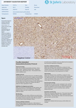
  • 1. Figure: Immunohistochemical analysis of paraffin embedded Human uterus tissue. 1: α- tubulin (Acetyl Lys40) Monoclonal Antibody(4A8) was diluted at 1:200 (4 degree Celsius,overnight). 2: Sodium citrate pH 6.0 was used for antibody retrieval (>98 degree Celsius,20min). 3: Secondary antibody was diluted at 1:200 (room temperature, 30min). Negative control was used by secondary antibody only. Report Number 97537-a Host Mouse Application IHC-P Clonality Monoclonal Model Number STJ97537 Clone ID 4A8 Antibody Name Anti-α-tubulin (Acetyl Lys40) antibody Testing Species HUMAN Testing Tissue UTERUS ANTIBODY VALIDATION REPORT a. (A small amount of distilled water was added into the incubation box to prevent evaporation of antibody). 51. Secondary antibody incubation a. Slides were washed 3 times, with PBS on a shaker for 5min. Shortly after the slides were dried the corresponding secondary antibody solution was added (HRP labelled), covering the tissues, and incubated at room temperature for 30min. b. 52. DAB staining a. Slides were washed 3 times, with PBS on a shaker for 5min. b. Shortly after, the slides were dried and fresh DAB staining buffer was added inside the circles. The staining time was adjusted under a microscope. Yellow-brown colour represented a positive result. Slides were washed with water to stop the staining. c. 53. Haematoxylin staining a. Haematoxylin was used to counter-staining for 1min, and then the slides were washed with water. 1% Hydrochloric acid and alcohol was added for several seconds and then washed with water. Ammonia was used to reveal blue colour, and then flushed with water. b. 54. Desolation and Clearing i. Slides were incubated sequentially into: 75% alcohol 5min, 85% alcohol 5min, Anhydrous ethanol - 5min, Anhydrous ethanol - 5min & Xylene - 5min. Shortly after slides were dried and neutral gum was used to seal the slides. ii. 55. Visualization a. Results were validated with microscope, and the slides were scanned. Paraffin-Embedded Immunohistochemistry Protocol 45. 46. Tissue processing a. Slides were incubated sequentially into Xylene; 15min – Xylene, 15min - Anhydrous ethanol, 5min - Anhydrous ethanol, 5min - 85% alcohol, 5min - 75% alcohol & 5min – wash in distilled water. b. 47. Antigen retrieval a. Tissue slides were incubated with citric acid (PH6.0) antigen retrieval buffer and microwaved for antigen retrieval (heated until boiled and then stopped heating) for 8min. Slides were then heated with medium power for 7min. During this process slides were kept from drying out. After cooling down at room temperature, slides were washed with PBS on shaker for 5min, repeated for 3 times. b. 48. Inhibition of endogenous peroxidase a. Slides were placed in 3% Hydrogen peroxide solution, and incubated for 10 min at room temperature without light exposure. Slides were then washed 3 times with PBS on a shaker for 5mins. b. 49. BSA Blocking a. Shortly after slides were dried, a PAP pen was used to draw circles around the tissue sections (and to prevent draining of the antibody solution). Inside the circles, BSA was used to cover the tissue evenly, blocking for 30min. b. 50. Primary antibody incubation After blocking solution was removed a 1:200 solution of primary antibody/PBS was added on the slide, and incubated overnight at 4°C. St John's Laboratory Ltd. www.stjohnslabs.com
  • 2. Figure: Immunohistochemical analysis of paraffin embedded Human liver tissue. 1: α-tubulin (Acetyl Lys40) Monoclonal Antibody(4A8) was diluted at 1:200 (4 degree Celsius,overnight). 2: Sodium citrate pH 6.0 was used for antibody retrieval (>98 degree Celsius,20min). 3: Secondary antibody was diluted at 1:200 (room temperature, 30min). Negative control was used by secondary antibody only. Report Number 97537-b Host Mouse Application IHC-P Clonality Monoclonal Model Number STJ97537 Clone ID 4A8 Antibody Name Anti-α-tubulin (Acetyl Lys40) antibody Testing Species HUMAN Testing Tissue LIVER ANTIBODY VALIDATION REPORT a. (A small amount of distilled water was added into the incubation box to prevent evaporation of antibody). 40. Secondary antibody incubation a. Slides were washed 3 times, with PBS on a shaker for 5min. Shortly after the slides were dried the corresponding secondary antibody solution was added (HRP labelled), covering the tissues, and incubated at room temperature for 30min. b. 41. DAB staining a. Slides were washed 3 times, with PBS on a shaker for 5min. b. Shortly after, the slides were dried and fresh DAB staining buffer was added inside the circles. The staining time was adjusted under a microscope. Yellow-brown colour represented a positive result. Slides were washed with water to stop the staining. c. 42. Haematoxylin staining a. Haematoxylin was used to counter-staining for 1min, and then the slides were washed with water. 1% Hydrochloric acid and alcohol was added for several seconds and then washed with water. Ammonia was used to reveal blue colour, and then flushed with water. b. 43. Desolation and Clearing i. Slides were incubated sequentially into: 75% alcohol 5min, 85% alcohol 5min, Anhydrous ethanol - 5min, Anhydrous ethanol - 5min & Xylene - 5min. Shortly after slides were dried and neutral gum was used to seal the slides. ii. 44. Visualization a. Results were validated with microscope, and the slides were scanned. Paraffin-Embedded Immunohistochemistry Protocol 34. 35. Tissue processing a. Slides were incubated sequentially into Xylene; 15min – Xylene, 15min - Anhydrous ethanol, 5min - Anhydrous ethanol, 5min - 85% alcohol, 5min - 75% alcohol & 5min – wash in distilled water. b. 36. Antigen retrieval a. Tissue slides were incubated with citric acid (PH6.0) antigen retrieval buffer and microwaved for antigen retrieval (heated until boiled and then stopped heating) for 8min. Slides were then heated with medium power for 7min. During this process slides were kept from drying out. After cooling down at room temperature, slides were washed with PBS on shaker for 5min, repeated for 3 times. b. 37. Inhibition of endogenous peroxidase a. Slides were placed in 3% Hydrogen peroxide solution, and incubated for 10 min at room temperature without light exposure. Slides were then washed 3 times with PBS on a shaker for 5mins. b. 38. BSA Blocking a. Shortly after slides were dried, a PAP pen was used to draw circles around the tissue sections (and to prevent draining of the antibody solution). Inside the circles, BSA was used to cover the tissue evenly, blocking for 30min. b. 39. Primary antibody incubation After blocking solution was removed a 1:200 solution of primary antibody/PBS was added on the slide, and incubated overnight at 4°C. St John's Laboratory Ltd. www.stjohnslabs.com
  • 3. Figure: Immunohistochemical analysis of paraffin embedded Human liver cancer tissue. 1: α- tubulin (Acetyl Lys40) Monoclonal Antibody(4A8) was diluted at 1:200 (4 degree Celsius,overnight). 2: Sodium citrate pH 6.0 was used for antibody retrieval (>98 degree Celsius,20min). 3: Secondary antibody was diluted at 1:200 (room temperature, 30min). Negative control was used by secondary antibody only. Report Number 97537-c Host Mouse Application IHC-P Clonality Monoclonal Model Number STJ97537 Clone ID 4A8 Antibody Name Anti-α-tubulin (Acetyl Lys40) antibody Testing Species HUMAN Testing Tissue LIVER CANCER ANTIBODY VALIDATION REPORT a. (A small amount of distilled water was added into the incubation box to prevent evaporation of antibody). 29. Secondary antibody incubation a. Slides were washed 3 times, with PBS on a shaker for 5min. Shortly after the slides were dried the corresponding secondary antibody solution was added (HRP labelled), covering the tissues, and incubated at room temperature for 30min. b. 30. DAB staining a. Slides were washed 3 times, with PBS on a shaker for 5min. b. Shortly after, the slides were dried and fresh DAB staining buffer was added inside the circles. The staining time was adjusted under a microscope. Yellow-brown colour represented a positive result. Slides were washed with water to stop the staining. c. 31. Haematoxylin staining a. Haematoxylin was used to counter-staining for 1min, and then the slides were washed with water. 1% Hydrochloric acid and alcohol was added for several seconds and then washed with water. Ammonia was used to reveal blue colour, and then flushed with water. b. 32. Desolation and Clearing i. Slides were incubated sequentially into: 75% alcohol 5min, 85% alcohol 5min, Anhydrous ethanol - 5min, Anhydrous ethanol - 5min & Xylene - 5min. Shortly after slides were dried and neutral gum was used to seal the slides. ii. 33. Visualization a. Results were validated with microscope, and the slides were scanned. Paraffin-Embedded Immunohistochemistry Protocol 23. 24. Tissue processing a. Slides were incubated sequentially into Xylene; 15min – Xylene, 15min - Anhydrous ethanol, 5min - Anhydrous ethanol, 5min - 85% alcohol, 5min - 75% alcohol & 5min – wash in distilled water. b. 25. Antigen retrieval a. Tissue slides were incubated with citric acid (PH6.0) antigen retrieval buffer and microwaved for antigen retrieval (heated until boiled and then stopped heating) for 8min. Slides were then heated with medium power for 7min. During this process slides were kept from drying out. After cooling down at room temperature, slides were washed with PBS on shaker for 5min, repeated for 3 times. b. 26. Inhibition of endogenous peroxidase a. Slides were placed in 3% Hydrogen peroxide solution, and incubated for 10 min at room temperature without light exposure. Slides were then washed 3 times with PBS on a shaker for 5mins. b. 27. BSA Blocking a. Shortly after slides were dried, a PAP pen was used to draw circles around the tissue sections (and to prevent draining of the antibody solution). Inside the circles, BSA was used to cover the tissue evenly, blocking for 30min. b. 28. Primary antibody incubation After blocking solution was removed a 1:200 solution of primary antibody/PBS was added on the slide, and incubated overnight at 4°C. St John's Laboratory Ltd. www.stjohnslabs.com
  • 4. Figure: Immunohistochemical analysis of paraffin embedded Human stomach cancer tissue. 1: α-tubulin (Acetyl Lys40) Monoclonal Antibody(4A8) was diluted at 1:200 (4 degree Celsius,overnight). 2: Sodium citrate pH 6.0 was used for antibody retrieval (>98 degree Celsius,20min). 3: Secondary antibody was diluted at 1:200 (room temperature, 30min). Negative control was used by secondary antibody only. Report Number 97537-d Host Mouse Application IHC-P Clonality Monoclonal Model Number STJ97537 Clone ID 4A8 Antibody Name Anti-α-tubulin (Acetyl Lys40) antibody Testing Species HUMAN Testing Tissue STOMACH CANCER ANTIBODY VALIDATION REPORT a. (A small amount of distilled water was added into the incubation box to prevent evaporation of antibody). 18. Secondary antibody incubation a. Slides were washed 3 times, with PBS on a shaker for 5min. Shortly after the slides were dried the corresponding secondary antibody solution was added (HRP labelled), covering the tissues, and incubated at room temperature for 30min. b. 19. DAB staining a. Slides were washed 3 times, with PBS on a shaker for 5min. b. Shortly after, the slides were dried and fresh DAB staining buffer was added inside the circles. The staining time was adjusted under a microscope. Yellow-brown colour represented a positive result. Slides were washed with water to stop the staining. c. 20. Haematoxylin staining a. Haematoxylin was used to counter-staining for 1min, and then the slides were washed with water. 1% Hydrochloric acid and alcohol was added for several seconds and then washed with water. Ammonia was used to reveal blue colour, and then flushed with water. b. 21. Desolation and Clearing i. Slides were incubated sequentially into: 75% alcohol 5min, 85% alcohol 5min, Anhydrous ethanol - 5min, Anhydrous ethanol - 5min & Xylene - 5min. Shortly after slides were dried and neutral gum was used to seal the slides. ii. 22. Visualization a. Results were validated with microscope, and the slides were scanned. Paraffin-Embedded Immunohistochemistry Protocol 12. 13. Tissue processing a. Slides were incubated sequentially into Xylene; 15min – Xylene, 15min - Anhydrous ethanol, 5min - Anhydrous ethanol, 5min - 85% alcohol, 5min - 75% alcohol & 5min – wash in distilled water. b. 14. Antigen retrieval a. Tissue slides were incubated with citric acid (PH6.0) antigen retrieval buffer and microwaved for antigen retrieval (heated until boiled and then stopped heating) for 8min. Slides were then heated with medium power for 7min. During this process slides were kept from drying out. After cooling down at room temperature, slides were washed with PBS on shaker for 5min, repeated for 3 times. b. 15. Inhibition of endogenous peroxidase a. Slides were placed in 3% Hydrogen peroxide solution, and incubated for 10 min at room temperature without light exposure. Slides were then washed 3 times with PBS on a shaker for 5mins. b. 16. BSA Blocking a. Shortly after slides were dried, a PAP pen was used to draw circles around the tissue sections (and to prevent draining of the antibody solution). Inside the circles, BSA was used to cover the tissue evenly, blocking for 30min. b. 17. Primary antibody incubation After blocking solution was removed a 1:200 solution of primary antibody/PBS was added on the slide, and incubated overnight at 4°C. St John's Laboratory Ltd. www.stjohnslabs.com
  • 5. Figure: Immunohistochemical analysis of paraffin embedded Rat heart tissue. 1: α-tubulin (Acetyl Lys40) Monoclonal Antibody(4A8) was diluted at 1:200 (4 degree Celsius,overnight). 2: Sodium citrate pH 6.0 was used for antibody retrieval (>98 degree Celsius,20min). 3: Secondary antibody was diluted at 1:200 (room temperature, 30min). Negative control was used by secondary antibody only. Report Number 97537-e Host Mouse Application IHC-P Clonality Monoclonal Model Number STJ97537 Clone ID 4A8 Antibody Name Anti-α-tubulin (Acetyl Lys40) antibody Testing Species RAT Testing Tissue HEART ANTIBODY VALIDATION REPORT a. (A small amount of distilled water was added into the incubation box to prevent evaporation of antibody). 7. Secondary antibody incubation a. Slides were washed 3 times, with PBS on a shaker for 5min. Shortly after the slides were dried the corresponding secondary antibody solution was added (HRP labelled), covering the tissues, and incubated at room temperature for 30min. b. 8. DAB staining a. Slides were washed 3 times, with PBS on a shaker for 5min. b. Shortly after, the slides were dried and fresh DAB staining buffer was added inside the circles. The staining time was adjusted under a microscope. Yellow-brown colour represented a positive result. Slides were washed with water to stop the staining. c. 9. Haematoxylin staining a. Haematoxylin was used to counter-staining for 1min, and then the slides were washed with water. 1% Hydrochloric acid and alcohol was added for several seconds and then washed with water. Ammonia was used to reveal blue colour, and then flushed with water. b. 10. Desolation and Clearing i. Slides were incubated sequentially into: 75% alcohol 5min, 85% alcohol 5min, Anhydrous ethanol - 5min, Anhydrous ethanol - 5min & Xylene - 5min. Shortly after slides were dried and neutral gum was used to seal the slides. ii. 11. Visualization a. Results were validated with microscope, and the slides were scanned. Paraffin-Embedded Immunohistochemistry Protocol 1. 2. Tissue processing a. Slides were incubated sequentially into Xylene; 15min – Xylene, 15min - Anhydrous ethanol, 5min - Anhydrous ethanol, 5min - 85% alcohol, 5min - 75% alcohol & 5min – wash in distilled water. b. 3. Antigen retrieval a. Tissue slides were incubated with citric acid (PH6.0) antigen retrieval buffer and microwaved for antigen retrieval (heated until boiled and then stopped heating) for 8min. Slides were then heated with medium power for 7min. During this process slides were kept from drying out. After cooling down at room temperature, slides were washed with PBS on shaker for 5min, repeated for 3 times. b. 4. Inhibition of endogenous peroxidase a. Slides were placed in 3% Hydrogen peroxide solution, and incubated for 10 min at room temperature without light exposure. Slides were then washed 3 times with PBS on a shaker for 5mins. b. 5. BSA Blocking a. Shortly after slides were dried, a PAP pen was used to draw circles around the tissue sections (and to prevent draining of the antibody solution). Inside the circles, BSA was used to cover the tissue evenly, blocking for 30min. b. 6. Primary antibody incubation After blocking solution was removed a 1:200 solution of primary antibody/PBS was added on the slide, and incubated overnight at 4°C. St John's Laboratory Ltd. www.stjohnslabs.com
  • 6. Figure: Immunohistochemical analysis of paraffin embedded Rat kidney tissue. 1: α-tubulin (Acetyl Lys40) Monoclonal Antibody(4A8) was diluted at 1:200 (4 degree Celsius,overnight). 2: Sodium citrate pH 6.0 was used for antibody retrieval (>98 degree Celsius,20min). 3: Secondary antibody was diluted at 1:200 (room temperature, 30min). Negative control was used by secondary antibody only. Report Number 97537-f Host Mouse Application IHC-P Clonality Monoclonal Model Number STJ97537 Clone ID 4A8 Antibody Name Anti-α-tubulin (Acetyl Lys40) antibody Testing Species RAT Testing Tissue KIDNEY ANTIBODY VALIDATION REPORT c. (A small amount of distilled water was added into the incubation box to prevent evaporation of antibody). 56. Secondary antibody incubation a. Slides were washed 3 times, with PBS on a shaker for 5min. Shortly after the slides were dried the corresponding secondary antibody solution was added (HRP labelled), covering the tissues, and incubated at room temperature for 30min. b. 57. DAB staining a. Slides were washed 3 times, with PBS on a shaker for 5min. b. Shortly after, the slides were dried and fresh DAB staining buffer was added inside the circles. The staining time was adjusted under a microscope. Yellow-brown colour represented a positive result. Slides were washed with water to stop the staining. c. 58. Haematoxylin staining a. Haematoxylin was used to counter-staining for 1min, and then the slides were washed with water. 1% Hydrochloric acid and alcohol was added for several seconds and then washed with water. Ammonia was used to reveal blue colour, and then flushed with water. b. 59. Desolation and Clearing i. Slides were incubated sequentially into: 75% alcohol 5min, 85% alcohol 5min, Anhydrous ethanol - 5min, Anhydrous ethanol - 5min & Xylene - 5min. Shortly after slides were dried and neutral gum was used to seal the slides. ii. 60. Visualization a. Results were validated with microscope, and the slides were scanned. Paraffin-Embedded Immunohistochemistry Protocol 61. 62. Tissue processing a. Slides were incubated sequentially into Xylene; 15min – Xylene, 15min - Anhydrous ethanol, 5min - Anhydrous ethanol, 5min - 85% alcohol, 5min - 75% alcohol & 5min – wash in distilled water. b. 63. Antigen retrieval a. Tissue slides were incubated with citric acid (PH6.0) antigen retrieval buffer and microwaved for antigen retrieval (heated until boiled and then stopped heating) for 8min. Slides were then heated with medium power for 7min. During this process slides were kept from drying out. After cooling down at room temperature, slides were washed with PBS on shaker for 5min, repeated for 3 times. b. 64. Inhibition of endogenous peroxidase a. Slides were placed in 3% Hydrogen peroxide solution, and incubated for 10 min at room temperature without light exposure. Slides were then washed 3 times with PBS on a shaker for 5mins. b. 65. BSA Blocking a. Shortly after slides were dried, a PAP pen was used to draw circles around the tissue sections (and to prevent draining of the antibody solution). Inside the circles, BSA was used to cover the tissue evenly, blocking for 30min. b. 66. Primary antibody incubation After blocking solution was removed a 1:200 solution of primary antibody/PBS was added on the slide, and incubated overnight at 4°C. St John's Laboratory Ltd. www.stjohnslabs.com
  • 7. Figure: Immunohistochemical analysis of paraffin embedded Mouse heart tissue. 1: α-tubulin (Acetyl Lys40) Monoclonal Antibody(4A8) was diluted at 1:200 (4 degree Celsius,overnight). 2: Sodium citrate pH 6.0 was used for antibody retrieval (>98 degree Celsius,20min). 3: Secondary antibody was diluted at 1:200 (room temperature, 30min). Negative control was used by secondary antibody only. Report Number 97537-g Host Mouse Application IHC-P Clonality Monoclonal Model Number STJ97537 Clone ID 4A8 Antibody Name Anti-α-tubulin (Acetyl Lys40) antibody Testing Species MOUSE Testing Tissue HEART ANTIBODY VALIDATION REPORT b. (A small amount of distilled water was added into the incubation box to prevent evaporation of antibody). 67. Secondary antibody incubation a. Slides were washed 3 times, with PBS on a shaker for 5min. Shortly after the slides were dried the corresponding secondary antibody solution was added (HRP labelled), covering the tissues, and incubated at room temperature for 30min. b. 68. DAB staining a. Slides were washed 3 times, with PBS on a shaker for 5min. b. Shortly after, the slides were dried and fresh DAB staining buffer was added inside the circles. The staining time was adjusted under a microscope. Yellow-brown colour represented a positive result. Slides were washed with water to stop the staining. c. 69. Haematoxylin staining a. Haematoxylin was used to counter-staining for 1min, and then the slides were washed with water. 1% Hydrochloric acid and alcohol was added for several seconds and then washed with water. Ammonia was used to reveal blue colour, and then flushed with water. b. 70. Desolation and Clearing i. Slides were incubated sequentially into: 75% alcohol 5min, 85% alcohol 5min, Anhydrous ethanol - 5min, Anhydrous ethanol - 5min & Xylene - 5min. Shortly after slides were dried and neutral gum was used to seal the slides. ii. 71. Visualization a. Results were validated with microscope, and the slides were scanned. Paraffin-Embedded Immunohistochemistry Protocol 72. 73. Tissue processing a. Slides were incubated sequentially into Xylene; 15min – Xylene, 15min - Anhydrous ethanol, 5min - Anhydrous ethanol, 5min - 85% alcohol, 5min - 75% alcohol & 5min – wash in distilled water. b. 74. Antigen retrieval a. Tissue slides were incubated with citric acid (PH6.0) antigen retrieval buffer and microwaved for antigen retrieval (heated until boiled and then stopped heating) for 8min. Slides were then heated with medium power for 7min. During this process slides were kept from drying out. After cooling down at room temperature, slides were washed with PBS on shaker for 5min, repeated for 3 times. b. 75. Inhibition of endogenous peroxidase a. Slides were placed in 3% Hydrogen peroxide solution, and incubated for 10 min at room temperature without light exposure. Slides were then washed 3 times with PBS on a shaker for 5mins. b. 76. BSA Blocking a. Shortly after slides were dried, a PAP pen was used to draw circles around the tissue sections (and to prevent draining of the antibody solution). Inside the circles, BSA was used to cover the tissue evenly, blocking for 30min. b. 77. Primary antibody incubation After blocking solution was removed a 1:200 solution of primary antibody/PBS was added on the slide, and incubated overnight at 4°C. St John's Laboratory Ltd. www.stjohnslabs.com
  • 8. Figure: Immunohistochemical analysis of paraffin embedded Mouse kidney tissue. 1: α- tubulin (Acetyl Lys40) Monoclonal Antibody(4A8) was diluted at 1:200 (4 degree Celsius,overnight). 2: Sodium citrate pH 6.0 was used for antibody retrieval (>98 degree Celsius,20min). 3: Secondary antibody was diluted at 1:200 (room temperature, 30min). Negative control was used by secondary antibody only. Report Number 97537-h Host Mouse Application IHC-P Clonality Monoclonal Model Number STJ97537 Clone ID 4A8 Antibody Name Anti-α-tubulin (Acetyl Lys40) antibody Testing Species MOUSE Testing Tissue KIDNEY ANTIBODY VALIDATION REPORT b. (A small amount of distilled water was added into the incubation box to prevent evaporation of antibody). 78. Secondary antibody incubation a. Slides were washed 3 times, with PBS on a shaker for 5min. Shortly after the slides were dried the corresponding secondary antibody solution was added (HRP labelled), covering the tissues, and incubated at room temperature for 30min. b. 79. DAB staining a. Slides were washed 3 times, with PBS on a shaker for 5min. b. Shortly after, the slides were dried and fresh DAB staining buffer was added inside the circles. The staining time was adjusted under a microscope. Yellow-brown colour represented a positive result. Slides were washed with water to stop the staining. c. 80. Haematoxylin staining a. Haematoxylin was used to counter-staining for 1min, and then the slides were washed with water. 1% Hydrochloric acid and alcohol was added for several seconds and then washed with water. Ammonia was used to reveal blue colour, and then flushed with water. b. 81. Desolation and Clearing i. Slides were incubated sequentially into: 75% alcohol 5min, 85% alcohol 5min, Anhydrous ethanol - 5min, Anhydrous ethanol - 5min & Xylene - 5min. Shortly after slides were dried and neutral gum was used to seal the slides. ii. 82. Visualization a. Results were validated with microscope, and the slides were scanned. Paraffin-Embedded Immunohistochemistry Protocol 83. 84. Tissue processing a. Slides were incubated sequentially into Xylene; 15min – Xylene, 15min - Anhydrous ethanol, 5min - Anhydrous ethanol, 5min - 85% alcohol, 5min - 75% alcohol & 5min – wash in distilled water. b. 85. Antigen retrieval a. Tissue slides were incubated with citric acid (PH6.0) antigen retrieval buffer and microwaved for antigen retrieval (heated until boiled and then stopped heating) for 8min. Slides were then heated with medium power for 7min. During this process slides were kept from drying out. After cooling down at room temperature, slides were washed with PBS on shaker for 5min, repeated for 3 times. b. 86. Inhibition of endogenous peroxidase a. Slides were placed in 3% Hydrogen peroxide solution, and incubated for 10 min at room temperature without light exposure. Slides were then washed 3 times with PBS on a shaker for 5mins. b. 87. BSA Blocking a. Shortly after slides were dried, a PAP pen was used to draw circles around the tissue sections (and to prevent draining of the antibody solution). Inside the circles, BSA was used to cover the tissue evenly, blocking for 30min. b. 88. Primary antibody incubation After blocking solution was removed a 1:200 solution of primary antibody/PBS was added on the slide, and incubated overnight at 4°C. St John's Laboratory Ltd. www.stjohnslabs.com