SlideShare a Scribd company logo
1 of 6
Download to read offline
ANTIBODY VALIDATION REPORT
Report Number 97420-a
Application Immunofluorescence
Model Number STJ97420
Antibody Name Anti-Catenin-β antibody
Host Mouse
Clonality Monoclonal
Clone ID 4F2
Species HUMAN Tissue STOMACH CANCER
Image
Description
Immunofluorescence analysis of Human
stomach cancer tissue. 1: Catenin-β
Monoclonal Antibody(4F2)(red) was
diluted at 1:200 (4 degree
Celsius,overnight). 2: Cy3 labled
Secondary antibody was diluted at
1:300 (room temperature, 50min).3:
Picture B: DAPI(blue) 10min. Picture
A:Target. Picture B: DAPI. Picture C:
merge of A+B.
Primary Antibody Incubation
After blocking solution was removed a 1:200 primary antibody/PBS
solution was added on the slide, and incubated overnight at 4°C (a
small amount of distilled water was added into the incubation box to
prevent evaporation of antibody).
Secondary Antibody Incubation
slides were washed with PBS on a shaker for 5min, and repeated 3
times. Shortly after the slides were dried and corresponding
secondary antibody solution was added (HRP labelled), covering the
tissues, and incubated at room temperature for 50min.
DAPI Counter-Staining
slides were washed with PBS on a shaker for 5min, repeated 3 times
and then dried. DAPI staining solution was added inside the PAP
circles and incubated for 10 min at room temperature without light
exposure.
Mounting
Slides were washed with PBS on a shaker for 5min, and repeated 3
times. Shortly after slides were dried, anti-quench mountings were
used to mount slides.
Visualization
The slides were observed and placed under a NIKON inverted
fluorescence microscope (Ultra violet excitation 330-380nm,
emission 420nm; FITC green excitation 465-495nm, emission 515-
555 nm; CY3 red excitation 510-560nm, emission 590nm)
Immunofluorescence Protocol
Tissue Processing
Slides were incubated sequentially into: Xylene - 15min, Anhydrous
ethanol – 15 min, Anhydrous ethanol – 5 min, 85% alcohol – 5 min,
75% alcohol – 5 min & washed with distilled water – 5 min.
Antigen Retrieval
Tissue slides were incubated with citric acid (PH6.0) antigen
retrieval buffer, and microwaved for antigen retrieval (heated until
boiled and then stop heating) for 8min. Slides were then heated with
medium power for 7min. During this process slides are kept from
drying out. After cooling down at room temperature, slides were
washed with PBS on a shaker for 5min, and repeated 3 times.
Anti-Quench
shortly after slides were dried, a PAP pen was used to draw circles
around the tissues (to prevent draining of the antibody). Inside the
circles, anti-quench mountings were added and incubated for 5 min,
and then flushed with water for 10min.
BSA Blocking
Inside the circles, BSA was used to cover the tissue evenly, blocking
for 30min.
St John's Laboratory Ltd.
www.stjohnslabs.com
ANTIBODY VALIDATION REPORT
Report Number 97420-b
Application Immunofluorescence
Model Number STJ97420
Antibody Name Anti-Catenin-β antibody
Host Mouse
Clonality Monoclonal
Clone ID 4F2
Species HUMAN Tissue STOMACH CANCER
Image
Description
Immunofluorescence analysis of Human
stomach cancer tissue. 1: Catenin-β
Monoclonal Antibody(4F2)(red) was
diluted at 1:200 (4 degree
Celsius,overnight). 2: Cy3 labled
Secondary antibody was diluted at
1:300 (room temperature, 50min).3:
Picture B: DAPI(blue) 10min. Picture
A:Target. Picture B: DAPI. Picture C:
merge of A+B.
Primary Antibody Incubation
After blocking solution was removed a 1:200 primary antibody/PBS
solution was added on the slide, and incubated overnight at 4°C (a
small amount of distilled water was added into the incubation box to
prevent evaporation of antibody).
Secondary Antibody Incubation
slides were washed with PBS on a shaker for 5min, and repeated 3
times. Shortly after the slides were dried and corresponding
secondary antibody solution was added (HRP labelled), covering the
tissues, and incubated at room temperature for 50min.
DAPI Counter-Staining
slides were washed with PBS on a shaker for 5min, repeated 3 times
and then dried. DAPI staining solution was added inside the PAP
circles and incubated for 10 min at room temperature without light
exposure.
Mounting
Slides were washed with PBS on a shaker for 5min, and repeated 3
times. Shortly after slides were dried, anti-quench mountings were
used to mount slides.
Visualization
The slides were observed and placed under a NIKON inverted
fluorescence microscope (Ultra violet excitation 330-380nm,
emission 420nm; FITC green excitation 465-495nm, emission 515-
555 nm; CY3 red excitation 510-560nm, emission 590nm)
Immunofluorescence Protocol
Tissue Processing
Slides were incubated sequentially into: Xylene - 15min, Anhydrous
ethanol – 15 min, Anhydrous ethanol – 5 min, 85% alcohol – 5 min,
75% alcohol – 5 min & washed with distilled water – 5 min.
Antigen Retrieval
Tissue slides were incubated with citric acid (PH6.0) antigen
retrieval buffer, and microwaved for antigen retrieval (heated until
boiled and then stop heating) for 8min. Slides were then heated with
medium power for 7min. During this process slides are kept from
drying out. After cooling down at room temperature, slides were
washed with PBS on a shaker for 5min, and repeated 3 times.
Anti-Quench
shortly after slides were dried, a PAP pen was used to draw circles
around the tissues (to prevent draining of the antibody). Inside the
circles, anti-quench mountings were added and incubated for 5 min,
and then flushed with water for 10min.
BSA Blocking
Inside the circles, BSA was used to cover the tissue evenly, blocking
for 30min.
St John's Laboratory Ltd.
www.stjohnslabs.com
ANTIBODY VALIDATION REPORT
Report Number 97420-c
Application Immunofluorescence
Model Number STJ97420
Antibody Name Anti-Catenin-β antibody
Host Mouse
Clonality Monoclonal
Clone ID 4F2
Species MOUSE Tissue SPLEEN
Image
Description
Immunofluorescence analysis of Mouse
spleen tissue. 1: Catenin-β Monoclonal
Antibody(4F2)(red) was diluted at
1:200 (4 degree Celsius,overnight). 2:
Cy3 labled Secondary antibody was
diluted at 1:300 (room temperature,
50min).3: Picture B: DAPI(blue) 10min.
Picture A:Target. Picture B: DAPI.
Picture C: merge of A+B.
Primary Antibody Incubation
After blocking solution was removed a 1:200 primary antibody/PBS
solution was added on the slide, and incubated overnight at 4°C (a
small amount of distilled water was added into the incubation box to
prevent evaporation of antibody).
Secondary Antibody Incubation
slides were washed with PBS on a shaker for 5min, and repeated 3
times. Shortly after the slides were dried and corresponding
secondary antibody solution was added (HRP labelled), covering the
tissues, and incubated at room temperature for 50min.
DAPI Counter-Staining
slides were washed with PBS on a shaker for 5min, repeated 3 times
and then dried. DAPI staining solution was added inside the PAP
circles and incubated for 10 min at room temperature without light
exposure.
Mounting
Slides were washed with PBS on a shaker for 5min, and repeated 3
times. Shortly after slides were dried, anti-quench mountings were
used to mount slides.
Visualization
The slides were observed and placed under a NIKON inverted
fluorescence microscope (Ultra violet excitation 330-380nm,
emission 420nm; FITC green excitation 465-495nm, emission 515-
555 nm; CY3 red excitation 510-560nm, emission 590nm)
Immunofluorescence Protocol
Tissue Processing
Slides were incubated sequentially into: Xylene - 15min, Anhydrous
ethanol – 15 min, Anhydrous ethanol – 5 min, 85% alcohol – 5 min,
75% alcohol – 5 min & washed with distilled water – 5 min.
Antigen Retrieval
Tissue slides were incubated with citric acid (PH6.0) antigen
retrieval buffer, and microwaved for antigen retrieval (heated until
boiled and then stop heating) for 8min. Slides were then heated with
medium power for 7min. During this process slides are kept from
drying out. After cooling down at room temperature, slides were
washed with PBS on a shaker for 5min, and repeated 3 times.
Anti-Quench
shortly after slides were dried, a PAP pen was used to draw circles
around the tissues (to prevent draining of the antibody). Inside the
circles, anti-quench mountings were added and incubated for 5 min,
and then flushed with water for 10min.
BSA Blocking
Inside the circles, BSA was used to cover the tissue evenly, blocking
for 30min.
St John's Laboratory Ltd.
www.stjohnslabs.com
ANTIBODY VALIDATION REPORT
Report Number 97420-d
Application Immunofluorescence
Model Number STJ97420
Antibody Name Anti-Catenin-β antibody
Host Mouse
Clonality Monoclonal
Clone ID 4F2
Species MOUSE Tissue SPLEEN
Image
Description
Immunofluorescence analysis of Mouse
spleen tissue. 1: Catenin-β Monoclonal
Antibody(4F2)(red) was diluted at
1:200 (4 degree Celsius,overnight). 2:
Cy3 labled Secondary antibody was
diluted at 1:300 (room temperature,
50min).3: Picture B: DAPI(blue) 10min.
Picture A:Target. Picture B: DAPI.
Picture C: merge of A+B.
Primary Antibody Incubation
After blocking solution was removed a 1:200 primary antibody/PBS
solution was added on the slide, and incubated overnight at 4°C (a
small amount of distilled water was added into the incubation box to
prevent evaporation of antibody).
Secondary Antibody Incubation
slides were washed with PBS on a shaker for 5min, and repeated 3
times. Shortly after the slides were dried and corresponding
secondary antibody solution was added (HRP labelled), covering the
tissues, and incubated at room temperature for 50min.
DAPI Counter-Staining
slides were washed with PBS on a shaker for 5min, repeated 3 times
and then dried. DAPI staining solution was added inside the PAP
circles and incubated for 10 min at room temperature without light
exposure.
Mounting
Slides were washed with PBS on a shaker for 5min, and repeated 3
times. Shortly after slides were dried, anti-quench mountings were
used to mount slides.
Visualization
The slides were observed and placed under a NIKON inverted
fluorescence microscope (Ultra violet excitation 330-380nm,
emission 420nm; FITC green excitation 465-495nm, emission 515-
555 nm; CY3 red excitation 510-560nm, emission 590nm)
Immunofluorescence Protocol
Tissue Processing
Slides were incubated sequentially into: Xylene - 15min, Anhydrous
ethanol – 15 min, Anhydrous ethanol – 5 min, 85% alcohol – 5 min,
75% alcohol – 5 min & washed with distilled water – 5 min.
Antigen Retrieval
Tissue slides were incubated with citric acid (PH6.0) antigen
retrieval buffer, and microwaved for antigen retrieval (heated until
boiled and then stop heating) for 8min. Slides were then heated with
medium power for 7min. During this process slides are kept from
drying out. After cooling down at room temperature, slides were
washed with PBS on a shaker for 5min, and repeated 3 times.
Anti-Quench
shortly after slides were dried, a PAP pen was used to draw circles
around the tissues (to prevent draining of the antibody). Inside the
circles, anti-quench mountings were added and incubated for 5 min,
and then flushed with water for 10min.
BSA Blocking
Inside the circles, BSA was used to cover the tissue evenly, blocking
for 30min.
St John's Laboratory Ltd.
www.stjohnslabs.com
ANTIBODY VALIDATION REPORT
Report Number 97420-e
Application Immunofluorescence
Model Number STJ97420
Antibody Name Anti-Catenin-β antibody
Host Mouse
Clonality Monoclonal
Clone ID 4F2
Species MOUSE Tissue TESTIS
Image
Description
Immunofluorescence analysis of Mouse
testis tissue. 1: Catenin-β Monoclonal
Antibody(4F2)(red) was diluted at
1:200 (4 degree Celsius,overnight). 2:
Cy3 labled Secondary antibody was
diluted at 1:300 (room temperature,
50min).3: Picture B: DAPI(blue) 10min.
Picture A:Target. Picture B: DAPI.
Picture C: merge of A+B.
Primary Antibody Incubation
After blocking solution was removed a 1:200 primary antibody/PBS
solution was added on the slide, and incubated overnight at 4°C (a
small amount of distilled water was added into the incubation box to
prevent evaporation of antibody).
Secondary Antibody Incubation
slides were washed with PBS on a shaker for 5min, and repeated 3
times. Shortly after the slides were dried and corresponding
secondary antibody solution was added (HRP labelled), covering the
tissues, and incubated at room temperature for 50min.
DAPI Counter-Staining
slides were washed with PBS on a shaker for 5min, repeated 3 times
and then dried. DAPI staining solution was added inside the PAP
circles and incubated for 10 min at room temperature without light
exposure.
Mounting
Slides were washed with PBS on a shaker for 5min, and repeated 3
times. Shortly after slides were dried, anti-quench mountings were
used to mount slides.
Visualization
The slides were observed and placed under a NIKON inverted
fluorescence microscope (Ultra violet excitation 330-380nm,
emission 420nm; FITC green excitation 465-495nm, emission 515-
555 nm; CY3 red excitation 510-560nm, emission 590nm)
Immunofluorescence Protocol
Tissue Processing
Slides were incubated sequentially into: Xylene - 15min, Anhydrous
ethanol – 15 min, Anhydrous ethanol – 5 min, 85% alcohol – 5 min,
75% alcohol – 5 min & washed with distilled water – 5 min.
Antigen Retrieval
Tissue slides were incubated with citric acid (PH6.0) antigen
retrieval buffer, and microwaved for antigen retrieval (heated until
boiled and then stop heating) for 8min. Slides were then heated with
medium power for 7min. During this process slides are kept from
drying out. After cooling down at room temperature, slides were
washed with PBS on a shaker for 5min, and repeated 3 times.
Anti-Quench
shortly after slides were dried, a PAP pen was used to draw circles
around the tissues (to prevent draining of the antibody). Inside the
circles, anti-quench mountings were added and incubated for 5 min,
and then flushed with water for 10min.
BSA Blocking
Inside the circles, BSA was used to cover the tissue evenly, blocking
for 30min.
St John's Laboratory Ltd.
www.stjohnslabs.com
ANTIBODY VALIDATION REPORT
Report Number 97420-f
Application Immunofluorescence
Model Number STJ97420
Antibody Name Anti-Catenin-β antibody
Host Mouse
Clonality Monoclonal
Clone ID 4F2
Species MOUSE Tissue TESTIS
Image
Description
Immunofluorescence analysis of Mouse
testis tissue. 1: Catenin-β Monoclonal
Antibody(4F2)(red) was diluted at
1:200 (4 degree Celsius,overnight). 2:
Cy3 labled Secondary antibody was
diluted at 1:300 (room temperature,
50min).3: Picture B: DAPI(blue) 10min.
Picture A:Target. Picture B: DAPI.
Picture C: merge of A+B.
Primary Antibody Incubation
After blocking solution was removed a 1:200 primary antibody/PBS
solution was added on the slide, and incubated overnight at 4°C (a
small amount of distilled water was added into the incubation box to
prevent evaporation of antibody).
Secondary Antibody Incubation
slides were washed with PBS on a shaker for 5min, and repeated 3
times. Shortly after the slides were dried and corresponding
secondary antibody solution was added (HRP labelled), covering the
tissues, and incubated at room temperature for 50min.
DAPI Counter-Staining
slides were washed with PBS on a shaker for 5min, repeated 3 times
and then dried. DAPI staining solution was added inside the PAP
circles and incubated for 10 min at room temperature without light
exposure.
Mounting
Slides were washed with PBS on a shaker for 5min, and repeated 3
times. Shortly after slides were dried, anti-quench mountings were
used to mount slides.
Visualization
The slides were observed and placed under a NIKON inverted
fluorescence microscope (Ultra violet excitation 330-380nm,
emission 420nm; FITC green excitation 465-495nm, emission 515-
555 nm; CY3 red excitation 510-560nm, emission 590nm)
Immunofluorescence Protocol
Tissue Processing
Slides were incubated sequentially into: Xylene - 15min, Anhydrous
ethanol – 15 min, Anhydrous ethanol – 5 min, 85% alcohol – 5 min,
75% alcohol – 5 min & washed with distilled water – 5 min.
Antigen Retrieval
Tissue slides were incubated with citric acid (PH6.0) antigen
retrieval buffer, and microwaved for antigen retrieval (heated until
boiled and then stop heating) for 8min. Slides were then heated with
medium power for 7min. During this process slides are kept from
drying out. After cooling down at room temperature, slides were
washed with PBS on a shaker for 5min, and repeated 3 times.
Anti-Quench
shortly after slides were dried, a PAP pen was used to draw circles
around the tissues (to prevent draining of the antibody). Inside the
circles, anti-quench mountings were added and incubated for 5 min,
and then flushed with water for 10min.
BSA Blocking
Inside the circles, BSA was used to cover the tissue evenly, blocking
for 30min.
St John's Laboratory Ltd.
www.stjohnslabs.com

More Related Content

What's hot

What's hot (20)

Immunofluorescence Antibody Validation Report for Anti-CA IX Antibody (STJ96978)
Immunofluorescence Antibody Validation Report for Anti-CA IX Antibody (STJ96978)Immunofluorescence Antibody Validation Report for Anti-CA IX Antibody (STJ96978)
Immunofluorescence Antibody Validation Report for Anti-CA IX Antibody (STJ96978)
 
Immunofluorescence Antibody Validation Report for Anti-IκB β Antibody (STJ97539)
Immunofluorescence Antibody Validation Report for Anti-IκB β Antibody (STJ97539)Immunofluorescence Antibody Validation Report for Anti-IκB β Antibody (STJ97539)
Immunofluorescence Antibody Validation Report for Anti-IκB β Antibody (STJ97539)
 
Immunofluorescence Antibody Validation Report for Anti-ABCG2 Antibody (STJ97253)
Immunofluorescence Antibody Validation Report for Anti-ABCG2 Antibody (STJ97253)Immunofluorescence Antibody Validation Report for Anti-ABCG2 Antibody (STJ97253)
Immunofluorescence Antibody Validation Report for Anti-ABCG2 Antibody (STJ97253)
 
Immunofluorescence Antibody Validation Report for Anti-PR Antibody (STJ97365)
Immunofluorescence Antibody Validation Report for Anti-PR Antibody (STJ97365)Immunofluorescence Antibody Validation Report for Anti-PR Antibody (STJ97365)
Immunofluorescence Antibody Validation Report for Anti-PR Antibody (STJ97365)
 
Immunofluorescence Antibody Validation Report for Anti-Catalase Antibody (STJ...
Immunofluorescence Antibody Validation Report for Anti-Catalase Antibody (STJ...Immunofluorescence Antibody Validation Report for Anti-Catalase Antibody (STJ...
Immunofluorescence Antibody Validation Report for Anti-Catalase Antibody (STJ...
 
Immunofluorescence Antibody Validation Report for Anti-β I tubulin Antibody (...
Immunofluorescence Antibody Validation Report for Anti-β I tubulin Antibody (...Immunofluorescence Antibody Validation Report for Anti-β I tubulin Antibody (...
Immunofluorescence Antibody Validation Report for Anti-β I tubulin Antibody (...
 
Immunofluorescence Antibody Validation Report for Anti-Cytokeratin 6 Antibody...
Immunofluorescence Antibody Validation Report for Anti-Cytokeratin 6 Antibody...Immunofluorescence Antibody Validation Report for Anti-Cytokeratin 6 Antibody...
Immunofluorescence Antibody Validation Report for Anti-Cytokeratin 6 Antibody...
 
Immunofluorescence Antibody Validation Report for Anti-OPG Antibody (STJ94831)
Immunofluorescence Antibody Validation Report for Anti-OPG Antibody (STJ94831)Immunofluorescence Antibody Validation Report for Anti-OPG Antibody (STJ94831)
Immunofluorescence Antibody Validation Report for Anti-OPG Antibody (STJ94831)
 
Immunofluorescence Antibody Validation Report for Anti-Jagged1 Antibody (STJ9...
Immunofluorescence Antibody Validation Report for Anti-Jagged1 Antibody (STJ9...Immunofluorescence Antibody Validation Report for Anti-Jagged1 Antibody (STJ9...
Immunofluorescence Antibody Validation Report for Anti-Jagged1 Antibody (STJ9...
 
Immunofluorescence Antibody Validation Report for Anti-IL8 Antibody (STJ96518)
Immunofluorescence Antibody Validation Report for Anti-IL8 Antibody (STJ96518)Immunofluorescence Antibody Validation Report for Anti-IL8 Antibody (STJ96518)
Immunofluorescence Antibody Validation Report for Anti-IL8 Antibody (STJ96518)
 
Immunofluorescence Antibody Validation Report for Anti-TGFβ RII Antibody (STJ...
Immunofluorescence Antibody Validation Report for Anti-TGFβ RII Antibody (STJ...Immunofluorescence Antibody Validation Report for Anti-TGFβ RII Antibody (STJ...
Immunofluorescence Antibody Validation Report for Anti-TGFβ RII Antibody (STJ...
 
Immunofluorescence Antibody Validation Report for Anti-Survivin Antibody (STJ...
Immunofluorescence Antibody Validation Report for Anti-Survivin Antibody (STJ...Immunofluorescence Antibody Validation Report for Anti-Survivin Antibody (STJ...
Immunofluorescence Antibody Validation Report for Anti-Survivin Antibody (STJ...
 
Immunofluorescence Antibody Validation Report for Anti-XRCC4 Antibody (STJ96988)
Immunofluorescence Antibody Validation Report for Anti-XRCC4 Antibody (STJ96988)Immunofluorescence Antibody Validation Report for Anti-XRCC4 Antibody (STJ96988)
Immunofluorescence Antibody Validation Report for Anti-XRCC4 Antibody (STJ96988)
 
Immunofluorescence Antibody Validation Report for Anti-Gamma Tubulin Antibody...
Immunofluorescence Antibody Validation Report for Anti-Gamma Tubulin Antibody...Immunofluorescence Antibody Validation Report for Anti-Gamma Tubulin Antibody...
Immunofluorescence Antibody Validation Report for Anti-Gamma Tubulin Antibody...
 
Immunofluorescence Antibody Validation Report for Anti-Tubulin α Antibody (ST...
Immunofluorescence Antibody Validation Report for Anti-Tubulin α Antibody (ST...Immunofluorescence Antibody Validation Report for Anti-Tubulin α Antibody (ST...
Immunofluorescence Antibody Validation Report for Anti-Tubulin α Antibody (ST...
 
Immunofluorescence Antibody Validation Report for Anti-p38 Antibody (STJ94878)
Immunofluorescence Antibody Validation Report for Anti-p38 Antibody (STJ94878)Immunofluorescence Antibody Validation Report for Anti-p38 Antibody (STJ94878)
Immunofluorescence Antibody Validation Report for Anti-p38 Antibody (STJ94878)
 
Immunofluorescence Antibody Validation Report for Anti-HMG-1 Antibody (STJ96867)
Immunofluorescence Antibody Validation Report for Anti-HMG-1 Antibody (STJ96867)Immunofluorescence Antibody Validation Report for Anti-HMG-1 Antibody (STJ96867)
Immunofluorescence Antibody Validation Report for Anti-HMG-1 Antibody (STJ96867)
 
Immunofluorescence Antibody Validation Report for Anti-HSP90β Antibody (STJ97...
Immunofluorescence Antibody Validation Report for Anti-HSP90β Antibody (STJ97...Immunofluorescence Antibody Validation Report for Anti-HSP90β Antibody (STJ97...
Immunofluorescence Antibody Validation Report for Anti-HSP90β Antibody (STJ97...
 
Immunofluorescence Antibody Validation Report for Anti-Peroxiredoxin 1 Antibo...
Immunofluorescence Antibody Validation Report for Anti-Peroxiredoxin 1 Antibo...Immunofluorescence Antibody Validation Report for Anti-Peroxiredoxin 1 Antibo...
Immunofluorescence Antibody Validation Report for Anti-Peroxiredoxin 1 Antibo...
 
Immunofluorescence Antibody Validation Report for Anti-CD63 Antibody (STJ96890)
Immunofluorescence Antibody Validation Report for Anti-CD63 Antibody (STJ96890)Immunofluorescence Antibody Validation Report for Anti-CD63 Antibody (STJ96890)
Immunofluorescence Antibody Validation Report for Anti-CD63 Antibody (STJ96890)
 

Viewers also liked

Viewers also liked (18)

Immunofluorescence Antibody Validation Report for Anti-α skeletal muscle acti...
Immunofluorescence Antibody Validation Report for Anti-α skeletal muscle acti...Immunofluorescence Antibody Validation Report for Anti-α skeletal muscle acti...
Immunofluorescence Antibody Validation Report for Anti-α skeletal muscle acti...
 
Immunofluorescence Antibody Validation Report for Anti-Ub Antibody (STJ97236)
Immunofluorescence Antibody Validation Report for Anti-Ub Antibody (STJ97236)Immunofluorescence Antibody Validation Report for Anti-Ub Antibody (STJ97236)
Immunofluorescence Antibody Validation Report for Anti-Ub Antibody (STJ97236)
 
Immunofluorescence Antibody Validation Report for Anti-OCT1 Antibody (STJ96991)
Immunofluorescence Antibody Validation Report for Anti-OCT1 Antibody (STJ96991)Immunofluorescence Antibody Validation Report for Anti-OCT1 Antibody (STJ96991)
Immunofluorescence Antibody Validation Report for Anti-OCT1 Antibody (STJ96991)
 
Immunofluorescence Antibody Validation Report for Anti-CD15 Antibody (STJ97007)
Immunofluorescence Antibody Validation Report for Anti-CD15 Antibody (STJ97007)Immunofluorescence Antibody Validation Report for Anti-CD15 Antibody (STJ97007)
Immunofluorescence Antibody Validation Report for Anti-CD15 Antibody (STJ97007)
 
Immunofluorescence Antibody Validation Report for Anti-β-tubulin (HRP)Antibo...
Immunofluorescence Antibody Validation Report for Anti-β-tubulin  (HRP)Antibo...Immunofluorescence Antibody Validation Report for Anti-β-tubulin  (HRP)Antibo...
Immunofluorescence Antibody Validation Report for Anti-β-tubulin (HRP)Antibo...
 
Immunofluorescence Antibody Validation Report for Anti-HDAC1 Antibody (STJ97722)
Immunofluorescence Antibody Validation Report for Anti-HDAC1 Antibody (STJ97722)Immunofluorescence Antibody Validation Report for Anti-HDAC1 Antibody (STJ97722)
Immunofluorescence Antibody Validation Report for Anti-HDAC1 Antibody (STJ97722)
 
Immunofluorescence Antibody Validation Report for Anti-PPAR Delta Antibody (S...
Immunofluorescence Antibody Validation Report for Anti-PPAR Delta Antibody (S...Immunofluorescence Antibody Validation Report for Anti-PPAR Delta Antibody (S...
Immunofluorescence Antibody Validation Report for Anti-PPAR Delta Antibody (S...
 
Immunofluorescence Antibody Validation Report for Anti-MICU1 Antibody (STJ97447)
Immunofluorescence Antibody Validation Report for Anti-MICU1 Antibody (STJ97447)Immunofluorescence Antibody Validation Report for Anti-MICU1 Antibody (STJ97447)
Immunofluorescence Antibody Validation Report for Anti-MICU1 Antibody (STJ97447)
 
Immunofluorescence Antibody Validation Report for Anti-Cystatin C Antibody (S...
Immunofluorescence Antibody Validation Report for Anti-Cystatin C Antibody (S...Immunofluorescence Antibody Validation Report for Anti-Cystatin C Antibody (S...
Immunofluorescence Antibody Validation Report for Anti-Cystatin C Antibody (S...
 
Immunofluorescence Antibody Validation Report for Anti-Histone H4 (Acetyl Lys...
Immunofluorescence Antibody Validation Report for Anti-Histone H4 (Acetyl Lys...Immunofluorescence Antibody Validation Report for Anti-Histone H4 (Acetyl Lys...
Immunofluorescence Antibody Validation Report for Anti-Histone H4 (Acetyl Lys...
 
Immunofluorescence Antibody Validation Report for Anti-VE-Cadherin Antibody (...
Immunofluorescence Antibody Validation Report for Anti-VE-Cadherin Antibody (...Immunofluorescence Antibody Validation Report for Anti-VE-Cadherin Antibody (...
Immunofluorescence Antibody Validation Report for Anti-VE-Cadherin Antibody (...
 
Immunofluorescence Antibody Validation Report for Anti-HAO1 Antibody (STJ97395)
Immunofluorescence Antibody Validation Report for Anti-HAO1 Antibody (STJ97395)Immunofluorescence Antibody Validation Report for Anti-HAO1 Antibody (STJ97395)
Immunofluorescence Antibody Validation Report for Anti-HAO1 Antibody (STJ97395)
 
Antibody Market Research 2016
Antibody Market Research 2016Antibody Market Research 2016
Antibody Market Research 2016
 
Immunofluorescence Antibody Validation Report for Anti-p70 S6 kinase α Antibo...
Immunofluorescence Antibody Validation Report for Anti-p70 S6 kinase α Antibo...Immunofluorescence Antibody Validation Report for Anti-p70 S6 kinase α Antibo...
Immunofluorescence Antibody Validation Report for Anti-p70 S6 kinase α Antibo...
 
Immunofluorescence Antibody Validation Report for Anti-EFHD1 Antibody (STJ96987)
Immunofluorescence Antibody Validation Report for Anti-EFHD1 Antibody (STJ96987)Immunofluorescence Antibody Validation Report for Anti-EFHD1 Antibody (STJ96987)
Immunofluorescence Antibody Validation Report for Anti-EFHD1 Antibody (STJ96987)
 
Immunofluorescence Antibody Validation Report for Anti-p70 S6 kinase α Antibo...
Immunofluorescence Antibody Validation Report for Anti-p70 S6 kinase α Antibo...Immunofluorescence Antibody Validation Report for Anti-p70 S6 kinase α Antibo...
Immunofluorescence Antibody Validation Report for Anti-p70 S6 kinase α Antibo...
 
Immunofluorescence Antibody Validation Report for Anti-Cleaved PARP Antibody ...
Immunofluorescence Antibody Validation Report for Anti-Cleaved PARP Antibody ...Immunofluorescence Antibody Validation Report for Anti-Cleaved PARP Antibody ...
Immunofluorescence Antibody Validation Report for Anti-Cleaved PARP Antibody ...
 
Immunofluorescence Antibody Validation Report for Anti-Ubiquitin Antibody (ST...
Immunofluorescence Antibody Validation Report for Anti-Ubiquitin Antibody (ST...Immunofluorescence Antibody Validation Report for Anti-Ubiquitin Antibody (ST...
Immunofluorescence Antibody Validation Report for Anti-Ubiquitin Antibody (ST...
 

Similar to Immunofluorescence Antibody Validation Report for Anti-Catenin-β Antibody (STJ97420)

Similar to Immunofluorescence Antibody Validation Report for Anti-Catenin-β Antibody (STJ97420) (13)

Immunofluorescence Antibody Validation Report for Anti-β-actin Antibody (STJ9...
Immunofluorescence Antibody Validation Report for Anti-β-actin Antibody (STJ9...Immunofluorescence Antibody Validation Report for Anti-β-actin Antibody (STJ9...
Immunofluorescence Antibody Validation Report for Anti-β-actin Antibody (STJ9...
 
Immunofluorescence Antibody Validation Report for Anti-Amyloid-β Antibody (ST...
Immunofluorescence Antibody Validation Report for Anti-Amyloid-β Antibody (ST...Immunofluorescence Antibody Validation Report for Anti-Amyloid-β Antibody (ST...
Immunofluorescence Antibody Validation Report for Anti-Amyloid-β Antibody (ST...
 
Immunofluorescence Antibody Validation Report for Anti-α-tubulin Antibody (ST...
Immunofluorescence Antibody Validation Report for Anti-α-tubulin Antibody (ST...Immunofluorescence Antibody Validation Report for Anti-α-tubulin Antibody (ST...
Immunofluorescence Antibody Validation Report for Anti-α-tubulin Antibody (ST...
 
Immunofluorescence Antibody Validation Report for Anti-Transferrin Antibody (...
Immunofluorescence Antibody Validation Report for Anti-Transferrin Antibody (...Immunofluorescence Antibody Validation Report for Anti-Transferrin Antibody (...
Immunofluorescence Antibody Validation Report for Anti-Transferrin Antibody (...
 
Immunofluorescence Antibody Validation Report for Anti-CD4 Antibody (STJ96972)
Immunofluorescence Antibody Validation Report for Anti-CD4 Antibody (STJ96972)Immunofluorescence Antibody Validation Report for Anti-CD4 Antibody (STJ96972)
Immunofluorescence Antibody Validation Report for Anti-CD4 Antibody (STJ96972)
 
Immunofluorescence Antibody Validation Report for Anti-Lamin B1 Antibody (STJ...
Immunofluorescence Antibody Validation Report for Anti-Lamin B1 Antibody (STJ...Immunofluorescence Antibody Validation Report for Anti-Lamin B1 Antibody (STJ...
Immunofluorescence Antibody Validation Report for Anti-Lamin B1 Antibody (STJ...
 
Immunofluorescence Antibody Validation Report for Anti-Ki 67 Antibody (STJ96966)
Immunofluorescence Antibody Validation Report for Anti-Ki 67 Antibody (STJ96966)Immunofluorescence Antibody Validation Report for Anti-Ki 67 Antibody (STJ96966)
Immunofluorescence Antibody Validation Report for Anti-Ki 67 Antibody (STJ96966)
 
Immunofluorescence Antibody Validation Report for Anti-CYCS Antibody (STJ98953)
Immunofluorescence Antibody Validation Report for Anti-CYCS Antibody (STJ98953)Immunofluorescence Antibody Validation Report for Anti-CYCS Antibody (STJ98953)
Immunofluorescence Antibody Validation Report for Anti-CYCS Antibody (STJ98953)
 
Immunofluorescence Antibody Validation Report for Anti-Actin β Antibody (STJ9...
Immunofluorescence Antibody Validation Report for Anti-Actin β Antibody (STJ9...Immunofluorescence Antibody Validation Report for Anti-Actin β Antibody (STJ9...
Immunofluorescence Antibody Validation Report for Anti-Actin β Antibody (STJ9...
 
Immunofluorescence Antibody Validation Report for Anti-TIMP-1 Antibody (STJ96...
Immunofluorescence Antibody Validation Report for Anti-TIMP-1 Antibody (STJ96...Immunofluorescence Antibody Validation Report for Anti-TIMP-1 Antibody (STJ96...
Immunofluorescence Antibody Validation Report for Anti-TIMP-1 Antibody (STJ96...
 
Immunofluorescence Antibody Validation Report for Anti-GAPDH Antibody (STJ96931)
Immunofluorescence Antibody Validation Report for Anti-GAPDH Antibody (STJ96931)Immunofluorescence Antibody Validation Report for Anti-GAPDH Antibody (STJ96931)
Immunofluorescence Antibody Validation Report for Anti-GAPDH Antibody (STJ96931)
 
Immunofluorescence Antibody Validation Report for Anti-HSP70 Antibody (STJ96944)
Immunofluorescence Antibody Validation Report for Anti-HSP70 Antibody (STJ96944)Immunofluorescence Antibody Validation Report for Anti-HSP70 Antibody (STJ96944)
Immunofluorescence Antibody Validation Report for Anti-HSP70 Antibody (STJ96944)
 
Immunofluorescence Antibody Validation Report for Anti-IDE Antibody (STJ96985)
Immunofluorescence Antibody Validation Report for Anti-IDE Antibody (STJ96985)Immunofluorescence Antibody Validation Report for Anti-IDE Antibody (STJ96985)
Immunofluorescence Antibody Validation Report for Anti-IDE Antibody (STJ96985)
 

More from St John's Laboratory Ltd

Western Blot Customer Review Anti Glucocorticoid Receptor antibody (STJ97101)
Western Blot Customer Review Anti Glucocorticoid Receptor antibody (STJ97101)Western Blot Customer Review Anti Glucocorticoid Receptor antibody (STJ97101)
Western Blot Customer Review Anti Glucocorticoid Receptor antibody (STJ97101)
St John's Laboratory Ltd
 
Immunohistochemistry Antibody Validation Report for Anti-Caspase-8 Antibody (...
Immunohistochemistry Antibody Validation Report for Anti-Caspase-8 Antibody (...Immunohistochemistry Antibody Validation Report for Anti-Caspase-8 Antibody (...
Immunohistochemistry Antibody Validation Report for Anti-Caspase-8 Antibody (...
St John's Laboratory Ltd
 

More from St John's Laboratory Ltd (20)

Immunohistochemistry Customer Review Anti-Smo antibody (STJ95710)
Immunohistochemistry Customer Review Anti-Smo antibody (STJ95710)Immunohistochemistry Customer Review Anti-Smo antibody (STJ95710)
Immunohistochemistry Customer Review Anti-Smo antibody (STJ95710)
 
Western Blot Customer Review Anti Glucocorticoid Receptor antibody (STJ97101)
Western Blot Customer Review Anti Glucocorticoid Receptor antibody (STJ97101)Western Blot Customer Review Anti Glucocorticoid Receptor antibody (STJ97101)
Western Blot Customer Review Anti Glucocorticoid Receptor antibody (STJ97101)
 
Western Blot Customer Review Anti-Phospho-Cofilin (S3) Antibody (STJ90230)
Western Blot Customer Review Anti-Phospho-Cofilin (S3) Antibody (STJ90230)Western Blot Customer Review Anti-Phospho-Cofilin (S3) Antibody (STJ90230)
Western Blot Customer Review Anti-Phospho-Cofilin (S3) Antibody (STJ90230)
 
Scientist of the Month - May Byron Baron
Scientist of the Month - May Byron BaronScientist of the Month - May Byron Baron
Scientist of the Month - May Byron Baron
 
Western Blot Customer Review Anti-WAVE2 Antibody (STJ96263)
Western Blot Customer Review Anti-WAVE2 Antibody (STJ96263)Western Blot Customer Review Anti-WAVE2 Antibody (STJ96263)
Western Blot Customer Review Anti-WAVE2 Antibody (STJ96263)
 
Western Blot Customer Review Anti-Endophilin I Antibody (STJ92924)
Western Blot Customer Review Anti-Endophilin I Antibody (STJ92924)Western Blot Customer Review Anti-Endophilin I Antibody (STJ92924)
Western Blot Customer Review Anti-Endophilin I Antibody (STJ92924)
 
Western Blot Customer Review Anti-β-tubulin Antibody (STJ97037)
Western Blot Customer Review Anti-β-tubulin Antibody (STJ97037)Western Blot Customer Review Anti-β-tubulin Antibody (STJ97037)
Western Blot Customer Review Anti-β-tubulin Antibody (STJ97037)
 
Immunohistochemistry Antibody Validation Report for Anti-Caspase-8 Antibody (...
Immunohistochemistry Antibody Validation Report for Anti-Caspase-8 Antibody (...Immunohistochemistry Antibody Validation Report for Anti-Caspase-8 Antibody (...
Immunohistochemistry Antibody Validation Report for Anti-Caspase-8 Antibody (...
 
Immunohistochemistry Antibody Validation Report for Anti-Gamma Tubulin Antibo...
Immunohistochemistry Antibody Validation Report for Anti-Gamma Tubulin Antibo...Immunohistochemistry Antibody Validation Report for Anti-Gamma Tubulin Antibo...
Immunohistochemistry Antibody Validation Report for Anti-Gamma Tubulin Antibo...
 
Immunohistochemistry Antibody Validation Report for Anti-Epsilon Tubulin Anti...
Immunohistochemistry Antibody Validation Report for Anti-Epsilon Tubulin Anti...Immunohistochemistry Antibody Validation Report for Anti-Epsilon Tubulin Anti...
Immunohistochemistry Antibody Validation Report for Anti-Epsilon Tubulin Anti...
 
Immunohistochemistry Antibody Validation Report for Anti-Luciferase Antibody ...
Immunohistochemistry Antibody Validation Report for Anti-Luciferase Antibody ...Immunohistochemistry Antibody Validation Report for Anti-Luciferase Antibody ...
Immunohistochemistry Antibody Validation Report for Anti-Luciferase Antibody ...
 
Immunohistochemistry Antibody Validation Report for Anti-LC3A Antibody (STJ97...
Immunohistochemistry Antibody Validation Report for Anti-LC3A Antibody (STJ97...Immunohistochemistry Antibody Validation Report for Anti-LC3A Antibody (STJ97...
Immunohistochemistry Antibody Validation Report for Anti-LC3A Antibody (STJ97...
 
Immunohistochemistry Antibody Validation Report for Anti-CHOP Antibody (STJ97...
Immunohistochemistry Antibody Validation Report for Anti-CHOP Antibody (STJ97...Immunohistochemistry Antibody Validation Report for Anti-CHOP Antibody (STJ97...
Immunohistochemistry Antibody Validation Report for Anti-CHOP Antibody (STJ97...
 
Immunohistochemistry Antibody Validation Report for Anti-phospho-MLKL (S358) ...
Immunohistochemistry Antibody Validation Report for Anti-phospho-MLKL (S358) ...Immunohistochemistry Antibody Validation Report for Anti-phospho-MLKL (S358) ...
Immunohistochemistry Antibody Validation Report for Anti-phospho-MLKL (S358) ...
 
Immunohistochemistry Antibody Validation Report for Anti-ERK1 Antibody (STJ97...
Immunohistochemistry Antibody Validation Report for Anti-ERK1 Antibody (STJ97...Immunohistochemistry Antibody Validation Report for Anti-ERK1 Antibody (STJ97...
Immunohistochemistry Antibody Validation Report for Anti-ERK1 Antibody (STJ97...
 
Immunohistochemistry Antibody Validation Report for Anti-PDGFRα Antibody (STJ...
Immunohistochemistry Antibody Validation Report for Anti-PDGFRα Antibody (STJ...Immunohistochemistry Antibody Validation Report for Anti-PDGFRα Antibody (STJ...
Immunohistochemistry Antibody Validation Report for Anti-PDGFRα Antibody (STJ...
 
Immunohistochemistry Antibody Validation Report for Anti-Caspase-1 Antibody (...
Immunohistochemistry Antibody Validation Report for Anti-Caspase-1 Antibody (...Immunohistochemistry Antibody Validation Report for Anti-Caspase-1 Antibody (...
Immunohistochemistry Antibody Validation Report for Anti-Caspase-1 Antibody (...
 
Immunohistochemistry Antibody Validation Report for Anti-Amyloid-β Antibody (...
Immunohistochemistry Antibody Validation Report for Anti-Amyloid-β Antibody (...Immunohistochemistry Antibody Validation Report for Anti-Amyloid-β Antibody (...
Immunohistochemistry Antibody Validation Report for Anti-Amyloid-β Antibody (...
 
Immunohistochemistry Antibody Validation Report for Anti-Bcl-2 Antibody (STJ9...
Immunohistochemistry Antibody Validation Report for Anti-Bcl-2 Antibody (STJ9...Immunohistochemistry Antibody Validation Report for Anti-Bcl-2 Antibody (STJ9...
Immunohistochemistry Antibody Validation Report for Anti-Bcl-2 Antibody (STJ9...
 
Immunohistochemistry Antibody Validation Report for Anti-α-SMA Antibody (STJ9...
Immunohistochemistry Antibody Validation Report for Anti-α-SMA Antibody (STJ9...Immunohistochemistry Antibody Validation Report for Anti-α-SMA Antibody (STJ9...
Immunohistochemistry Antibody Validation Report for Anti-α-SMA Antibody (STJ9...
 

Recently uploaded

Labelling Requirements and Label Claims for Dietary Supplements and Recommend...
Labelling Requirements and Label Claims for Dietary Supplements and Recommend...Labelling Requirements and Label Claims for Dietary Supplements and Recommend...
Labelling Requirements and Label Claims for Dietary Supplements and Recommend...
Lokesh Kothari
 
Pests of cotton_Borer_Pests_Binomics_Dr.UPR.pdf
Pests of cotton_Borer_Pests_Binomics_Dr.UPR.pdfPests of cotton_Borer_Pests_Binomics_Dr.UPR.pdf
Pests of cotton_Borer_Pests_Binomics_Dr.UPR.pdf
PirithiRaju
 
Asymmetry in the atmosphere of the ultra-hot Jupiter WASP-76 b
Asymmetry in the atmosphere of the ultra-hot Jupiter WASP-76 bAsymmetry in the atmosphere of the ultra-hot Jupiter WASP-76 b
Asymmetry in the atmosphere of the ultra-hot Jupiter WASP-76 b
Sérgio Sacani
 
Biogenic Sulfur Gases as Biosignatures on Temperate Sub-Neptune Waterworlds
Biogenic Sulfur Gases as Biosignatures on Temperate Sub-Neptune WaterworldsBiogenic Sulfur Gases as Biosignatures on Temperate Sub-Neptune Waterworlds
Biogenic Sulfur Gases as Biosignatures on Temperate Sub-Neptune Waterworlds
Sérgio Sacani
 
Seismic Method Estimate velocity from seismic data.pptx
Seismic Method Estimate velocity from seismic  data.pptxSeismic Method Estimate velocity from seismic  data.pptx
Seismic Method Estimate velocity from seismic data.pptx
AlMamun560346
 
Pests of cotton_Sucking_Pests_Dr.UPR.pdf
Pests of cotton_Sucking_Pests_Dr.UPR.pdfPests of cotton_Sucking_Pests_Dr.UPR.pdf
Pests of cotton_Sucking_Pests_Dr.UPR.pdf
PirithiRaju
 
Pests of mustard_Identification_Management_Dr.UPR.pdf
Pests of mustard_Identification_Management_Dr.UPR.pdfPests of mustard_Identification_Management_Dr.UPR.pdf
Pests of mustard_Identification_Management_Dr.UPR.pdf
PirithiRaju
 

Recently uploaded (20)

Pulmonary drug delivery system M.pharm -2nd sem P'ceutics
Pulmonary drug delivery system M.pharm -2nd sem P'ceuticsPulmonary drug delivery system M.pharm -2nd sem P'ceutics
Pulmonary drug delivery system M.pharm -2nd sem P'ceutics
 
9654467111 Call Girls In Raj Nagar Delhi Short 1500 Night 6000
9654467111 Call Girls In Raj Nagar Delhi Short 1500 Night 60009654467111 Call Girls In Raj Nagar Delhi Short 1500 Night 6000
9654467111 Call Girls In Raj Nagar Delhi Short 1500 Night 6000
 
Labelling Requirements and Label Claims for Dietary Supplements and Recommend...
Labelling Requirements and Label Claims for Dietary Supplements and Recommend...Labelling Requirements and Label Claims for Dietary Supplements and Recommend...
Labelling Requirements and Label Claims for Dietary Supplements and Recommend...
 
FAIRSpectra - Enabling the FAIRification of Spectroscopy and Spectrometry
FAIRSpectra - Enabling the FAIRification of Spectroscopy and SpectrometryFAIRSpectra - Enabling the FAIRification of Spectroscopy and Spectrometry
FAIRSpectra - Enabling the FAIRification of Spectroscopy and Spectrometry
 
Pests of cotton_Borer_Pests_Binomics_Dr.UPR.pdf
Pests of cotton_Borer_Pests_Binomics_Dr.UPR.pdfPests of cotton_Borer_Pests_Binomics_Dr.UPR.pdf
Pests of cotton_Borer_Pests_Binomics_Dr.UPR.pdf
 
High Profile 🔝 8250077686 📞 Call Girls Service in GTB Nagar🍑
High Profile 🔝 8250077686 📞 Call Girls Service in GTB Nagar🍑High Profile 🔝 8250077686 📞 Call Girls Service in GTB Nagar🍑
High Profile 🔝 8250077686 📞 Call Girls Service in GTB Nagar🍑
 
Botany 4th semester file By Sumit Kumar yadav.pdf
Botany 4th semester file By Sumit Kumar yadav.pdfBotany 4th semester file By Sumit Kumar yadav.pdf
Botany 4th semester file By Sumit Kumar yadav.pdf
 
❤Jammu Kashmir Call Girls 8617697112 Personal Whatsapp Number 💦✅.
❤Jammu Kashmir Call Girls 8617697112 Personal Whatsapp Number 💦✅.❤Jammu Kashmir Call Girls 8617697112 Personal Whatsapp Number 💦✅.
❤Jammu Kashmir Call Girls 8617697112 Personal Whatsapp Number 💦✅.
 
High Class Escorts in Hyderabad ₹7.5k Pick Up & Drop With Cash Payment 969456...
High Class Escorts in Hyderabad ₹7.5k Pick Up & Drop With Cash Payment 969456...High Class Escorts in Hyderabad ₹7.5k Pick Up & Drop With Cash Payment 969456...
High Class Escorts in Hyderabad ₹7.5k Pick Up & Drop With Cash Payment 969456...
 
Botany 4th semester series (krishna).pdf
Botany 4th semester series (krishna).pdfBotany 4th semester series (krishna).pdf
Botany 4th semester series (krishna).pdf
 
Asymmetry in the atmosphere of the ultra-hot Jupiter WASP-76 b
Asymmetry in the atmosphere of the ultra-hot Jupiter WASP-76 bAsymmetry in the atmosphere of the ultra-hot Jupiter WASP-76 b
Asymmetry in the atmosphere of the ultra-hot Jupiter WASP-76 b
 
Factory Acceptance Test( FAT).pptx .
Factory Acceptance Test( FAT).pptx       .Factory Acceptance Test( FAT).pptx       .
Factory Acceptance Test( FAT).pptx .
 
Biogenic Sulfur Gases as Biosignatures on Temperate Sub-Neptune Waterworlds
Biogenic Sulfur Gases as Biosignatures on Temperate Sub-Neptune WaterworldsBiogenic Sulfur Gases as Biosignatures on Temperate Sub-Neptune Waterworlds
Biogenic Sulfur Gases as Biosignatures on Temperate Sub-Neptune Waterworlds
 
Seismic Method Estimate velocity from seismic data.pptx
Seismic Method Estimate velocity from seismic  data.pptxSeismic Method Estimate velocity from seismic  data.pptx
Seismic Method Estimate velocity from seismic data.pptx
 
Nanoparticles synthesis and characterization​ ​
Nanoparticles synthesis and characterization​  ​Nanoparticles synthesis and characterization​  ​
Nanoparticles synthesis and characterization​ ​
 
Zoology 4th semester series (krishna).pdf
Zoology 4th semester series (krishna).pdfZoology 4th semester series (krishna).pdf
Zoology 4th semester series (krishna).pdf
 
GBSN - Microbiology (Unit 2)
GBSN - Microbiology (Unit 2)GBSN - Microbiology (Unit 2)
GBSN - Microbiology (Unit 2)
 
Pests of cotton_Sucking_Pests_Dr.UPR.pdf
Pests of cotton_Sucking_Pests_Dr.UPR.pdfPests of cotton_Sucking_Pests_Dr.UPR.pdf
Pests of cotton_Sucking_Pests_Dr.UPR.pdf
 
Kochi ❤CALL GIRL 84099*07087 ❤CALL GIRLS IN Kochi ESCORT SERVICE❤CALL GIRL
Kochi ❤CALL GIRL 84099*07087 ❤CALL GIRLS IN Kochi ESCORT SERVICE❤CALL GIRLKochi ❤CALL GIRL 84099*07087 ❤CALL GIRLS IN Kochi ESCORT SERVICE❤CALL GIRL
Kochi ❤CALL GIRL 84099*07087 ❤CALL GIRLS IN Kochi ESCORT SERVICE❤CALL GIRL
 
Pests of mustard_Identification_Management_Dr.UPR.pdf
Pests of mustard_Identification_Management_Dr.UPR.pdfPests of mustard_Identification_Management_Dr.UPR.pdf
Pests of mustard_Identification_Management_Dr.UPR.pdf
 

Immunofluorescence Antibody Validation Report for Anti-Catenin-β Antibody (STJ97420)

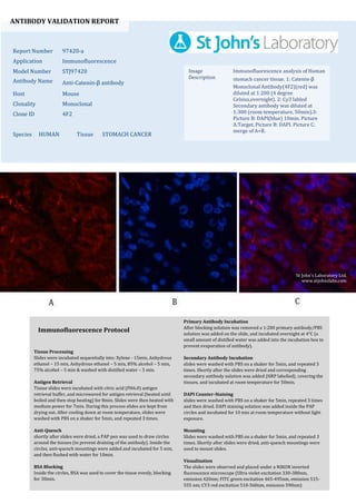
  • 1. ANTIBODY VALIDATION REPORT Report Number 97420-a Application Immunofluorescence Model Number STJ97420 Antibody Name Anti-Catenin-β antibody Host Mouse Clonality Monoclonal Clone ID 4F2 Species HUMAN Tissue STOMACH CANCER Image Description Immunofluorescence analysis of Human stomach cancer tissue. 1: Catenin-β Monoclonal Antibody(4F2)(red) was diluted at 1:200 (4 degree Celsius,overnight). 2: Cy3 labled Secondary antibody was diluted at 1:300 (room temperature, 50min).3: Picture B: DAPI(blue) 10min. Picture A:Target. Picture B: DAPI. Picture C: merge of A+B. Primary Antibody Incubation After blocking solution was removed a 1:200 primary antibody/PBS solution was added on the slide, and incubated overnight at 4°C (a small amount of distilled water was added into the incubation box to prevent evaporation of antibody). Secondary Antibody Incubation slides were washed with PBS on a shaker for 5min, and repeated 3 times. Shortly after the slides were dried and corresponding secondary antibody solution was added (HRP labelled), covering the tissues, and incubated at room temperature for 50min. DAPI Counter-Staining slides were washed with PBS on a shaker for 5min, repeated 3 times and then dried. DAPI staining solution was added inside the PAP circles and incubated for 10 min at room temperature without light exposure. Mounting Slides were washed with PBS on a shaker for 5min, and repeated 3 times. Shortly after slides were dried, anti-quench mountings were used to mount slides. Visualization The slides were observed and placed under a NIKON inverted fluorescence microscope (Ultra violet excitation 330-380nm, emission 420nm; FITC green excitation 465-495nm, emission 515- 555 nm; CY3 red excitation 510-560nm, emission 590nm) Immunofluorescence Protocol Tissue Processing Slides were incubated sequentially into: Xylene - 15min, Anhydrous ethanol – 15 min, Anhydrous ethanol – 5 min, 85% alcohol – 5 min, 75% alcohol – 5 min & washed with distilled water – 5 min. Antigen Retrieval Tissue slides were incubated with citric acid (PH6.0) antigen retrieval buffer, and microwaved for antigen retrieval (heated until boiled and then stop heating) for 8min. Slides were then heated with medium power for 7min. During this process slides are kept from drying out. After cooling down at room temperature, slides were washed with PBS on a shaker for 5min, and repeated 3 times. Anti-Quench shortly after slides were dried, a PAP pen was used to draw circles around the tissues (to prevent draining of the antibody). Inside the circles, anti-quench mountings were added and incubated for 5 min, and then flushed with water for 10min. BSA Blocking Inside the circles, BSA was used to cover the tissue evenly, blocking for 30min. St John's Laboratory Ltd. www.stjohnslabs.com
  • 2. ANTIBODY VALIDATION REPORT Report Number 97420-b Application Immunofluorescence Model Number STJ97420 Antibody Name Anti-Catenin-β antibody Host Mouse Clonality Monoclonal Clone ID 4F2 Species HUMAN Tissue STOMACH CANCER Image Description Immunofluorescence analysis of Human stomach cancer tissue. 1: Catenin-β Monoclonal Antibody(4F2)(red) was diluted at 1:200 (4 degree Celsius,overnight). 2: Cy3 labled Secondary antibody was diluted at 1:300 (room temperature, 50min).3: Picture B: DAPI(blue) 10min. Picture A:Target. Picture B: DAPI. Picture C: merge of A+B. Primary Antibody Incubation After blocking solution was removed a 1:200 primary antibody/PBS solution was added on the slide, and incubated overnight at 4°C (a small amount of distilled water was added into the incubation box to prevent evaporation of antibody). Secondary Antibody Incubation slides were washed with PBS on a shaker for 5min, and repeated 3 times. Shortly after the slides were dried and corresponding secondary antibody solution was added (HRP labelled), covering the tissues, and incubated at room temperature for 50min. DAPI Counter-Staining slides were washed with PBS on a shaker for 5min, repeated 3 times and then dried. DAPI staining solution was added inside the PAP circles and incubated for 10 min at room temperature without light exposure. Mounting Slides were washed with PBS on a shaker for 5min, and repeated 3 times. Shortly after slides were dried, anti-quench mountings were used to mount slides. Visualization The slides were observed and placed under a NIKON inverted fluorescence microscope (Ultra violet excitation 330-380nm, emission 420nm; FITC green excitation 465-495nm, emission 515- 555 nm; CY3 red excitation 510-560nm, emission 590nm) Immunofluorescence Protocol Tissue Processing Slides were incubated sequentially into: Xylene - 15min, Anhydrous ethanol – 15 min, Anhydrous ethanol – 5 min, 85% alcohol – 5 min, 75% alcohol – 5 min & washed with distilled water – 5 min. Antigen Retrieval Tissue slides were incubated with citric acid (PH6.0) antigen retrieval buffer, and microwaved for antigen retrieval (heated until boiled and then stop heating) for 8min. Slides were then heated with medium power for 7min. During this process slides are kept from drying out. After cooling down at room temperature, slides were washed with PBS on a shaker for 5min, and repeated 3 times. Anti-Quench shortly after slides were dried, a PAP pen was used to draw circles around the tissues (to prevent draining of the antibody). Inside the circles, anti-quench mountings were added and incubated for 5 min, and then flushed with water for 10min. BSA Blocking Inside the circles, BSA was used to cover the tissue evenly, blocking for 30min. St John's Laboratory Ltd. www.stjohnslabs.com
  • 3. ANTIBODY VALIDATION REPORT Report Number 97420-c Application Immunofluorescence Model Number STJ97420 Antibody Name Anti-Catenin-β antibody Host Mouse Clonality Monoclonal Clone ID 4F2 Species MOUSE Tissue SPLEEN Image Description Immunofluorescence analysis of Mouse spleen tissue. 1: Catenin-β Monoclonal Antibody(4F2)(red) was diluted at 1:200 (4 degree Celsius,overnight). 2: Cy3 labled Secondary antibody was diluted at 1:300 (room temperature, 50min).3: Picture B: DAPI(blue) 10min. Picture A:Target. Picture B: DAPI. Picture C: merge of A+B. Primary Antibody Incubation After blocking solution was removed a 1:200 primary antibody/PBS solution was added on the slide, and incubated overnight at 4°C (a small amount of distilled water was added into the incubation box to prevent evaporation of antibody). Secondary Antibody Incubation slides were washed with PBS on a shaker for 5min, and repeated 3 times. Shortly after the slides were dried and corresponding secondary antibody solution was added (HRP labelled), covering the tissues, and incubated at room temperature for 50min. DAPI Counter-Staining slides were washed with PBS on a shaker for 5min, repeated 3 times and then dried. DAPI staining solution was added inside the PAP circles and incubated for 10 min at room temperature without light exposure. Mounting Slides were washed with PBS on a shaker for 5min, and repeated 3 times. Shortly after slides were dried, anti-quench mountings were used to mount slides. Visualization The slides were observed and placed under a NIKON inverted fluorescence microscope (Ultra violet excitation 330-380nm, emission 420nm; FITC green excitation 465-495nm, emission 515- 555 nm; CY3 red excitation 510-560nm, emission 590nm) Immunofluorescence Protocol Tissue Processing Slides were incubated sequentially into: Xylene - 15min, Anhydrous ethanol – 15 min, Anhydrous ethanol – 5 min, 85% alcohol – 5 min, 75% alcohol – 5 min & washed with distilled water – 5 min. Antigen Retrieval Tissue slides were incubated with citric acid (PH6.0) antigen retrieval buffer, and microwaved for antigen retrieval (heated until boiled and then stop heating) for 8min. Slides were then heated with medium power for 7min. During this process slides are kept from drying out. After cooling down at room temperature, slides were washed with PBS on a shaker for 5min, and repeated 3 times. Anti-Quench shortly after slides were dried, a PAP pen was used to draw circles around the tissues (to prevent draining of the antibody). Inside the circles, anti-quench mountings were added and incubated for 5 min, and then flushed with water for 10min. BSA Blocking Inside the circles, BSA was used to cover the tissue evenly, blocking for 30min. St John's Laboratory Ltd. www.stjohnslabs.com
  • 4. ANTIBODY VALIDATION REPORT Report Number 97420-d Application Immunofluorescence Model Number STJ97420 Antibody Name Anti-Catenin-β antibody Host Mouse Clonality Monoclonal Clone ID 4F2 Species MOUSE Tissue SPLEEN Image Description Immunofluorescence analysis of Mouse spleen tissue. 1: Catenin-β Monoclonal Antibody(4F2)(red) was diluted at 1:200 (4 degree Celsius,overnight). 2: Cy3 labled Secondary antibody was diluted at 1:300 (room temperature, 50min).3: Picture B: DAPI(blue) 10min. Picture A:Target. Picture B: DAPI. Picture C: merge of A+B. Primary Antibody Incubation After blocking solution was removed a 1:200 primary antibody/PBS solution was added on the slide, and incubated overnight at 4°C (a small amount of distilled water was added into the incubation box to prevent evaporation of antibody). Secondary Antibody Incubation slides were washed with PBS on a shaker for 5min, and repeated 3 times. Shortly after the slides were dried and corresponding secondary antibody solution was added (HRP labelled), covering the tissues, and incubated at room temperature for 50min. DAPI Counter-Staining slides were washed with PBS on a shaker for 5min, repeated 3 times and then dried. DAPI staining solution was added inside the PAP circles and incubated for 10 min at room temperature without light exposure. Mounting Slides were washed with PBS on a shaker for 5min, and repeated 3 times. Shortly after slides were dried, anti-quench mountings were used to mount slides. Visualization The slides were observed and placed under a NIKON inverted fluorescence microscope (Ultra violet excitation 330-380nm, emission 420nm; FITC green excitation 465-495nm, emission 515- 555 nm; CY3 red excitation 510-560nm, emission 590nm) Immunofluorescence Protocol Tissue Processing Slides were incubated sequentially into: Xylene - 15min, Anhydrous ethanol – 15 min, Anhydrous ethanol – 5 min, 85% alcohol – 5 min, 75% alcohol – 5 min & washed with distilled water – 5 min. Antigen Retrieval Tissue slides were incubated with citric acid (PH6.0) antigen retrieval buffer, and microwaved for antigen retrieval (heated until boiled and then stop heating) for 8min. Slides were then heated with medium power for 7min. During this process slides are kept from drying out. After cooling down at room temperature, slides were washed with PBS on a shaker for 5min, and repeated 3 times. Anti-Quench shortly after slides were dried, a PAP pen was used to draw circles around the tissues (to prevent draining of the antibody). Inside the circles, anti-quench mountings were added and incubated for 5 min, and then flushed with water for 10min. BSA Blocking Inside the circles, BSA was used to cover the tissue evenly, blocking for 30min. St John's Laboratory Ltd. www.stjohnslabs.com
  • 5. ANTIBODY VALIDATION REPORT Report Number 97420-e Application Immunofluorescence Model Number STJ97420 Antibody Name Anti-Catenin-β antibody Host Mouse Clonality Monoclonal Clone ID 4F2 Species MOUSE Tissue TESTIS Image Description Immunofluorescence analysis of Mouse testis tissue. 1: Catenin-β Monoclonal Antibody(4F2)(red) was diluted at 1:200 (4 degree Celsius,overnight). 2: Cy3 labled Secondary antibody was diluted at 1:300 (room temperature, 50min).3: Picture B: DAPI(blue) 10min. Picture A:Target. Picture B: DAPI. Picture C: merge of A+B. Primary Antibody Incubation After blocking solution was removed a 1:200 primary antibody/PBS solution was added on the slide, and incubated overnight at 4°C (a small amount of distilled water was added into the incubation box to prevent evaporation of antibody). Secondary Antibody Incubation slides were washed with PBS on a shaker for 5min, and repeated 3 times. Shortly after the slides were dried and corresponding secondary antibody solution was added (HRP labelled), covering the tissues, and incubated at room temperature for 50min. DAPI Counter-Staining slides were washed with PBS on a shaker for 5min, repeated 3 times and then dried. DAPI staining solution was added inside the PAP circles and incubated for 10 min at room temperature without light exposure. Mounting Slides were washed with PBS on a shaker for 5min, and repeated 3 times. Shortly after slides were dried, anti-quench mountings were used to mount slides. Visualization The slides were observed and placed under a NIKON inverted fluorescence microscope (Ultra violet excitation 330-380nm, emission 420nm; FITC green excitation 465-495nm, emission 515- 555 nm; CY3 red excitation 510-560nm, emission 590nm) Immunofluorescence Protocol Tissue Processing Slides were incubated sequentially into: Xylene - 15min, Anhydrous ethanol – 15 min, Anhydrous ethanol – 5 min, 85% alcohol – 5 min, 75% alcohol – 5 min & washed with distilled water – 5 min. Antigen Retrieval Tissue slides were incubated with citric acid (PH6.0) antigen retrieval buffer, and microwaved for antigen retrieval (heated until boiled and then stop heating) for 8min. Slides were then heated with medium power for 7min. During this process slides are kept from drying out. After cooling down at room temperature, slides were washed with PBS on a shaker for 5min, and repeated 3 times. Anti-Quench shortly after slides were dried, a PAP pen was used to draw circles around the tissues (to prevent draining of the antibody). Inside the circles, anti-quench mountings were added and incubated for 5 min, and then flushed with water for 10min. BSA Blocking Inside the circles, BSA was used to cover the tissue evenly, blocking for 30min. St John's Laboratory Ltd. www.stjohnslabs.com
  • 6. ANTIBODY VALIDATION REPORT Report Number 97420-f Application Immunofluorescence Model Number STJ97420 Antibody Name Anti-Catenin-β antibody Host Mouse Clonality Monoclonal Clone ID 4F2 Species MOUSE Tissue TESTIS Image Description Immunofluorescence analysis of Mouse testis tissue. 1: Catenin-β Monoclonal Antibody(4F2)(red) was diluted at 1:200 (4 degree Celsius,overnight). 2: Cy3 labled Secondary antibody was diluted at 1:300 (room temperature, 50min).3: Picture B: DAPI(blue) 10min. Picture A:Target. Picture B: DAPI. Picture C: merge of A+B. Primary Antibody Incubation After blocking solution was removed a 1:200 primary antibody/PBS solution was added on the slide, and incubated overnight at 4°C (a small amount of distilled water was added into the incubation box to prevent evaporation of antibody). Secondary Antibody Incubation slides were washed with PBS on a shaker for 5min, and repeated 3 times. Shortly after the slides were dried and corresponding secondary antibody solution was added (HRP labelled), covering the tissues, and incubated at room temperature for 50min. DAPI Counter-Staining slides were washed with PBS on a shaker for 5min, repeated 3 times and then dried. DAPI staining solution was added inside the PAP circles and incubated for 10 min at room temperature without light exposure. Mounting Slides were washed with PBS on a shaker for 5min, and repeated 3 times. Shortly after slides were dried, anti-quench mountings were used to mount slides. Visualization The slides were observed and placed under a NIKON inverted fluorescence microscope (Ultra violet excitation 330-380nm, emission 420nm; FITC green excitation 465-495nm, emission 515- 555 nm; CY3 red excitation 510-560nm, emission 590nm) Immunofluorescence Protocol Tissue Processing Slides were incubated sequentially into: Xylene - 15min, Anhydrous ethanol – 15 min, Anhydrous ethanol – 5 min, 85% alcohol – 5 min, 75% alcohol – 5 min & washed with distilled water – 5 min. Antigen Retrieval Tissue slides were incubated with citric acid (PH6.0) antigen retrieval buffer, and microwaved for antigen retrieval (heated until boiled and then stop heating) for 8min. Slides were then heated with medium power for 7min. During this process slides are kept from drying out. After cooling down at room temperature, slides were washed with PBS on a shaker for 5min, and repeated 3 times. Anti-Quench shortly after slides were dried, a PAP pen was used to draw circles around the tissues (to prevent draining of the antibody). Inside the circles, anti-quench mountings were added and incubated for 5 min, and then flushed with water for 10min. BSA Blocking Inside the circles, BSA was used to cover the tissue evenly, blocking for 30min. St John's Laboratory Ltd. www.stjohnslabs.com