Immunohistochemistry Antibody Validation Report for Anti-Cleaved PARP Antibody (STJ97047)

Involved in the base excision repair (BER) pathway, by catalyzing the poly(ADP-ribosyl)ation of a limited number of acceptor proteins involved in chromatin architecture and in DNA metabolism. This modification follows DNA damages and appears as an obligatory step in a detection/signaling pathway leading to the reparation of DNA strand breaks. Mediates the poly(ADP-ribosyl)ation of APLF and CHFR. Positively regulates the transcription of MTUS1 and negatively regulates the transcription of MTUS2/TIP150. With EEF1A1 and TXK, forms a complex that acts as a T-helper 1 (Th1) cell-specific transcription factor and binds the promoter of IFN-gamma to directly regulate its transcription, and is thus involved importantly in Th1 cytokine production. Required for PARP9 and DTX3L recruitment to DNA damage sites. PARP1-dependent PARP9-DTX3L-mediated ubiquitination promotes the rapid and specific recruitment of 53BP1/TP53BP1, UIMC1/RAP80, and BRCA1 to DNA damage sites. Anti-Cleaved PARP-http://www.stjohnslabs.com/cleaved-parp-antibody-p-98682 Join our Antibody Validation Project - http://www.stjohnslabs.com/services/antibody-validation

Recommended
Recommended
More Related Content
What's hot
What's hot (20)
Viewers also liked
Viewers also liked (20)
Similar to Immunohistochemistry Antibody Validation Report for Anti-Cleaved PARP Antibody (STJ97047)
Similar to Immunohistochemistry Antibody Validation Report for Anti-Cleaved PARP Antibody (STJ97047) (20)
More from St John's Laboratory Ltd
More from St John's Laboratory Ltd (20)
Recently uploaded
Recently uploaded (20)
Immunohistochemistry Antibody Validation Report for Anti-Cleaved PARP Antibody (STJ97047)
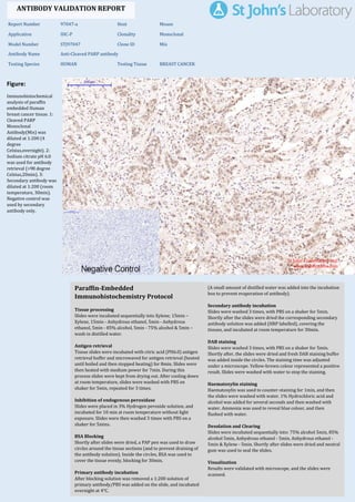
- 1. Figure: Immunohistochemical analysis of paraffin embedded Human breast cancer tissue. 1: Cleaved PARP Monoclonal Antibody(Mix) was diluted at 1:200 (4 degree Celsius,overnight). 2: Sodium citrate pH 6.0 was used for antibody retrieval (>98 degree Celsius,20min). 3: Secondary antibody was diluted at 1:200 (room temperature, 30min). Negative control was used by secondary antibody only. Report Number 97047-a Host Mouse Application IHC-P Clonality Monoclonal Model Number STJ97047 Clone ID Mix Antibody Name Anti-Cleaved PARP antibody Testing Species HUMAN Testing Tissue BREAST CANCER ANTIBODY VALIDATION REPORT b. (A small amount of distilled water was added into the incubation box to prevent evaporation of antibody). 18. Secondary antibody incubation a. Slides were washed 3 times, with PBS on a shaker for 5min. Shortly after the slides were dried the corresponding secondary antibody solution was added (HRP labelled), covering the tissues, and incubated at room temperature for 30min. b. 19. DAB staining a. Slides were washed 3 times, with PBS on a shaker for 5min. b. Shortly after, the slides were dried and fresh DAB staining buffer was added inside the circles. The staining time was adjusted under a microscope. Yellow-brown colour represented a positive result. Slides were washed with water to stop the staining. c. 20. Haematoxylin staining a. Haematoxylin was used to counter-staining for 1min, and then the slides were washed with water. 1% Hydrochloric acid and alcohol was added for several seconds and then washed with water. Ammonia was used to reveal blue colour, and then flushed with water. b. 21. Desolation and Clearing i. Slides were incubated sequentially into: 75% alcohol 5min, 85% alcohol 5min, Anhydrous ethanol - 5min, Anhydrous ethanol - 5min & Xylene - 5min. Shortly after slides were dried and neutral gum was used to seal the slides. ii. 22. Visualization a. Results were validated with microscope, and the slides were scanned. Paraffin-Embedded Immunohistochemistry Protocol 12. 13. Tissue processing a. Slides were incubated sequentially into Xylene; 15min – Xylene, 15min - Anhydrous ethanol, 5min - Anhydrous ethanol, 5min - 85% alcohol, 5min - 75% alcohol & 5min – wash in distilled water. b. 14. Antigen retrieval a. Tissue slides were incubated with citric acid (PH6.0) antigen retrieval buffer and microwaved for antigen retrieval (heated until boiled and then stopped heating) for 8min. Slides were then heated with medium power for 7min. During this process slides were kept from drying out. After cooling down at room temperature, slides were washed with PBS on shaker for 5min, repeated for 3 times. b. 15. Inhibition of endogenous peroxidase a. Slides were placed in 3% Hydrogen peroxide solution, and incubated for 10 min at room temperature without light exposure. Slides were then washed 3 times with PBS on a shaker for 5mins. b. 16. BSA Blocking a. Shortly after slides were dried, a PAP pen was used to draw circles around the tissue sections (and to prevent draining of the antibody solution). Inside the circles, BSA was used to cover the tissue evenly, blocking for 30min. b. 17. Primary antibody incubation After blocking solution was removed a 1:200 solution of primary antibody/PBS was added on the slide, and incubated overnight at 4°C. St John's Laboratory Ltd. www.stjohnslabs.com
- 2. Figure: Immunohistochemical analysis of paraffin embedded Human liver cancer tissue. 1: Cleaved PARP Monoclonal Antibody(Mix) was diluted at 1:200 (4 degree Celsius,overnight). 2: Sodium citrate pH 6.0 was used for antibody retrieval (>98 degree Celsius,20min). 3: Secondary antibody was diluted at 1:200 (room temperature, 30min). Negative control was used by secondary antibody only. Report Number 97047-b Host Mouse Application IHC-P Clonality Monoclonal Model Number STJ97047 Clone ID Mix Antibody Name Anti-Cleaved PARP antibody Testing Species HUMAN Testing Tissue LIVER CANCER ANTIBODY VALIDATION REPORT b. (A small amount of distilled water was added into the incubation box to prevent evaporation of antibody). 7. Secondary antibody incubation a. Slides were washed 3 times, with PBS on a shaker for 5min. Shortly after the slides were dried the corresponding secondary antibody solution was added (HRP labelled), covering the tissues, and incubated at room temperature for 30min. b. 8. DAB staining a. Slides were washed 3 times, with PBS on a shaker for 5min. b. Shortly after, the slides were dried and fresh DAB staining buffer was added inside the circles. The staining time was adjusted under a microscope. Yellow-brown colour represented a positive result. Slides were washed with water to stop the staining. c. 9. Haematoxylin staining a. Haematoxylin was used to counter-staining for 1min, and then the slides were washed with water. 1% Hydrochloric acid and alcohol was added for several seconds and then washed with water. Ammonia was used to reveal blue colour, and then flushed with water. b. 10. Desolation and Clearing i. Slides were incubated sequentially into: 75% alcohol 5min, 85% alcohol 5min, Anhydrous ethanol - 5min, Anhydrous ethanol - 5min & Xylene - 5min. Shortly after slides were dried and neutral gum was used to seal the slides. ii. 11. Visualization a. Results were validated with microscope, and the slides were scanned. Paraffin-Embedded Immunohistochemistry Protocol 1. 2. Tissue processing a. Slides were incubated sequentially into Xylene; 15min – Xylene, 15min - Anhydrous ethanol, 5min - Anhydrous ethanol, 5min - 85% alcohol, 5min - 75% alcohol & 5min – wash in distilled water. b. 3. Antigen retrieval a. Tissue slides were incubated with citric acid (PH6.0) antigen retrieval buffer and microwaved for antigen retrieval (heated until boiled and then stopped heating) for 8min. Slides were then heated with medium power for 7min. During this process slides were kept from drying out. After cooling down at room temperature, slides were washed with PBS on shaker for 5min, repeated for 3 times. b. 4. Inhibition of endogenous peroxidase a. Slides were placed in 3% Hydrogen peroxide solution, and incubated for 10 min at room temperature without light exposure. Slides were then washed 3 times with PBS on a shaker for 5mins. b. 5. BSA Blocking a. Shortly after slides were dried, a PAP pen was used to draw circles around the tissue sections (and to prevent draining of the antibody solution). Inside the circles, BSA was used to cover the tissue evenly, blocking for 30min. b. 6. Primary antibody incubation After blocking solution was removed a 1:200 solution of primary antibody/PBS was added on the slide, and incubated overnight at 4°C. St John's Laboratory Ltd. www.stjohnslabs.com
- 3. Figure: Immunohistochemical analysis of paraffin embedded Human lung tissue. 1: Cleaved PARP Monoclonal Antibody(Mix) was diluted at 1:200 (4 degree Celsius,overnight). 2: Sodium citrate pH 6.0 was used for antibody retrieval (>98 degree Celsius,20min). 3: Secondary antibody was diluted at 1:200 (room temperature, 30min). Negative control was used by secondary antibody only. Report Number 97047-c Host Mouse Application IHC-P Clonality Monoclonal Model Number STJ97047 Clone ID Mix Antibody Name Anti-Cleaved PARP antibody Testing Species HUMAN Testing Tissue LUNG ANTIBODY VALIDATION REPORT b. (A small amount of distilled water was added into the incubation box to prevent evaporation of antibody). 23. Secondary antibody incubation a. Slides were washed 3 times, with PBS on a shaker for 5min. Shortly after the slides were dried the corresponding secondary antibody solution was added (HRP labelled), covering the tissues, and incubated at room temperature for 30min. b. 24. DAB staining a. Slides were washed 3 times, with PBS on a shaker for 5min. b. Shortly after, the slides were dried and fresh DAB staining buffer was added inside the circles. The staining time was adjusted under a microscope. Yellow-brown colour represented a positive result. Slides were washed with water to stop the staining. c. 25. Haematoxylin staining a. Haematoxylin was used to counter-staining for 1min, and then the slides were washed with water. 1% Hydrochloric acid and alcohol was added for several seconds and then washed with water. Ammonia was used to reveal blue colour, and then flushed with water. b. 26. Desolation and Clearing i. Slides were incubated sequentially into: 75% alcohol 5min, 85% alcohol 5min, Anhydrous ethanol - 5min, Anhydrous ethanol - 5min & Xylene - 5min. Shortly after slides were dried and neutral gum was used to seal the slides. ii. 27. Visualization a. Results were validated with microscope, and the slides were scanned. Paraffin-Embedded Immunohistochemistry Protocol 28. 29. Tissue processing a. Slides were incubated sequentially into Xylene; 15min – Xylene, 15min - Anhydrous ethanol, 5min - Anhydrous ethanol, 5min - 85% alcohol, 5min - 75% alcohol & 5min – wash in distilled water. b. 30. Antigen retrieval a. Tissue slides were incubated with citric acid (PH6.0) antigen retrieval buffer and microwaved for antigen retrieval (heated until boiled and then stopped heating) for 8min. Slides were then heated with medium power for 7min. During this process slides were kept from drying out. After cooling down at room temperature, slides were washed with PBS on shaker for 5min, repeated for 3 times. b. 31. Inhibition of endogenous peroxidase a. Slides were placed in 3% Hydrogen peroxide solution, and incubated for 10 min at room temperature without light exposure. Slides were then washed 3 times with PBS on a shaker for 5mins. b. 32. BSA Blocking a. Shortly after slides were dried, a PAP pen was used to draw circles around the tissue sections (and to prevent draining of the antibody solution). Inside the circles, BSA was used to cover the tissue evenly, blocking for 30min. b. 33. Primary antibody incubation After blocking solution was removed a 1:200 solution of primary antibody/PBS was added on the slide, and incubated overnight at 4°C. St John's Laboratory Ltd. www.stjohnslabs.com
- 4. Figure: Immunohistochemical analysis of paraffin embedded Human lung cancer tissue. 1: Cleaved PARP Monoclonal Antibody(Mix) was diluted at 1:200 (4 degree Celsius,overnight). 2: Sodium citrate pH 6.0 was used for antibody retrieval (>98 degree Celsius,20min). 3: Secondary antibody was diluted at 1:200 (room temperature, 30min). Negative control was used by secondary antibody only. Report Number 97047-d Host Mouse Application IHC-P Clonality Monoclonal Model Number STJ97047 Clone ID Mix Antibody Name Anti-Cleaved PARP antibody Testing Species HUMAN Testing Tissue LUNG CANCER ANTIBODY VALIDATION REPORT b. (A small amount of distilled water was added into the incubation box to prevent evaporation of antibody). 34. Secondary antibody incubation a. Slides were washed 3 times, with PBS on a shaker for 5min. Shortly after the slides were dried the corresponding secondary antibody solution was added (HRP labelled), covering the tissues, and incubated at room temperature for 30min. b. 35. DAB staining a. Slides were washed 3 times, with PBS on a shaker for 5min. b. Shortly after, the slides were dried and fresh DAB staining buffer was added inside the circles. The staining time was adjusted under a microscope. Yellow-brown colour represented a positive result. Slides were washed with water to stop the staining. c. 36. Haematoxylin staining a. Haematoxylin was used to counter-staining for 1min, and then the slides were washed with water. 1% Hydrochloric acid and alcohol was added for several seconds and then washed with water. Ammonia was used to reveal blue colour, and then flushed with water. b. 37. Desolation and Clearing i. Slides were incubated sequentially into: 75% alcohol 5min, 85% alcohol 5min, Anhydrous ethanol - 5min, Anhydrous ethanol - 5min & Xylene - 5min. Shortly after slides were dried and neutral gum was used to seal the slides. ii. 38. Visualization a. Results were validated with microscope, and the slides were scanned. Paraffin-Embedded Immunohistochemistry Protocol 39. 40. Tissue processing a. Slides were incubated sequentially into Xylene; 15min – Xylene, 15min - Anhydrous ethanol, 5min - Anhydrous ethanol, 5min - 85% alcohol, 5min - 75% alcohol & 5min – wash in distilled water. b. 41. Antigen retrieval a. Tissue slides were incubated with citric acid (PH6.0) antigen retrieval buffer and microwaved for antigen retrieval (heated until boiled and then stopped heating) for 8min. Slides were then heated with medium power for 7min. During this process slides were kept from drying out. After cooling down at room temperature, slides were washed with PBS on shaker for 5min, repeated for 3 times. b. 42. Inhibition of endogenous peroxidase a. Slides were placed in 3% Hydrogen peroxide solution, and incubated for 10 min at room temperature without light exposure. Slides were then washed 3 times with PBS on a shaker for 5mins. b. 43. BSA Blocking a. Shortly after slides were dried, a PAP pen was used to draw circles around the tissue sections (and to prevent draining of the antibody solution). Inside the circles, BSA was used to cover the tissue evenly, blocking for 30min. b. 44. Primary antibody incubation After blocking solution was removed a 1:200 solution of primary antibody/PBS was added on the slide, and incubated overnight at 4°C. St John's Laboratory Ltd. www.stjohnslabs.com
- 5. Figure: Immunohistochemical analysis of paraffin embedded Human kidney tissue. 1: Cleaved PARP Monoclonal Antibody(Mix) was diluted at 1:200 (4 degree Celsius,overnight). 2: Sodium citrate pH 6.0 was used for antibody retrieval (>98 degree Celsius,20min). 3: Secondary antibody was diluted at 1:200 (room temperature, 30min). Negative control was used by secondary antibody only. Report Number 97047-e Host Mouse Application IHC-P Clonality Monoclonal Model Number STJ97047 Clone ID Mix Antibody Name Anti-Cleaved PARP antibody Testing Species HUMAN Testing Tissue KIDNEY ANTIBODY VALIDATION REPORT b. (A small amount of distilled water was added into the incubation box to prevent evaporation of antibody). 45. Secondary antibody incubation a. Slides were washed 3 times, with PBS on a shaker for 5min. Shortly after the slides were dried the corresponding secondary antibody solution was added (HRP labelled), covering the tissues, and incubated at room temperature for 30min. b. 46. DAB staining a. Slides were washed 3 times, with PBS on a shaker for 5min. b. Shortly after, the slides were dried and fresh DAB staining buffer was added inside the circles. The staining time was adjusted under a microscope. Yellow-brown colour represented a positive result. Slides were washed with water to stop the staining. c. 47. Haematoxylin staining a. Haematoxylin was used to counter-staining for 1min, and then the slides were washed with water. 1% Hydrochloric acid and alcohol was added for several seconds and then washed with water. Ammonia was used to reveal blue colour, and then flushed with water. b. 48. Desolation and Clearing i. Slides were incubated sequentially into: 75% alcohol 5min, 85% alcohol 5min, Anhydrous ethanol - 5min, Anhydrous ethanol - 5min & Xylene - 5min. Shortly after slides were dried and neutral gum was used to seal the slides. ii. 49. Visualization a. Results were validated with microscope, and the slides were scanned. Paraffin-Embedded Immunohistochemistry Protocol 50. 51. Tissue processing a. Slides were incubated sequentially into Xylene; 15min – Xylene, 15min - Anhydrous ethanol, 5min - Anhydrous ethanol, 5min - 85% alcohol, 5min - 75% alcohol & 5min – wash in distilled water. b. 52. Antigen retrieval a. Tissue slides were incubated with citric acid (PH6.0) antigen retrieval buffer and microwaved for antigen retrieval (heated until boiled and then stopped heating) for 8min. Slides were then heated with medium power for 7min. During this process slides were kept from drying out. After cooling down at room temperature, slides were washed with PBS on shaker for 5min, repeated for 3 times. b. 53. Inhibition of endogenous peroxidase a. Slides were placed in 3% Hydrogen peroxide solution, and incubated for 10 min at room temperature without light exposure. Slides were then washed 3 times with PBS on a shaker for 5mins. b. 54. BSA Blocking a. Shortly after slides were dried, a PAP pen was used to draw circles around the tissue sections (and to prevent draining of the antibody solution). Inside the circles, BSA was used to cover the tissue evenly, blocking for 30min. b. 55. Primary antibody incubation After blocking solution was removed a 1:200 solution of primary antibody/PBS was added on the slide, and incubated overnight at 4°C. St John's Laboratory Ltd. www.stjohnslabs.com