SlideShare a Scribd company logo
1 of 7
Download to read offline
Figure:
Immunohistochemical
analysis of paraffin
embedded Human
Tonsil tissue. 1: IGF-IR
Polyclonal Antibody
was diluted at 1:200 (4
degree
Celsius,overnight). 2:
Sodium citrate pH 6.0
was used for antibody
retrieval (>98 degree
Celsius,20min). 3:
Secondary antibody was
diluted at 1:200 (room
temperature, 30min).
Negative control was
used by secondary
antibody only.
Report Number 93647-a Host Rabbit
Application IHC-P Clonality Polyclonal
Model Number STJ93647 Clone ID NA
Antibody Name Anti-IGF-IR antibody
Testing Species HUMAN Testing Tissue TONSIL
ANTIBODY VALIDATION REPORT
a. (A small amount of distilled water was added into the incubation
box to prevent evaporation of antibody).
62. Secondary antibody incubation
a. Slides were washed 3 times, with PBS on a shaker for 5min.
Shortly after the slides were dried the corresponding secondary
antibody solution was added (HRP labelled), covering the
tissues, and incubated at room temperature for 30min.
b.
63. DAB staining
a. Slides were washed 3 times, with PBS on a shaker for 5min.
b. Shortly after, the slides were dried and fresh DAB staining buffer
was added inside the circles. The staining time was adjusted
under a microscope. Yellow-brown colour represented a positive
result. Slides were washed with water to stop the staining.
c.
64. Haematoxylin staining
a. Haematoxylin was used to counter-staining for 1min, and then
the slides were washed with water. 1% Hydrochloric acid and
alcohol was added for several seconds and then washed with
water. Ammonia was used to reveal blue colour, and then
flushed with water.
b.
65. Desolation and Clearing
i. Slides were incubated sequentially into: 75% alcohol 5min, 85%
alcohol 5min, Anhydrous ethanol - 5min, Anhydrous ethanol -
5min & Xylene - 5min. Shortly after slides were dried and neutral
gum was used to seal the slides.
ii.
66. Visualization
a. Results were validated with microscope, and the slides were
scanned.
Paraffin-Embedded
Immunohistochemistry Protocol
56.
57. Tissue processing
a. Slides were incubated sequentially into Xylene; 15min –
Xylene, 15min - Anhydrous ethanol, 5min - Anhydrous
ethanol, 5min - 85% alcohol, 5min - 75% alcohol & 5min –
wash in distilled water.
b.
58. Antigen retrieval
a. Tissue slides were incubated with citric acid (PH6.0) antigen
retrieval buffer and microwaved for antigen retrieval (heated
until boiled and then stopped heating) for 8min. Slides were
then heated with medium power for 7min. During this
process slides were kept from drying out. After cooling down
at room temperature, slides were washed with PBS on
shaker for 5min, repeated for 3 times.
b.
59. Inhibition of endogenous peroxidase
a. Slides were placed in 3% Hydrogen peroxide solution, and
incubated for 10 min at room temperature without light
exposure. Slides were then washed 3 times with PBS on a
shaker for 5mins.
b.
60. BSA Blocking
a. Shortly after slides were dried, a PAP pen was used to draw
circles around the tissue sections (and to prevent draining of
the antibody solution). Inside the circles, BSA was used to
cover the tissue evenly, blocking for 30min.
b.
61. Primary antibody incubation
After blocking solution was removed a 1:200 solution of
primary antibody/PBS was added on the slide, and incubated
overnight at 4°C.
St John's Laboratory Ltd.
www.stjohnslabs.com
Figure:
Immunohistochemical
analysis of paraffin
embedded Human liver
tissue. 1: IGF-IR
Polyclonal Antibody
was diluted at 1:200 (4
degree
Celsius,overnight). 2:
Sodium citrate pH 6.0
was used for antibody
retrieval (>98 degree
Celsius,20min). 3:
Secondary antibody was
diluted at 1:200 (room
temperature, 30min).
Negative control was
used by secondary
antibody only.
Report Number 93647-b Host Rabbit
Application IHC-P Clonality Polyclonal
Model Number STJ93647 Clone ID NA
Antibody Name Anti-IGF-IR antibody
Testing Species HUMAN Testing Tissue LIVER
ANTIBODY VALIDATION REPORT
a. (A small amount of distilled water was added into the incubation
box to prevent evaporation of antibody).
51. Secondary antibody incubation
a. Slides were washed 3 times, with PBS on a shaker for 5min.
Shortly after the slides were dried the corresponding secondary
antibody solution was added (HRP labelled), covering the
tissues, and incubated at room temperature for 30min.
b.
52. DAB staining
a. Slides were washed 3 times, with PBS on a shaker for 5min.
b. Shortly after, the slides were dried and fresh DAB staining buffer
was added inside the circles. The staining time was adjusted
under a microscope. Yellow-brown colour represented a positive
result. Slides were washed with water to stop the staining.
c.
53. Haematoxylin staining
a. Haematoxylin was used to counter-staining for 1min, and then
the slides were washed with water. 1% Hydrochloric acid and
alcohol was added for several seconds and then washed with
water. Ammonia was used to reveal blue colour, and then
flushed with water.
b.
54. Desolation and Clearing
i. Slides were incubated sequentially into: 75% alcohol 5min, 85%
alcohol 5min, Anhydrous ethanol - 5min, Anhydrous ethanol -
5min & Xylene - 5min. Shortly after slides were dried and neutral
gum was used to seal the slides.
ii.
55. Visualization
a. Results were validated with microscope, and the slides were
scanned.
Paraffin-Embedded
Immunohistochemistry Protocol
45.
46. Tissue processing
a. Slides were incubated sequentially into Xylene; 15min –
Xylene, 15min - Anhydrous ethanol, 5min - Anhydrous
ethanol, 5min - 85% alcohol, 5min - 75% alcohol & 5min –
wash in distilled water.
b.
47. Antigen retrieval
a. Tissue slides were incubated with citric acid (PH6.0) antigen
retrieval buffer and microwaved for antigen retrieval (heated
until boiled and then stopped heating) for 8min. Slides were
then heated with medium power for 7min. During this
process slides were kept from drying out. After cooling down
at room temperature, slides were washed with PBS on
shaker for 5min, repeated for 3 times.
b.
48. Inhibition of endogenous peroxidase
a. Slides were placed in 3% Hydrogen peroxide solution, and
incubated for 10 min at room temperature without light
exposure. Slides were then washed 3 times with PBS on a
shaker for 5mins.
b.
49. BSA Blocking
a. Shortly after slides were dried, a PAP pen was used to draw
circles around the tissue sections (and to prevent draining of
the antibody solution). Inside the circles, BSA was used to
cover the tissue evenly, blocking for 30min.
b.
50. Primary antibody incubation
After blocking solution was removed a 1:200 solution of
primary antibody/PBS was added on the slide, and incubated
overnight at 4°C.
St John's Laboratory Ltd.
www.stjohnslabs.com
Figure:
Immunohistochemical
analysis of paraffin
embedded Human liver
cancer tissue. 1: IGF-IR
Polyclonal Antibody
was diluted at 1:200 (4
degree
Celsius,overnight). 2:
Sodium citrate pH 6.0
was used for antibody
retrieval (>98 degree
Celsius,20min). 3:
Secondary antibody was
diluted at 1:200 (room
temperature, 30min).
Negative control was
used by secondary
antibody only.
Report Number 93647-c Host Rabbit
Application IHC-P Clonality Polyclonal
Model Number STJ93647 Clone ID NA
Antibody Name Anti-IGF-IR antibody
Testing Species HUMAN Testing Tissue LIVER CANCER
ANTIBODY VALIDATION REPORT
a. (A small amount of distilled water was added into the incubation
box to prevent evaporation of antibody).
40. Secondary antibody incubation
a. Slides were washed 3 times, with PBS on a shaker for 5min.
Shortly after the slides were dried the corresponding secondary
antibody solution was added (HRP labelled), covering the
tissues, and incubated at room temperature for 30min.
b.
41. DAB staining
a. Slides were washed 3 times, with PBS on a shaker for 5min.
b. Shortly after, the slides were dried and fresh DAB staining buffer
was added inside the circles. The staining time was adjusted
under a microscope. Yellow-brown colour represented a positive
result. Slides were washed with water to stop the staining.
c.
42. Haematoxylin staining
a. Haematoxylin was used to counter-staining for 1min, and then
the slides were washed with water. 1% Hydrochloric acid and
alcohol was added for several seconds and then washed with
water. Ammonia was used to reveal blue colour, and then
flushed with water.
b.
43. Desolation and Clearing
i. Slides were incubated sequentially into: 75% alcohol 5min, 85%
alcohol 5min, Anhydrous ethanol - 5min, Anhydrous ethanol -
5min & Xylene - 5min. Shortly after slides were dried and neutral
gum was used to seal the slides.
ii.
44. Visualization
a. Results were validated with microscope, and the slides were
scanned.
Paraffin-Embedded
Immunohistochemistry Protocol
34.
35. Tissue processing
a. Slides were incubated sequentially into Xylene; 15min –
Xylene, 15min - Anhydrous ethanol, 5min - Anhydrous
ethanol, 5min - 85% alcohol, 5min - 75% alcohol & 5min –
wash in distilled water.
b.
36. Antigen retrieval
a. Tissue slides were incubated with citric acid (PH6.0) antigen
retrieval buffer and microwaved for antigen retrieval (heated
until boiled and then stopped heating) for 8min. Slides were
then heated with medium power for 7min. During this
process slides were kept from drying out. After cooling down
at room temperature, slides were washed with PBS on
shaker for 5min, repeated for 3 times.
b.
37. Inhibition of endogenous peroxidase
a. Slides were placed in 3% Hydrogen peroxide solution, and
incubated for 10 min at room temperature without light
exposure. Slides were then washed 3 times with PBS on a
shaker for 5mins.
b.
38. BSA Blocking
a. Shortly after slides were dried, a PAP pen was used to draw
circles around the tissue sections (and to prevent draining of
the antibody solution). Inside the circles, BSA was used to
cover the tissue evenly, blocking for 30min.
b.
39. Primary antibody incubation
After blocking solution was removed a 1:200 solution of
primary antibody/PBS was added on the slide, and incubated
overnight at 4°C.
St John's Laboratory Ltd.
www.stjohnslabs.com
Figure:
Immunohistochemical
analysis of paraffin
embedded Human
stomach cancer tissue.
1: IGF-IR Polyclonal
Antibody was diluted at
1:200 (4 degree
Celsius,overnight). 2:
Sodium citrate pH 6.0
was used for antibody
retrieval (>98 degree
Celsius,20min). 3:
Secondary antibody was
diluted at 1:200 (room
temperature, 30min).
Negative control was
used by secondary
antibody only.
Report Number 93647-d Host Rabbit
Application IHC-P Clonality Polyclonal
Model Number STJ93647 Clone ID NA
Antibody Name Anti-IGF-IR antibody
Testing Species HUMAN Testing Tissue STOMACH CANCER
ANTIBODY VALIDATION REPORT
a. (A small amount of distilled water was added into the incubation
box to prevent evaporation of antibody).
29. Secondary antibody incubation
a. Slides were washed 3 times, with PBS on a shaker for 5min.
Shortly after the slides were dried the corresponding secondary
antibody solution was added (HRP labelled), covering the
tissues, and incubated at room temperature for 30min.
b.
30. DAB staining
a. Slides were washed 3 times, with PBS on a shaker for 5min.
b. Shortly after, the slides were dried and fresh DAB staining buffer
was added inside the circles. The staining time was adjusted
under a microscope. Yellow-brown colour represented a positive
result. Slides were washed with water to stop the staining.
c.
31. Haematoxylin staining
a. Haematoxylin was used to counter-staining for 1min, and then
the slides were washed with water. 1% Hydrochloric acid and
alcohol was added for several seconds and then washed with
water. Ammonia was used to reveal blue colour, and then
flushed with water.
b.
32. Desolation and Clearing
i. Slides were incubated sequentially into: 75% alcohol 5min, 85%
alcohol 5min, Anhydrous ethanol - 5min, Anhydrous ethanol -
5min & Xylene - 5min. Shortly after slides were dried and neutral
gum was used to seal the slides.
ii.
33. Visualization
a. Results were validated with microscope, and the slides were
scanned.
Paraffin-Embedded
Immunohistochemistry Protocol
23.
24. Tissue processing
a. Slides were incubated sequentially into Xylene; 15min –
Xylene, 15min - Anhydrous ethanol, 5min - Anhydrous
ethanol, 5min - 85% alcohol, 5min - 75% alcohol & 5min –
wash in distilled water.
b.
25. Antigen retrieval
a. Tissue slides were incubated with citric acid (PH6.0) antigen
retrieval buffer and microwaved for antigen retrieval (heated
until boiled and then stopped heating) for 8min. Slides were
then heated with medium power for 7min. During this
process slides were kept from drying out. After cooling down
at room temperature, slides were washed with PBS on
shaker for 5min, repeated for 3 times.
b.
26. Inhibition of endogenous peroxidase
a. Slides were placed in 3% Hydrogen peroxide solution, and
incubated for 10 min at room temperature without light
exposure. Slides were then washed 3 times with PBS on a
shaker for 5mins.
b.
27. BSA Blocking
a. Shortly after slides were dried, a PAP pen was used to draw
circles around the tissue sections (and to prevent draining of
the antibody solution). Inside the circles, BSA was used to
cover the tissue evenly, blocking for 30min.
b.
28. Primary antibody incubation
After blocking solution was removed a 1:200 solution of
primary antibody/PBS was added on the slide, and incubated
overnight at 4°C.
St John's Laboratory Ltd.
www.stjohnslabs.com
Figure:
Immunohistochemical
analysis of paraffin
embedded Rat kidney
tissue. 1: IGF-IR
Polyclonal Antibody
was diluted at 1:200 (4
degree
Celsius,overnight). 2:
Sodium citrate pH 6.0
was used for antibody
retrieval (>98 degree
Celsius,20min). 3:
Secondary antibody was
diluted at 1:200 (room
temperature, 30min).
Negative control was
used by secondary
antibody only.
Report Number 93647-e Host Rabbit
Application IHC-P Clonality Polyclonal
Model Number STJ93647 Clone ID NA
Antibody Name Anti-IGF-IR antibody
Testing Species RAT Testing Tissue KIDNEY
ANTIBODY VALIDATION REPORT
a. (A small amount of distilled water was added into the incubation
box to prevent evaporation of antibody).
18. Secondary antibody incubation
a. Slides were washed 3 times, with PBS on a shaker for 5min.
Shortly after the slides were dried the corresponding secondary
antibody solution was added (HRP labelled), covering the
tissues, and incubated at room temperature for 30min.
b.
19. DAB staining
a. Slides were washed 3 times, with PBS on a shaker for 5min.
b. Shortly after, the slides were dried and fresh DAB staining buffer
was added inside the circles. The staining time was adjusted
under a microscope. Yellow-brown colour represented a positive
result. Slides were washed with water to stop the staining.
c.
20. Haematoxylin staining
a. Haematoxylin was used to counter-staining for 1min, and then
the slides were washed with water. 1% Hydrochloric acid and
alcohol was added for several seconds and then washed with
water. Ammonia was used to reveal blue colour, and then
flushed with water.
b.
21. Desolation and Clearing
i. Slides were incubated sequentially into: 75% alcohol 5min, 85%
alcohol 5min, Anhydrous ethanol - 5min, Anhydrous ethanol -
5min & Xylene - 5min. Shortly after slides were dried and neutral
gum was used to seal the slides.
ii.
22. Visualization
a. Results were validated with microscope, and the slides were
scanned.
Paraffin-Embedded
Immunohistochemistry Protocol
12.
13. Tissue processing
a. Slides were incubated sequentially into Xylene; 15min –
Xylene, 15min - Anhydrous ethanol, 5min - Anhydrous
ethanol, 5min - 85% alcohol, 5min - 75% alcohol & 5min –
wash in distilled water.
b.
14. Antigen retrieval
a. Tissue slides were incubated with citric acid (PH6.0) antigen
retrieval buffer and microwaved for antigen retrieval (heated
until boiled and then stopped heating) for 8min. Slides were
then heated with medium power for 7min. During this
process slides were kept from drying out. After cooling down
at room temperature, slides were washed with PBS on
shaker for 5min, repeated for 3 times.
b.
15. Inhibition of endogenous peroxidase
a. Slides were placed in 3% Hydrogen peroxide solution, and
incubated for 10 min at room temperature without light
exposure. Slides were then washed 3 times with PBS on a
shaker for 5mins.
b.
16. BSA Blocking
a. Shortly after slides were dried, a PAP pen was used to draw
circles around the tissue sections (and to prevent draining of
the antibody solution). Inside the circles, BSA was used to
cover the tissue evenly, blocking for 30min.
b.
17. Primary antibody incubation
After blocking solution was removed a 1:200 solution of
primary antibody/PBS was added on the slide, and incubated
overnight at 4°C.
St John's Laboratory Ltd.
www.stjohnslabs.com
Figure:
Immunohistochemical
analysis of paraffin
embedded Mouse testis
tissue. 1: IGF-IR
Polyclonal Antibody
was diluted at 1:200 (4
degree
Celsius,overnight). 2:
Sodium citrate pH 6.0
was used for antibody
retrieval (>98 degree
Celsius,20min). 3:
Secondary antibody was
diluted at 1:200 (room
temperature, 30min).
Negative control was
used by secondary
antibody only.
Report Number 93647-f Host Rabbit
Application IHC-P Clonality Polyclonal
Model Number STJ93647 Clone ID NA
Antibody Name Anti-IGF-IR antibody
Testing Species MOUSE Testing Tissue TESTIS
ANTIBODY VALIDATION REPORT
a. (A small amount of distilled water was added into the incubation
box to prevent evaporation of antibody).
7. Secondary antibody incubation
a. Slides were washed 3 times, with PBS on a shaker for 5min.
Shortly after the slides were dried the corresponding secondary
antibody solution was added (HRP labelled), covering the
tissues, and incubated at room temperature for 30min.
b.
8. DAB staining
a. Slides were washed 3 times, with PBS on a shaker for 5min.
b. Shortly after, the slides were dried and fresh DAB staining buffer
was added inside the circles. The staining time was adjusted
under a microscope. Yellow-brown colour represented a positive
result. Slides were washed with water to stop the staining.
c.
9. Haematoxylin staining
a. Haematoxylin was used to counter-staining for 1min, and then
the slides were washed with water. 1% Hydrochloric acid and
alcohol was added for several seconds and then washed with
water. Ammonia was used to reveal blue colour, and then
flushed with water.
b.
10. Desolation and Clearing
i. Slides were incubated sequentially into: 75% alcohol 5min, 85%
alcohol 5min, Anhydrous ethanol - 5min, Anhydrous ethanol -
5min & Xylene - 5min. Shortly after slides were dried and neutral
gum was used to seal the slides.
ii.
11. Visualization
a. Results were validated with microscope, and the slides were
scanned.
Paraffin-Embedded
Immunohistochemistry Protocol
1.
2. Tissue processing
a. Slides were incubated sequentially into Xylene; 15min –
Xylene, 15min - Anhydrous ethanol, 5min - Anhydrous
ethanol, 5min - 85% alcohol, 5min - 75% alcohol & 5min –
wash in distilled water.
b.
3. Antigen retrieval
a. Tissue slides were incubated with citric acid (PH6.0) antigen
retrieval buffer and microwaved for antigen retrieval (heated
until boiled and then stopped heating) for 8min. Slides were
then heated with medium power for 7min. During this
process slides were kept from drying out. After cooling down
at room temperature, slides were washed with PBS on
shaker for 5min, repeated for 3 times.
b.
4. Inhibition of endogenous peroxidase
a. Slides were placed in 3% Hydrogen peroxide solution, and
incubated for 10 min at room temperature without light
exposure. Slides were then washed 3 times with PBS on a
shaker for 5mins.
b.
5. BSA Blocking
a. Shortly after slides were dried, a PAP pen was used to draw
circles around the tissue sections (and to prevent draining of
the antibody solution). Inside the circles, BSA was used to
cover the tissue evenly, blocking for 30min.
b.
6. Primary antibody incubation
After blocking solution was removed a 1:200 solution of
primary antibody/PBS was added on the slide, and incubated
overnight at 4°C.
St John's Laboratory Ltd.
www.stjohnslabs.com
Figure:
Immunohistochemical
analysis of paraffin
embedded Mouse
kidney tissue. 1: IGF-IR
Polyclonal Antibody
was diluted at 1:200 (4
degree
Celsius,overnight). 2:
Sodium citrate pH 6.0
was used for antibody
retrieval (>98 degree
Celsius,20min). 3:
Secondary antibody was
diluted at 1:200 (room
temperature, 30min).
Negative control was
used by secondary
antibody only.
Report Number 93647-g Host Rabbit
Application IHC-P Clonality Polyclonal
Model Number STJ93647 Clone ID NA
Antibody Name Anti-IGF-IR antibody
Testing Species MOUSE Testing Tissue KIDNEY
ANTIBODY VALIDATION REPORT
c. (A small amount of distilled water was added into the incubation
box to prevent evaporation of antibody).
67. Secondary antibody incubation
a. Slides were washed 3 times, with PBS on a shaker for 5min.
Shortly after the slides were dried the corresponding secondary
antibody solution was added (HRP labelled), covering the
tissues, and incubated at room temperature for 30min.
b.
68. DAB staining
a. Slides were washed 3 times, with PBS on a shaker for 5min.
b. Shortly after, the slides were dried and fresh DAB staining buffer
was added inside the circles. The staining time was adjusted
under a microscope. Yellow-brown colour represented a positive
result. Slides were washed with water to stop the staining.
c.
69. Haematoxylin staining
a. Haematoxylin was used to counter-staining for 1min, and then
the slides were washed with water. 1% Hydrochloric acid and
alcohol was added for several seconds and then washed with
water. Ammonia was used to reveal blue colour, and then
flushed with water.
b.
70. Desolation and Clearing
i. Slides were incubated sequentially into: 75% alcohol 5min, 85%
alcohol 5min, Anhydrous ethanol - 5min, Anhydrous ethanol -
5min & Xylene - 5min. Shortly after slides were dried and neutral
gum was used to seal the slides.
ii.
71. Visualization
a. Results were validated with microscope, and the slides were
scanned.
Paraffin-Embedded
Immunohistochemistry Protocol
72.
73. Tissue processing
a. Slides were incubated sequentially into Xylene; 15min –
Xylene, 15min - Anhydrous ethanol, 5min - Anhydrous
ethanol, 5min - 85% alcohol, 5min - 75% alcohol & 5min –
wash in distilled water.
b.
74. Antigen retrieval
a. Tissue slides were incubated with citric acid (PH6.0) antigen
retrieval buffer and microwaved for antigen retrieval (heated
until boiled and then stopped heating) for 8min. Slides were
then heated with medium power for 7min. During this
process slides were kept from drying out. After cooling down
at room temperature, slides were washed with PBS on
shaker for 5min, repeated for 3 times.
b.
75. Inhibition of endogenous peroxidase
a. Slides were placed in 3% Hydrogen peroxide solution, and
incubated for 10 min at room temperature without light
exposure. Slides were then washed 3 times with PBS on a
shaker for 5mins.
b.
76. BSA Blocking
a. Shortly after slides were dried, a PAP pen was used to draw
circles around the tissue sections (and to prevent draining of
the antibody solution). Inside the circles, BSA was used to
cover the tissue evenly, blocking for 30min.
b.
77. Primary antibody incubation
After blocking solution was removed a 1:200 solution of
primary antibody/PBS was added on the slide, and incubated
overnight at 4°C.
St John's Laboratory Ltd.
www.stjohnslabs.com

More Related Content

What's hot

Immunohistochemistry Antibody Validation Report for Anti-Caspase-8 Antibody (...
Immunohistochemistry Antibody Validation Report for Anti-Caspase-8 Antibody (...Immunohistochemistry Antibody Validation Report for Anti-Caspase-8 Antibody (...
Immunohistochemistry Antibody Validation Report for Anti-Caspase-8 Antibody (...
St John's Laboratory Ltd
 
Immunohistochemistry Antibody Validation Report for Anti-Bcl-6 Antibody (STJ9...
Immunohistochemistry Antibody Validation Report for Anti-Bcl-6 Antibody (STJ9...Immunohistochemistry Antibody Validation Report for Anti-Bcl-6 Antibody (STJ9...
Immunohistochemistry Antibody Validation Report for Anti-Bcl-6 Antibody (STJ9...
St John's Laboratory Ltd
 

What's hot (20)

Immunohistochemistry Antibody Validation Report for Anti-Catalase Antibody (S...
Immunohistochemistry Antibody Validation Report for Anti-Catalase Antibody (S...Immunohistochemistry Antibody Validation Report for Anti-Catalase Antibody (S...
Immunohistochemistry Antibody Validation Report for Anti-Catalase Antibody (S...
 
Immunohistochemistry Antibody Validation Report for Anti-Caspase-8 Antibody (...
Immunohistochemistry Antibody Validation Report for Anti-Caspase-8 Antibody (...Immunohistochemistry Antibody Validation Report for Anti-Caspase-8 Antibody (...
Immunohistochemistry Antibody Validation Report for Anti-Caspase-8 Antibody (...
 
Immunohistochemistry Antibody Validation Report for Anti-HSP27 Antibody (STJ9...
Immunohistochemistry Antibody Validation Report for Anti-HSP27 Antibody (STJ9...Immunohistochemistry Antibody Validation Report for Anti-HSP27 Antibody (STJ9...
Immunohistochemistry Antibody Validation Report for Anti-HSP27 Antibody (STJ9...
 
Immunohistochemistry Antibody Validation Report for Anti-Bcl-6 Antibody (STJ9...
Immunohistochemistry Antibody Validation Report for Anti-Bcl-6 Antibody (STJ9...Immunohistochemistry Antibody Validation Report for Anti-Bcl-6 Antibody (STJ9...
Immunohistochemistry Antibody Validation Report for Anti-Bcl-6 Antibody (STJ9...
 
Immunohistochemistry Antibody Validation Report for Anti-NGF Antibody (STJ97260)
Immunohistochemistry Antibody Validation Report for Anti-NGF Antibody (STJ97260)Immunohistochemistry Antibody Validation Report for Anti-NGF Antibody (STJ97260)
Immunohistochemistry Antibody Validation Report for Anti-NGF Antibody (STJ97260)
 
Immunohistochemistry Antibody Validation Report for Anti-N-cadherin Antibody ...
Immunohistochemistry Antibody Validation Report for Anti-N-cadherin Antibody ...Immunohistochemistry Antibody Validation Report for Anti-N-cadherin Antibody ...
Immunohistochemistry Antibody Validation Report for Anti-N-cadherin Antibody ...
 
Immunohistochemistry Antibody Validation Report for Anti-Caspase-7 Antibody (...
Immunohistochemistry Antibody Validation Report for Anti-Caspase-7 Antibody (...Immunohistochemistry Antibody Validation Report for Anti-Caspase-7 Antibody (...
Immunohistochemistry Antibody Validation Report for Anti-Caspase-7 Antibody (...
 
Immunohistochemistry Antibody Validation Report for Anti-Luciferase Antibody ...
Immunohistochemistry Antibody Validation Report for Anti-Luciferase Antibody ...Immunohistochemistry Antibody Validation Report for Anti-Luciferase Antibody ...
Immunohistochemistry Antibody Validation Report for Anti-Luciferase Antibody ...
 
Immunohistochemistry Antibody Validation Report for Anti-PR Antibody (STJ97365)
Immunohistochemistry Antibody Validation Report for Anti-PR Antibody (STJ97365)Immunohistochemistry Antibody Validation Report for Anti-PR Antibody (STJ97365)
Immunohistochemistry Antibody Validation Report for Anti-PR Antibody (STJ97365)
 
Immunohistochemistry Antibody Validation Report for Anti-Phospho-CREB-1 (S133...
Immunohistochemistry Antibody Validation Report for Anti-Phospho-CREB-1 (S133...Immunohistochemistry Antibody Validation Report for Anti-Phospho-CREB-1 (S133...
Immunohistochemistry Antibody Validation Report for Anti-Phospho-CREB-1 (S133...
 
Immunohistochemistry Antibody Validation Report for Anti-FAS-L Antibody (STJ9...
Immunohistochemistry Antibody Validation Report for Anti-FAS-L Antibody (STJ9...Immunohistochemistry Antibody Validation Report for Anti-FAS-L Antibody (STJ9...
Immunohistochemistry Antibody Validation Report for Anti-FAS-L Antibody (STJ9...
 
Immunohistochemistry Antibody Validation Report for Anti-NOS3 Antibody (STJ94...
Immunohistochemistry Antibody Validation Report for Anti-NOS3 Antibody (STJ94...Immunohistochemistry Antibody Validation Report for Anti-NOS3 Antibody (STJ94...
Immunohistochemistry Antibody Validation Report for Anti-NOS3 Antibody (STJ94...
 
Immunohistochemistry Antibody Validation Report for Anti-CYCS Antibody (STJ98...
Immunohistochemistry Antibody Validation Report for Anti-CYCS Antibody (STJ98...Immunohistochemistry Antibody Validation Report for Anti-CYCS Antibody (STJ98...
Immunohistochemistry Antibody Validation Report for Anti-CYCS Antibody (STJ98...
 
Immunohistochemistry Antibody Validation Report for Anti-JAK1 Antibody (STJ93...
Immunohistochemistry Antibody Validation Report for Anti-JAK1 Antibody (STJ93...Immunohistochemistry Antibody Validation Report for Anti-JAK1 Antibody (STJ93...
Immunohistochemistry Antibody Validation Report for Anti-JAK1 Antibody (STJ93...
 
Immunohistochemistry Antibody Validation Report for Anti-Akt Antibody (STJ91550)
Immunohistochemistry Antibody Validation Report for Anti-Akt Antibody (STJ91550)Immunohistochemistry Antibody Validation Report for Anti-Akt Antibody (STJ91550)
Immunohistochemistry Antibody Validation Report for Anti-Akt Antibody (STJ91550)
 
Immunohistochemistry Antibody Validation Report for Anti-Caspase-3 Antibody (...
Immunohistochemistry Antibody Validation Report for Anti-Caspase-3 Antibody (...Immunohistochemistry Antibody Validation Report for Anti-Caspase-3 Antibody (...
Immunohistochemistry Antibody Validation Report for Anti-Caspase-3 Antibody (...
 
Immunohistochemistry Antibody Validation Report for Anti-Cyclophilin B Antibo...
Immunohistochemistry Antibody Validation Report for Anti-Cyclophilin B Antibo...Immunohistochemistry Antibody Validation Report for Anti-Cyclophilin B Antibo...
Immunohistochemistry Antibody Validation Report for Anti-Cyclophilin B Antibo...
 
Immunohistochemistry Antibody Validation Report for Anti-Synapsin I Antibody ...
Immunohistochemistry Antibody Validation Report for Anti-Synapsin I Antibody ...Immunohistochemistry Antibody Validation Report for Anti-Synapsin I Antibody ...
Immunohistochemistry Antibody Validation Report for Anti-Synapsin I Antibody ...
 
Immunohistochemistry Antibody Validation Report for Anti-JAK3 Antibody (STJ93...
Immunohistochemistry Antibody Validation Report for Anti-JAK3 Antibody (STJ93...Immunohistochemistry Antibody Validation Report for Anti-JAK3 Antibody (STJ93...
Immunohistochemistry Antibody Validation Report for Anti-JAK3 Antibody (STJ93...
 
Immunohistochemistry Antibody Validation Report for Anti-Phospho-NFκB-p65 (S2...
Immunohistochemistry Antibody Validation Report for Anti-Phospho-NFκB-p65 (S2...Immunohistochemistry Antibody Validation Report for Anti-Phospho-NFκB-p65 (S2...
Immunohistochemistry Antibody Validation Report for Anti-Phospho-NFκB-p65 (S2...
 

Viewers also liked

Immunohistochemistry Antibody Validation Report for Anti-MMP-2 Antibody (STJ9...
Immunohistochemistry Antibody Validation Report for Anti-MMP-2 Antibody (STJ9...Immunohistochemistry Antibody Validation Report for Anti-MMP-2 Antibody (STJ9...
Immunohistochemistry Antibody Validation Report for Anti-MMP-2 Antibody (STJ9...
St John's Laboratory Ltd
 
Immunohistochemistry Antibody Validation Report for Anti-JAK2 Antibody (STJ93...
Immunohistochemistry Antibody Validation Report for Anti-JAK2 Antibody (STJ93...Immunohistochemistry Antibody Validation Report for Anti-JAK2 Antibody (STJ93...
Immunohistochemistry Antibody Validation Report for Anti-JAK2 Antibody (STJ93...
St John's Laboratory Ltd
 
Immunohistochemistry Antibody Validation Report for Anti-FoxO1 Antibody (STJ9...
Immunohistochemistry Antibody Validation Report for Anti-FoxO1 Antibody (STJ9...Immunohistochemistry Antibody Validation Report for Anti-FoxO1 Antibody (STJ9...
Immunohistochemistry Antibody Validation Report for Anti-FoxO1 Antibody (STJ9...
St John's Laboratory Ltd
 

Viewers also liked (20)

Immunohistochemistry Antibody Validation Report for Anti-GSK3β Antibody (STJ9...
Immunohistochemistry Antibody Validation Report for Anti-GSK3β Antibody (STJ9...Immunohistochemistry Antibody Validation Report for Anti-GSK3β Antibody (STJ9...
Immunohistochemistry Antibody Validation Report for Anti-GSK3β Antibody (STJ9...
 
Immunohistochemistry Antibody Validation Report for Anti-MMP-2 Antibody (STJ9...
Immunohistochemistry Antibody Validation Report for Anti-MMP-2 Antibody (STJ9...Immunohistochemistry Antibody Validation Report for Anti-MMP-2 Antibody (STJ9...
Immunohistochemistry Antibody Validation Report for Anti-MMP-2 Antibody (STJ9...
 
Immunohistochemistry Antibody Validation Report for Anti-COL2A1 Antibody (STJ...
Immunohistochemistry Antibody Validation Report for Anti-COL2A1 Antibody (STJ...Immunohistochemistry Antibody Validation Report for Anti-COL2A1 Antibody (STJ...
Immunohistochemistry Antibody Validation Report for Anti-COL2A1 Antibody (STJ...
 
Immunohistochemistry Antibody Validation Report for Anti-JAK2 Antibody (STJ93...
Immunohistochemistry Antibody Validation Report for Anti-JAK2 Antibody (STJ93...Immunohistochemistry Antibody Validation Report for Anti-JAK2 Antibody (STJ93...
Immunohistochemistry Antibody Validation Report for Anti-JAK2 Antibody (STJ93...
 
Immunohistochemistry Antibody Validation Report for Anti-ERα Antibody (STJ92999)
Immunohistochemistry Antibody Validation Report for Anti-ERα Antibody (STJ92999)Immunohistochemistry Antibody Validation Report for Anti-ERα Antibody (STJ92999)
Immunohistochemistry Antibody Validation Report for Anti-ERα Antibody (STJ92999)
 
Immunohistochemistry Antibody Validation Report for Anti-Histone H2A.X Antibo...
Immunohistochemistry Antibody Validation Report for Anti-Histone H2A.X Antibo...Immunohistochemistry Antibody Validation Report for Anti-Histone H2A.X Antibo...
Immunohistochemistry Antibody Validation Report for Anti-Histone H2A.X Antibo...
 
Immunohistochemistry Antibody Validation Report for Anti-Cytokeratin 19 Antib...
Immunohistochemistry Antibody Validation Report for Anti-Cytokeratin 19 Antib...Immunohistochemistry Antibody Validation Report for Anti-Cytokeratin 19 Antib...
Immunohistochemistry Antibody Validation Report for Anti-Cytokeratin 19 Antib...
 
Immunohistochemistry Antibody Validation Report for Anti-FoxO1 Antibody (STJ9...
Immunohistochemistry Antibody Validation Report for Anti-FoxO1 Antibody (STJ9...Immunohistochemistry Antibody Validation Report for Anti-FoxO1 Antibody (STJ9...
Immunohistochemistry Antibody Validation Report for Anti-FoxO1 Antibody (STJ9...
 
Immunohistochemistry Antibody Validation Report for Anti-IκB-α Antibody (STJ9...
Immunohistochemistry Antibody Validation Report for Anti-IκB-α Antibody (STJ9...Immunohistochemistry Antibody Validation Report for Anti-IκB-α Antibody (STJ9...
Immunohistochemistry Antibody Validation Report for Anti-IκB-α Antibody (STJ9...
 
Immunohistochemistry Antibody Validation Report for Anti-Cyclin A Antibody (S...
Immunohistochemistry Antibody Validation Report for Anti-Cyclin A Antibody (S...Immunohistochemistry Antibody Validation Report for Anti-Cyclin A Antibody (S...
Immunohistochemistry Antibody Validation Report for Anti-Cyclin A Antibody (S...
 
Immunohistochemistry Antibody Validation Report for Anti-FAS Antibody (STJ93041)
Immunohistochemistry Antibody Validation Report for Anti-FAS Antibody (STJ93041)Immunohistochemistry Antibody Validation Report for Anti-FAS Antibody (STJ93041)
Immunohistochemistry Antibody Validation Report for Anti-FAS Antibody (STJ93041)
 
Immunohistochemistry Antibody Validation Report for Anti-Glut1 Antibody (STJ9...
Immunohistochemistry Antibody Validation Report for Anti-Glut1 Antibody (STJ9...Immunohistochemistry Antibody Validation Report for Anti-Glut1 Antibody (STJ9...
Immunohistochemistry Antibody Validation Report for Anti-Glut1 Antibody (STJ9...
 
Immunohistochemistry Antibody Validation Report for Anti-Connexin 43 Antibody...
Immunohistochemistry Antibody Validation Report for Anti-Connexin 43 Antibody...Immunohistochemistry Antibody Validation Report for Anti-Connexin 43 Antibody...
Immunohistochemistry Antibody Validation Report for Anti-Connexin 43 Antibody...
 
Immunohistochemistry Antibody Validation Report for Anti-JNK1/2/3 Antibody (S...
Immunohistochemistry Antibody Validation Report for Anti-JNK1/2/3 Antibody (S...Immunohistochemistry Antibody Validation Report for Anti-JNK1/2/3 Antibody (S...
Immunohistochemistry Antibody Validation Report for Anti-JNK1/2/3 Antibody (S...
 
Immunohistochemistry Antibody Validation Report for Anti-ERβ Antibody (STJ93002)
Immunohistochemistry Antibody Validation Report for Anti-ERβ Antibody (STJ93002)Immunohistochemistry Antibody Validation Report for Anti-ERβ Antibody (STJ93002)
Immunohistochemistry Antibody Validation Report for Anti-ERβ Antibody (STJ93002)
 
Immunohistochemistry Antibody Validation Report for Anti-Cytokeratin 18 Antib...
Immunohistochemistry Antibody Validation Report for Anti-Cytokeratin 18 Antib...Immunohistochemistry Antibody Validation Report for Anti-Cytokeratin 18 Antib...
Immunohistochemistry Antibody Validation Report for Anti-Cytokeratin 18 Antib...
 
Immunohistochemistry Antibody Validation Report for Anti-Histone H3 Antibody ...
Immunohistochemistry Antibody Validation Report for Anti-Histone H3 Antibody ...Immunohistochemistry Antibody Validation Report for Anti-Histone H3 Antibody ...
Immunohistochemistry Antibody Validation Report for Anti-Histone H3 Antibody ...
 
Immunohistochemistry Antibody Validation Report for Anti-MIF Antibody (STJ94126)
Immunohistochemistry Antibody Validation Report for Anti-MIF Antibody (STJ94126)Immunohistochemistry Antibody Validation Report for Anti-MIF Antibody (STJ94126)
Immunohistochemistry Antibody Validation Report for Anti-MIF Antibody (STJ94126)
 
Immunohistochemistry Antibody Validation Report for Anti-c-Src Antibody (STJ9...
Immunohistochemistry Antibody Validation Report for Anti-c-Src Antibody (STJ9...Immunohistochemistry Antibody Validation Report for Anti-c-Src Antibody (STJ9...
Immunohistochemistry Antibody Validation Report for Anti-c-Src Antibody (STJ9...
 
Immunohistochemistry Antibody Validation Report for Anti-Flk-1 Antibody (STJ9...
Immunohistochemistry Antibody Validation Report for Anti-Flk-1 Antibody (STJ9...Immunohistochemistry Antibody Validation Report for Anti-Flk-1 Antibody (STJ9...
Immunohistochemistry Antibody Validation Report for Anti-Flk-1 Antibody (STJ9...
 

Similar to Immunohistochemistry Antibody Validation Report for Anti-IGF-IR Antibody (STJ93647)

Immunohistochemistry Antibody Validation Report for Anti-p53 Antibody (STJ94893)
Immunohistochemistry Antibody Validation Report for Anti-p53 Antibody (STJ94893)Immunohistochemistry Antibody Validation Report for Anti-p53 Antibody (STJ94893)
Immunohistochemistry Antibody Validation Report for Anti-p53 Antibody (STJ94893)
St John's Laboratory Ltd
 
Immunohistochemistry Antibody Validation Report for Anti-NFκB-p65 Antibody (S...
Immunohistochemistry Antibody Validation Report for Anti-NFκB-p65 Antibody (S...Immunohistochemistry Antibody Validation Report for Anti-NFκB-p65 Antibody (S...
Immunohistochemistry Antibody Validation Report for Anti-NFκB-p65 Antibody (S...
St John's Laboratory Ltd
 
Immunohistochemistry Antibody Validation Report for Anti-PKC Antibody (STJ95117)
Immunohistochemistry Antibody Validation Report for Anti-PKC Antibody (STJ95117)Immunohistochemistry Antibody Validation Report for Anti-PKC Antibody (STJ95117)
Immunohistochemistry Antibody Validation Report for Anti-PKC Antibody (STJ95117)
St John's Laboratory Ltd
 
Immunohistochemistry Antibody Validation Report for Anti-YAP Antibody (STJ96289)
Immunohistochemistry Antibody Validation Report for Anti-YAP Antibody (STJ96289)Immunohistochemistry Antibody Validation Report for Anti-YAP Antibody (STJ96289)
Immunohistochemistry Antibody Validation Report for Anti-YAP Antibody (STJ96289)
St John's Laboratory Ltd
 

Similar to Immunohistochemistry Antibody Validation Report for Anti-IGF-IR Antibody (STJ93647) (20)

Immunohistochemistry Antibody Validation Report for Anti-p53 Antibody (STJ94893)
Immunohistochemistry Antibody Validation Report for Anti-p53 Antibody (STJ94893)Immunohistochemistry Antibody Validation Report for Anti-p53 Antibody (STJ94893)
Immunohistochemistry Antibody Validation Report for Anti-p53 Antibody (STJ94893)
 
Immunohistochemistry Antibody Validation Report for Anti-p70 S6 kinase α Anti...
Immunohistochemistry Antibody Validation Report for Anti-p70 S6 kinase α Anti...Immunohistochemistry Antibody Validation Report for Anti-p70 S6 kinase α Anti...
Immunohistochemistry Antibody Validation Report for Anti-p70 S6 kinase α Anti...
 
Immunohistochemistry Antibody Validation Report for Anti-VE-Cadherin Antibody...
Immunohistochemistry Antibody Validation Report for Anti-VE-Cadherin Antibody...Immunohistochemistry Antibody Validation Report for Anti-VE-Cadherin Antibody...
Immunohistochemistry Antibody Validation Report for Anti-VE-Cadherin Antibody...
 
Immunohistochemistry Antibody Validation Report for Anti-NFκB-p65 Antibody (S...
Immunohistochemistry Antibody Validation Report for Anti-NFκB-p65 Antibody (S...Immunohistochemistry Antibody Validation Report for Anti-NFκB-p65 Antibody (S...
Immunohistochemistry Antibody Validation Report for Anti-NFκB-p65 Antibody (S...
 
Immunohistochemistry Antibody Validation Report for Anti-CD63 Antibody (STJ96...
Immunohistochemistry Antibody Validation Report for Anti-CD63 Antibody (STJ96...Immunohistochemistry Antibody Validation Report for Anti-CD63 Antibody (STJ96...
Immunohistochemistry Antibody Validation Report for Anti-CD63 Antibody (STJ96...
 
Immunohistochemistry Antibody Validation Report for Anti-SIRT1 Antibody (STJ9...
Immunohistochemistry Antibody Validation Report for Anti-SIRT1 Antibody (STJ9...Immunohistochemistry Antibody Validation Report for Anti-SIRT1 Antibody (STJ9...
Immunohistochemistry Antibody Validation Report for Anti-SIRT1 Antibody (STJ9...
 
Immunohistochemistry Antibody Validation Report for Anti-Lamin B1 Antibody (S...
Immunohistochemistry Antibody Validation Report for Anti-Lamin B1 Antibody (S...Immunohistochemistry Antibody Validation Report for Anti-Lamin B1 Antibody (S...
Immunohistochemistry Antibody Validation Report for Anti-Lamin B1 Antibody (S...
 
Immunohistochemistry Antibody Validation Report for Anti-Rho A Antibody (STJ9...
Immunohistochemistry Antibody Validation Report for Anti-Rho A Antibody (STJ9...Immunohistochemistry Antibody Validation Report for Anti-Rho A Antibody (STJ9...
Immunohistochemistry Antibody Validation Report for Anti-Rho A Antibody (STJ9...
 
Immunohistochemistry Antibody Validation Report for Anti-MAP LC3β Antibody (S...
Immunohistochemistry Antibody Validation Report for Anti-MAP LC3β Antibody (S...Immunohistochemistry Antibody Validation Report for Anti-MAP LC3β Antibody (S...
Immunohistochemistry Antibody Validation Report for Anti-MAP LC3β Antibody (S...
 
Immunohistochemistry Antibody Validation Report for Anti-AR Antibody (STJ97233)
Immunohistochemistry Antibody Validation Report for Anti-AR Antibody (STJ97233)Immunohistochemistry Antibody Validation Report for Anti-AR Antibody (STJ97233)
Immunohistochemistry Antibody Validation Report for Anti-AR Antibody (STJ97233)
 
Immunohistochemistry Antibody Validation Report for Anti-HAO1 Antibody (STJ97...
Immunohistochemistry Antibody Validation Report for Anti-HAO1 Antibody (STJ97...Immunohistochemistry Antibody Validation Report for Anti-HAO1 Antibody (STJ97...
Immunohistochemistry Antibody Validation Report for Anti-HAO1 Antibody (STJ97...
 
Immunohistochemistry Antibody Validation Report for Anti-Rock-1 Antibody (STJ...
Immunohistochemistry Antibody Validation Report for Anti-Rock-1 Antibody (STJ...Immunohistochemistry Antibody Validation Report for Anti-Rock-1 Antibody (STJ...
Immunohistochemistry Antibody Validation Report for Anti-Rock-1 Antibody (STJ...
 
Immunohistochemistry Antibody Validation Report for Anti-Cleaved PARP Antibod...
Immunohistochemistry Antibody Validation Report for Anti-Cleaved PARP Antibod...Immunohistochemistry Antibody Validation Report for Anti-Cleaved PARP Antibod...
Immunohistochemistry Antibody Validation Report for Anti-Cleaved PARP Antibod...
 
Immunohistochemistry Antibody Validation Report for Anti-p70 S6 kinase α Anti...
Immunohistochemistry Antibody Validation Report for Anti-p70 S6 kinase α Anti...Immunohistochemistry Antibody Validation Report for Anti-p70 S6 kinase α Anti...
Immunohistochemistry Antibody Validation Report for Anti-p70 S6 kinase α Anti...
 
Immunohistochemistry Antibody Validation Report for Anti-PKC Antibody (STJ95117)
Immunohistochemistry Antibody Validation Report for Anti-PKC Antibody (STJ95117)Immunohistochemistry Antibody Validation Report for Anti-PKC Antibody (STJ95117)
Immunohistochemistry Antibody Validation Report for Anti-PKC Antibody (STJ95117)
 
Immunohistochemistry Antibody Validation Report for Anti-Survivin Antibody (S...
Immunohistochemistry Antibody Validation Report for Anti-Survivin Antibody (S...Immunohistochemistry Antibody Validation Report for Anti-Survivin Antibody (S...
Immunohistochemistry Antibody Validation Report for Anti-Survivin Antibody (S...
 
Immunohistochemistry Antibody Validation Report for Anti-BRCA1 Antibody (STJ9...
Immunohistochemistry Antibody Validation Report for Anti-BRCA1 Antibody (STJ9...Immunohistochemistry Antibody Validation Report for Anti-BRCA1 Antibody (STJ9...
Immunohistochemistry Antibody Validation Report for Anti-BRCA1 Antibody (STJ9...
 
Immunohistochemistry Antibody Validation Report for Anti-Active Caspase-3 Ant...
Immunohistochemistry Antibody Validation Report for Anti-Active Caspase-3 Ant...Immunohistochemistry Antibody Validation Report for Anti-Active Caspase-3 Ant...
Immunohistochemistry Antibody Validation Report for Anti-Active Caspase-3 Ant...
 
Immunohistochemistry Antibody Validation Report for Anti-YAP Antibody (STJ96289)
Immunohistochemistry Antibody Validation Report for Anti-YAP Antibody (STJ96289)Immunohistochemistry Antibody Validation Report for Anti-YAP Antibody (STJ96289)
Immunohistochemistry Antibody Validation Report for Anti-YAP Antibody (STJ96289)
 
Immunohistochemistry Antibody Validation Report for Anti-GFAP Antibody (STJ96...
Immunohistochemistry Antibody Validation Report for Anti-GFAP Antibody (STJ96...Immunohistochemistry Antibody Validation Report for Anti-GFAP Antibody (STJ96...
Immunohistochemistry Antibody Validation Report for Anti-GFAP Antibody (STJ96...
 

More from St John's Laboratory Ltd

Western Blot Customer Review Anti Glucocorticoid Receptor antibody (STJ97101)
Western Blot Customer Review Anti Glucocorticoid Receptor antibody (STJ97101)Western Blot Customer Review Anti Glucocorticoid Receptor antibody (STJ97101)
Western Blot Customer Review Anti Glucocorticoid Receptor antibody (STJ97101)
St John's Laboratory Ltd
 

More from St John's Laboratory Ltd (20)

Immunohistochemistry Customer Review Anti-Smo antibody (STJ95710)
Immunohistochemistry Customer Review Anti-Smo antibody (STJ95710)Immunohistochemistry Customer Review Anti-Smo antibody (STJ95710)
Immunohistochemistry Customer Review Anti-Smo antibody (STJ95710)
 
Western Blot Customer Review Anti Glucocorticoid Receptor antibody (STJ97101)
Western Blot Customer Review Anti Glucocorticoid Receptor antibody (STJ97101)Western Blot Customer Review Anti Glucocorticoid Receptor antibody (STJ97101)
Western Blot Customer Review Anti Glucocorticoid Receptor antibody (STJ97101)
 
Western Blot Customer Review Anti-Phospho-Cofilin (S3) Antibody (STJ90230)
Western Blot Customer Review Anti-Phospho-Cofilin (S3) Antibody (STJ90230)Western Blot Customer Review Anti-Phospho-Cofilin (S3) Antibody (STJ90230)
Western Blot Customer Review Anti-Phospho-Cofilin (S3) Antibody (STJ90230)
 
Scientist of the Month - May Byron Baron
Scientist of the Month - May Byron BaronScientist of the Month - May Byron Baron
Scientist of the Month - May Byron Baron
 
Western Blot Customer Review Anti-WAVE2 Antibody (STJ96263)
Western Blot Customer Review Anti-WAVE2 Antibody (STJ96263)Western Blot Customer Review Anti-WAVE2 Antibody (STJ96263)
Western Blot Customer Review Anti-WAVE2 Antibody (STJ96263)
 
Western Blot Customer Review Anti-Endophilin I Antibody (STJ92924)
Western Blot Customer Review Anti-Endophilin I Antibody (STJ92924)Western Blot Customer Review Anti-Endophilin I Antibody (STJ92924)
Western Blot Customer Review Anti-Endophilin I Antibody (STJ92924)
 
Western Blot Customer Review Anti-β-tubulin Antibody (STJ97037)
Western Blot Customer Review Anti-β-tubulin Antibody (STJ97037)Western Blot Customer Review Anti-β-tubulin Antibody (STJ97037)
Western Blot Customer Review Anti-β-tubulin Antibody (STJ97037)
 
Immunohistochemistry Antibody Validation Report for Anti-Gamma Tubulin Antibo...
Immunohistochemistry Antibody Validation Report for Anti-Gamma Tubulin Antibo...Immunohistochemistry Antibody Validation Report for Anti-Gamma Tubulin Antibo...
Immunohistochemistry Antibody Validation Report for Anti-Gamma Tubulin Antibo...
 
Immunohistochemistry Antibody Validation Report for Anti-Epsilon Tubulin Anti...
Immunohistochemistry Antibody Validation Report for Anti-Epsilon Tubulin Anti...Immunohistochemistry Antibody Validation Report for Anti-Epsilon Tubulin Anti...
Immunohistochemistry Antibody Validation Report for Anti-Epsilon Tubulin Anti...
 
Immunohistochemistry Antibody Validation Report for Anti-LC3A Antibody (STJ97...
Immunohistochemistry Antibody Validation Report for Anti-LC3A Antibody (STJ97...Immunohistochemistry Antibody Validation Report for Anti-LC3A Antibody (STJ97...
Immunohistochemistry Antibody Validation Report for Anti-LC3A Antibody (STJ97...
 
Immunohistochemistry Antibody Validation Report for Anti-CHOP Antibody (STJ97...
Immunohistochemistry Antibody Validation Report for Anti-CHOP Antibody (STJ97...Immunohistochemistry Antibody Validation Report for Anti-CHOP Antibody (STJ97...
Immunohistochemistry Antibody Validation Report for Anti-CHOP Antibody (STJ97...
 
Immunohistochemistry Antibody Validation Report for Anti-phospho-MLKL (S358) ...
Immunohistochemistry Antibody Validation Report for Anti-phospho-MLKL (S358) ...Immunohistochemistry Antibody Validation Report for Anti-phospho-MLKL (S358) ...
Immunohistochemistry Antibody Validation Report for Anti-phospho-MLKL (S358) ...
 
Immunohistochemistry Antibody Validation Report for Anti-ERK1 Antibody (STJ97...
Immunohistochemistry Antibody Validation Report for Anti-ERK1 Antibody (STJ97...Immunohistochemistry Antibody Validation Report for Anti-ERK1 Antibody (STJ97...
Immunohistochemistry Antibody Validation Report for Anti-ERK1 Antibody (STJ97...
 
Immunohistochemistry Antibody Validation Report for Anti-PDGFRα Antibody (STJ...
Immunohistochemistry Antibody Validation Report for Anti-PDGFRα Antibody (STJ...Immunohistochemistry Antibody Validation Report for Anti-PDGFRα Antibody (STJ...
Immunohistochemistry Antibody Validation Report for Anti-PDGFRα Antibody (STJ...
 
Immunohistochemistry Antibody Validation Report for Anti-Caspase-1 Antibody (...
Immunohistochemistry Antibody Validation Report for Anti-Caspase-1 Antibody (...Immunohistochemistry Antibody Validation Report for Anti-Caspase-1 Antibody (...
Immunohistochemistry Antibody Validation Report for Anti-Caspase-1 Antibody (...
 
Immunohistochemistry Antibody Validation Report for Anti-Amyloid-β Antibody (...
Immunohistochemistry Antibody Validation Report for Anti-Amyloid-β Antibody (...Immunohistochemistry Antibody Validation Report for Anti-Amyloid-β Antibody (...
Immunohistochemistry Antibody Validation Report for Anti-Amyloid-β Antibody (...
 
Immunohistochemistry Antibody Validation Report for Anti-Bcl-2 Antibody (STJ9...
Immunohistochemistry Antibody Validation Report for Anti-Bcl-2 Antibody (STJ9...Immunohistochemistry Antibody Validation Report for Anti-Bcl-2 Antibody (STJ9...
Immunohistochemistry Antibody Validation Report for Anti-Bcl-2 Antibody (STJ9...
 
Immunohistochemistry Antibody Validation Report for Anti-α-SMA Antibody (STJ9...
Immunohistochemistry Antibody Validation Report for Anti-α-SMA Antibody (STJ9...Immunohistochemistry Antibody Validation Report for Anti-α-SMA Antibody (STJ9...
Immunohistochemistry Antibody Validation Report for Anti-α-SMA Antibody (STJ9...
 
Immunohistochemistry Antibody Validation Report for Anti-E2F-1 Antibody (STJ9...
Immunohistochemistry Antibody Validation Report for Anti-E2F-1 Antibody (STJ9...Immunohistochemistry Antibody Validation Report for Anti-E2F-1 Antibody (STJ9...
Immunohistochemistry Antibody Validation Report for Anti-E2F-1 Antibody (STJ9...
 
Immunohistochemistry Antibody Validation Report for Anti-Annexin I Antibody (...
Immunohistochemistry Antibody Validation Report for Anti-Annexin I Antibody (...Immunohistochemistry Antibody Validation Report for Anti-Annexin I Antibody (...
Immunohistochemistry Antibody Validation Report for Anti-Annexin I Antibody (...
 

Recently uploaded

The Mariana Trench remarkable geological features on Earth.pptx
The Mariana Trench remarkable geological features on Earth.pptxThe Mariana Trench remarkable geological features on Earth.pptx
The Mariana Trench remarkable geological features on Earth.pptx
seri bangash
 
Module for Grade 9 for Asynchronous/Distance learning
Module for Grade 9 for Asynchronous/Distance learningModule for Grade 9 for Asynchronous/Distance learning
Module for Grade 9 for Asynchronous/Distance learning
levieagacer
 
biology HL practice questions IB BIOLOGY
biology HL practice questions IB BIOLOGYbiology HL practice questions IB BIOLOGY
biology HL practice questions IB BIOLOGY
1301aanya
 
Conjugation, transduction and transformation
Conjugation, transduction and transformationConjugation, transduction and transformation
Conjugation, transduction and transformation
Areesha Ahmad
 

Recently uploaded (20)

Zoology 5th semester notes( Sumit_yadav).pdf
Zoology 5th semester notes( Sumit_yadav).pdfZoology 5th semester notes( Sumit_yadav).pdf
Zoology 5th semester notes( Sumit_yadav).pdf
 
Dr. E. Muralinath_ Blood indices_clinical aspects
Dr. E. Muralinath_ Blood indices_clinical  aspectsDr. E. Muralinath_ Blood indices_clinical  aspects
Dr. E. Muralinath_ Blood indices_clinical aspects
 
COMPUTING ANTI-DERIVATIVES (Integration by SUBSTITUTION)
COMPUTING ANTI-DERIVATIVES(Integration by SUBSTITUTION)COMPUTING ANTI-DERIVATIVES(Integration by SUBSTITUTION)
COMPUTING ANTI-DERIVATIVES (Integration by SUBSTITUTION)
 
FAIRSpectra - Enabling the FAIRification of Analytical Science
FAIRSpectra - Enabling the FAIRification of Analytical ScienceFAIRSpectra - Enabling the FAIRification of Analytical Science
FAIRSpectra - Enabling the FAIRification of Analytical Science
 
Bhiwandi Bhiwandi ❤CALL GIRL 7870993772 ❤CALL GIRLS ESCORT SERVICE In Bhiwan...
Bhiwandi Bhiwandi ❤CALL GIRL 7870993772 ❤CALL GIRLS  ESCORT SERVICE In Bhiwan...Bhiwandi Bhiwandi ❤CALL GIRL 7870993772 ❤CALL GIRLS  ESCORT SERVICE In Bhiwan...
Bhiwandi Bhiwandi ❤CALL GIRL 7870993772 ❤CALL GIRLS ESCORT SERVICE In Bhiwan...
 
Thyroid Physiology_Dr.E. Muralinath_ Associate Professor
Thyroid Physiology_Dr.E. Muralinath_ Associate ProfessorThyroid Physiology_Dr.E. Muralinath_ Associate Professor
Thyroid Physiology_Dr.E. Muralinath_ Associate Professor
 
The Mariana Trench remarkable geological features on Earth.pptx
The Mariana Trench remarkable geological features on Earth.pptxThe Mariana Trench remarkable geological features on Earth.pptx
The Mariana Trench remarkable geological features on Earth.pptx
 
Stages in the normal growth curve
Stages in the normal growth curveStages in the normal growth curve
Stages in the normal growth curve
 
module for grade 9 for distance learning
module for grade 9 for distance learningmodule for grade 9 for distance learning
module for grade 9 for distance learning
 
300003-World Science Day For Peace And Development.pptx
300003-World Science Day For Peace And Development.pptx300003-World Science Day For Peace And Development.pptx
300003-World Science Day For Peace And Development.pptx
 
Chemistry 5th semester paper 1st Notes.pdf
Chemistry 5th semester paper 1st Notes.pdfChemistry 5th semester paper 1st Notes.pdf
Chemistry 5th semester paper 1st Notes.pdf
 
pumpkin fruit fly, water melon fruit fly, cucumber fruit fly
pumpkin fruit fly, water melon fruit fly, cucumber fruit flypumpkin fruit fly, water melon fruit fly, cucumber fruit fly
pumpkin fruit fly, water melon fruit fly, cucumber fruit fly
 
Locating and isolating a gene, FISH, GISH, Chromosome walking and jumping, te...
Locating and isolating a gene, FISH, GISH, Chromosome walking and jumping, te...Locating and isolating a gene, FISH, GISH, Chromosome walking and jumping, te...
Locating and isolating a gene, FISH, GISH, Chromosome walking and jumping, te...
 
GBSN - Microbiology (Unit 2)
GBSN - Microbiology (Unit 2)GBSN - Microbiology (Unit 2)
GBSN - Microbiology (Unit 2)
 
Module for Grade 9 for Asynchronous/Distance learning
Module for Grade 9 for Asynchronous/Distance learningModule for Grade 9 for Asynchronous/Distance learning
Module for Grade 9 for Asynchronous/Distance learning
 
Proteomics: types, protein profiling steps etc.
Proteomics: types, protein profiling steps etc.Proteomics: types, protein profiling steps etc.
Proteomics: types, protein profiling steps etc.
 
biology HL practice questions IB BIOLOGY
biology HL practice questions IB BIOLOGYbiology HL practice questions IB BIOLOGY
biology HL practice questions IB BIOLOGY
 
GBSN - Microbiology (Unit 3)
GBSN - Microbiology (Unit 3)GBSN - Microbiology (Unit 3)
GBSN - Microbiology (Unit 3)
 
Conjugation, transduction and transformation
Conjugation, transduction and transformationConjugation, transduction and transformation
Conjugation, transduction and transformation
 
Factory Acceptance Test( FAT).pptx .
Factory Acceptance Test( FAT).pptx       .Factory Acceptance Test( FAT).pptx       .
Factory Acceptance Test( FAT).pptx .
 

Immunohistochemistry Antibody Validation Report for Anti-IGF-IR Antibody (STJ93647)

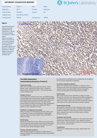
  • 1. Figure: Immunohistochemical analysis of paraffin embedded Human Tonsil tissue. 1: IGF-IR Polyclonal Antibody was diluted at 1:200 (4 degree Celsius,overnight). 2: Sodium citrate pH 6.0 was used for antibody retrieval (>98 degree Celsius,20min). 3: Secondary antibody was diluted at 1:200 (room temperature, 30min). Negative control was used by secondary antibody only. Report Number 93647-a Host Rabbit Application IHC-P Clonality Polyclonal Model Number STJ93647 Clone ID NA Antibody Name Anti-IGF-IR antibody Testing Species HUMAN Testing Tissue TONSIL ANTIBODY VALIDATION REPORT a. (A small amount of distilled water was added into the incubation box to prevent evaporation of antibody). 62. Secondary antibody incubation a. Slides were washed 3 times, with PBS on a shaker for 5min. Shortly after the slides were dried the corresponding secondary antibody solution was added (HRP labelled), covering the tissues, and incubated at room temperature for 30min. b. 63. DAB staining a. Slides were washed 3 times, with PBS on a shaker for 5min. b. Shortly after, the slides were dried and fresh DAB staining buffer was added inside the circles. The staining time was adjusted under a microscope. Yellow-brown colour represented a positive result. Slides were washed with water to stop the staining. c. 64. Haematoxylin staining a. Haematoxylin was used to counter-staining for 1min, and then the slides were washed with water. 1% Hydrochloric acid and alcohol was added for several seconds and then washed with water. Ammonia was used to reveal blue colour, and then flushed with water. b. 65. Desolation and Clearing i. Slides were incubated sequentially into: 75% alcohol 5min, 85% alcohol 5min, Anhydrous ethanol - 5min, Anhydrous ethanol - 5min & Xylene - 5min. Shortly after slides were dried and neutral gum was used to seal the slides. ii. 66. Visualization a. Results were validated with microscope, and the slides were scanned. Paraffin-Embedded Immunohistochemistry Protocol 56. 57. Tissue processing a. Slides were incubated sequentially into Xylene; 15min – Xylene, 15min - Anhydrous ethanol, 5min - Anhydrous ethanol, 5min - 85% alcohol, 5min - 75% alcohol & 5min – wash in distilled water. b. 58. Antigen retrieval a. Tissue slides were incubated with citric acid (PH6.0) antigen retrieval buffer and microwaved for antigen retrieval (heated until boiled and then stopped heating) for 8min. Slides were then heated with medium power for 7min. During this process slides were kept from drying out. After cooling down at room temperature, slides were washed with PBS on shaker for 5min, repeated for 3 times. b. 59. Inhibition of endogenous peroxidase a. Slides were placed in 3% Hydrogen peroxide solution, and incubated for 10 min at room temperature without light exposure. Slides were then washed 3 times with PBS on a shaker for 5mins. b. 60. BSA Blocking a. Shortly after slides were dried, a PAP pen was used to draw circles around the tissue sections (and to prevent draining of the antibody solution). Inside the circles, BSA was used to cover the tissue evenly, blocking for 30min. b. 61. Primary antibody incubation After blocking solution was removed a 1:200 solution of primary antibody/PBS was added on the slide, and incubated overnight at 4°C. St John's Laboratory Ltd. www.stjohnslabs.com
  • 2. Figure: Immunohistochemical analysis of paraffin embedded Human liver tissue. 1: IGF-IR Polyclonal Antibody was diluted at 1:200 (4 degree Celsius,overnight). 2: Sodium citrate pH 6.0 was used for antibody retrieval (>98 degree Celsius,20min). 3: Secondary antibody was diluted at 1:200 (room temperature, 30min). Negative control was used by secondary antibody only. Report Number 93647-b Host Rabbit Application IHC-P Clonality Polyclonal Model Number STJ93647 Clone ID NA Antibody Name Anti-IGF-IR antibody Testing Species HUMAN Testing Tissue LIVER ANTIBODY VALIDATION REPORT a. (A small amount of distilled water was added into the incubation box to prevent evaporation of antibody). 51. Secondary antibody incubation a. Slides were washed 3 times, with PBS on a shaker for 5min. Shortly after the slides were dried the corresponding secondary antibody solution was added (HRP labelled), covering the tissues, and incubated at room temperature for 30min. b. 52. DAB staining a. Slides were washed 3 times, with PBS on a shaker for 5min. b. Shortly after, the slides were dried and fresh DAB staining buffer was added inside the circles. The staining time was adjusted under a microscope. Yellow-brown colour represented a positive result. Slides were washed with water to stop the staining. c. 53. Haematoxylin staining a. Haematoxylin was used to counter-staining for 1min, and then the slides were washed with water. 1% Hydrochloric acid and alcohol was added for several seconds and then washed with water. Ammonia was used to reveal blue colour, and then flushed with water. b. 54. Desolation and Clearing i. Slides were incubated sequentially into: 75% alcohol 5min, 85% alcohol 5min, Anhydrous ethanol - 5min, Anhydrous ethanol - 5min & Xylene - 5min. Shortly after slides were dried and neutral gum was used to seal the slides. ii. 55. Visualization a. Results were validated with microscope, and the slides were scanned. Paraffin-Embedded Immunohistochemistry Protocol 45. 46. Tissue processing a. Slides were incubated sequentially into Xylene; 15min – Xylene, 15min - Anhydrous ethanol, 5min - Anhydrous ethanol, 5min - 85% alcohol, 5min - 75% alcohol & 5min – wash in distilled water. b. 47. Antigen retrieval a. Tissue slides were incubated with citric acid (PH6.0) antigen retrieval buffer and microwaved for antigen retrieval (heated until boiled and then stopped heating) for 8min. Slides were then heated with medium power for 7min. During this process slides were kept from drying out. After cooling down at room temperature, slides were washed with PBS on shaker for 5min, repeated for 3 times. b. 48. Inhibition of endogenous peroxidase a. Slides were placed in 3% Hydrogen peroxide solution, and incubated for 10 min at room temperature without light exposure. Slides were then washed 3 times with PBS on a shaker for 5mins. b. 49. BSA Blocking a. Shortly after slides were dried, a PAP pen was used to draw circles around the tissue sections (and to prevent draining of the antibody solution). Inside the circles, BSA was used to cover the tissue evenly, blocking for 30min. b. 50. Primary antibody incubation After blocking solution was removed a 1:200 solution of primary antibody/PBS was added on the slide, and incubated overnight at 4°C. St John's Laboratory Ltd. www.stjohnslabs.com
  • 3. Figure: Immunohistochemical analysis of paraffin embedded Human liver cancer tissue. 1: IGF-IR Polyclonal Antibody was diluted at 1:200 (4 degree Celsius,overnight). 2: Sodium citrate pH 6.0 was used for antibody retrieval (>98 degree Celsius,20min). 3: Secondary antibody was diluted at 1:200 (room temperature, 30min). Negative control was used by secondary antibody only. Report Number 93647-c Host Rabbit Application IHC-P Clonality Polyclonal Model Number STJ93647 Clone ID NA Antibody Name Anti-IGF-IR antibody Testing Species HUMAN Testing Tissue LIVER CANCER ANTIBODY VALIDATION REPORT a. (A small amount of distilled water was added into the incubation box to prevent evaporation of antibody). 40. Secondary antibody incubation a. Slides were washed 3 times, with PBS on a shaker for 5min. Shortly after the slides were dried the corresponding secondary antibody solution was added (HRP labelled), covering the tissues, and incubated at room temperature for 30min. b. 41. DAB staining a. Slides were washed 3 times, with PBS on a shaker for 5min. b. Shortly after, the slides were dried and fresh DAB staining buffer was added inside the circles. The staining time was adjusted under a microscope. Yellow-brown colour represented a positive result. Slides were washed with water to stop the staining. c. 42. Haematoxylin staining a. Haematoxylin was used to counter-staining for 1min, and then the slides were washed with water. 1% Hydrochloric acid and alcohol was added for several seconds and then washed with water. Ammonia was used to reveal blue colour, and then flushed with water. b. 43. Desolation and Clearing i. Slides were incubated sequentially into: 75% alcohol 5min, 85% alcohol 5min, Anhydrous ethanol - 5min, Anhydrous ethanol - 5min & Xylene - 5min. Shortly after slides were dried and neutral gum was used to seal the slides. ii. 44. Visualization a. Results were validated with microscope, and the slides were scanned. Paraffin-Embedded Immunohistochemistry Protocol 34. 35. Tissue processing a. Slides were incubated sequentially into Xylene; 15min – Xylene, 15min - Anhydrous ethanol, 5min - Anhydrous ethanol, 5min - 85% alcohol, 5min - 75% alcohol & 5min – wash in distilled water. b. 36. Antigen retrieval a. Tissue slides were incubated with citric acid (PH6.0) antigen retrieval buffer and microwaved for antigen retrieval (heated until boiled and then stopped heating) for 8min. Slides were then heated with medium power for 7min. During this process slides were kept from drying out. After cooling down at room temperature, slides were washed with PBS on shaker for 5min, repeated for 3 times. b. 37. Inhibition of endogenous peroxidase a. Slides were placed in 3% Hydrogen peroxide solution, and incubated for 10 min at room temperature without light exposure. Slides were then washed 3 times with PBS on a shaker for 5mins. b. 38. BSA Blocking a. Shortly after slides were dried, a PAP pen was used to draw circles around the tissue sections (and to prevent draining of the antibody solution). Inside the circles, BSA was used to cover the tissue evenly, blocking for 30min. b. 39. Primary antibody incubation After blocking solution was removed a 1:200 solution of primary antibody/PBS was added on the slide, and incubated overnight at 4°C. St John's Laboratory Ltd. www.stjohnslabs.com
  • 4. Figure: Immunohistochemical analysis of paraffin embedded Human stomach cancer tissue. 1: IGF-IR Polyclonal Antibody was diluted at 1:200 (4 degree Celsius,overnight). 2: Sodium citrate pH 6.0 was used for antibody retrieval (>98 degree Celsius,20min). 3: Secondary antibody was diluted at 1:200 (room temperature, 30min). Negative control was used by secondary antibody only. Report Number 93647-d Host Rabbit Application IHC-P Clonality Polyclonal Model Number STJ93647 Clone ID NA Antibody Name Anti-IGF-IR antibody Testing Species HUMAN Testing Tissue STOMACH CANCER ANTIBODY VALIDATION REPORT a. (A small amount of distilled water was added into the incubation box to prevent evaporation of antibody). 29. Secondary antibody incubation a. Slides were washed 3 times, with PBS on a shaker for 5min. Shortly after the slides were dried the corresponding secondary antibody solution was added (HRP labelled), covering the tissues, and incubated at room temperature for 30min. b. 30. DAB staining a. Slides were washed 3 times, with PBS on a shaker for 5min. b. Shortly after, the slides were dried and fresh DAB staining buffer was added inside the circles. The staining time was adjusted under a microscope. Yellow-brown colour represented a positive result. Slides were washed with water to stop the staining. c. 31. Haematoxylin staining a. Haematoxylin was used to counter-staining for 1min, and then the slides were washed with water. 1% Hydrochloric acid and alcohol was added for several seconds and then washed with water. Ammonia was used to reveal blue colour, and then flushed with water. b. 32. Desolation and Clearing i. Slides were incubated sequentially into: 75% alcohol 5min, 85% alcohol 5min, Anhydrous ethanol - 5min, Anhydrous ethanol - 5min & Xylene - 5min. Shortly after slides were dried and neutral gum was used to seal the slides. ii. 33. Visualization a. Results were validated with microscope, and the slides were scanned. Paraffin-Embedded Immunohistochemistry Protocol 23. 24. Tissue processing a. Slides were incubated sequentially into Xylene; 15min – Xylene, 15min - Anhydrous ethanol, 5min - Anhydrous ethanol, 5min - 85% alcohol, 5min - 75% alcohol & 5min – wash in distilled water. b. 25. Antigen retrieval a. Tissue slides were incubated with citric acid (PH6.0) antigen retrieval buffer and microwaved for antigen retrieval (heated until boiled and then stopped heating) for 8min. Slides were then heated with medium power for 7min. During this process slides were kept from drying out. After cooling down at room temperature, slides were washed with PBS on shaker for 5min, repeated for 3 times. b. 26. Inhibition of endogenous peroxidase a. Slides were placed in 3% Hydrogen peroxide solution, and incubated for 10 min at room temperature without light exposure. Slides were then washed 3 times with PBS on a shaker for 5mins. b. 27. BSA Blocking a. Shortly after slides were dried, a PAP pen was used to draw circles around the tissue sections (and to prevent draining of the antibody solution). Inside the circles, BSA was used to cover the tissue evenly, blocking for 30min. b. 28. Primary antibody incubation After blocking solution was removed a 1:200 solution of primary antibody/PBS was added on the slide, and incubated overnight at 4°C. St John's Laboratory Ltd. www.stjohnslabs.com
  • 5. Figure: Immunohistochemical analysis of paraffin embedded Rat kidney tissue. 1: IGF-IR Polyclonal Antibody was diluted at 1:200 (4 degree Celsius,overnight). 2: Sodium citrate pH 6.0 was used for antibody retrieval (>98 degree Celsius,20min). 3: Secondary antibody was diluted at 1:200 (room temperature, 30min). Negative control was used by secondary antibody only. Report Number 93647-e Host Rabbit Application IHC-P Clonality Polyclonal Model Number STJ93647 Clone ID NA Antibody Name Anti-IGF-IR antibody Testing Species RAT Testing Tissue KIDNEY ANTIBODY VALIDATION REPORT a. (A small amount of distilled water was added into the incubation box to prevent evaporation of antibody). 18. Secondary antibody incubation a. Slides were washed 3 times, with PBS on a shaker for 5min. Shortly after the slides were dried the corresponding secondary antibody solution was added (HRP labelled), covering the tissues, and incubated at room temperature for 30min. b. 19. DAB staining a. Slides were washed 3 times, with PBS on a shaker for 5min. b. Shortly after, the slides were dried and fresh DAB staining buffer was added inside the circles. The staining time was adjusted under a microscope. Yellow-brown colour represented a positive result. Slides were washed with water to stop the staining. c. 20. Haematoxylin staining a. Haematoxylin was used to counter-staining for 1min, and then the slides were washed with water. 1% Hydrochloric acid and alcohol was added for several seconds and then washed with water. Ammonia was used to reveal blue colour, and then flushed with water. b. 21. Desolation and Clearing i. Slides were incubated sequentially into: 75% alcohol 5min, 85% alcohol 5min, Anhydrous ethanol - 5min, Anhydrous ethanol - 5min & Xylene - 5min. Shortly after slides were dried and neutral gum was used to seal the slides. ii. 22. Visualization a. Results were validated with microscope, and the slides were scanned. Paraffin-Embedded Immunohistochemistry Protocol 12. 13. Tissue processing a. Slides were incubated sequentially into Xylene; 15min – Xylene, 15min - Anhydrous ethanol, 5min - Anhydrous ethanol, 5min - 85% alcohol, 5min - 75% alcohol & 5min – wash in distilled water. b. 14. Antigen retrieval a. Tissue slides were incubated with citric acid (PH6.0) antigen retrieval buffer and microwaved for antigen retrieval (heated until boiled and then stopped heating) for 8min. Slides were then heated with medium power for 7min. During this process slides were kept from drying out. After cooling down at room temperature, slides were washed with PBS on shaker for 5min, repeated for 3 times. b. 15. Inhibition of endogenous peroxidase a. Slides were placed in 3% Hydrogen peroxide solution, and incubated for 10 min at room temperature without light exposure. Slides were then washed 3 times with PBS on a shaker for 5mins. b. 16. BSA Blocking a. Shortly after slides were dried, a PAP pen was used to draw circles around the tissue sections (and to prevent draining of the antibody solution). Inside the circles, BSA was used to cover the tissue evenly, blocking for 30min. b. 17. Primary antibody incubation After blocking solution was removed a 1:200 solution of primary antibody/PBS was added on the slide, and incubated overnight at 4°C. St John's Laboratory Ltd. www.stjohnslabs.com
  • 6. Figure: Immunohistochemical analysis of paraffin embedded Mouse testis tissue. 1: IGF-IR Polyclonal Antibody was diluted at 1:200 (4 degree Celsius,overnight). 2: Sodium citrate pH 6.0 was used for antibody retrieval (>98 degree Celsius,20min). 3: Secondary antibody was diluted at 1:200 (room temperature, 30min). Negative control was used by secondary antibody only. Report Number 93647-f Host Rabbit Application IHC-P Clonality Polyclonal Model Number STJ93647 Clone ID NA Antibody Name Anti-IGF-IR antibody Testing Species MOUSE Testing Tissue TESTIS ANTIBODY VALIDATION REPORT a. (A small amount of distilled water was added into the incubation box to prevent evaporation of antibody). 7. Secondary antibody incubation a. Slides were washed 3 times, with PBS on a shaker for 5min. Shortly after the slides were dried the corresponding secondary antibody solution was added (HRP labelled), covering the tissues, and incubated at room temperature for 30min. b. 8. DAB staining a. Slides were washed 3 times, with PBS on a shaker for 5min. b. Shortly after, the slides were dried and fresh DAB staining buffer was added inside the circles. The staining time was adjusted under a microscope. Yellow-brown colour represented a positive result. Slides were washed with water to stop the staining. c. 9. Haematoxylin staining a. Haematoxylin was used to counter-staining for 1min, and then the slides were washed with water. 1% Hydrochloric acid and alcohol was added for several seconds and then washed with water. Ammonia was used to reveal blue colour, and then flushed with water. b. 10. Desolation and Clearing i. Slides were incubated sequentially into: 75% alcohol 5min, 85% alcohol 5min, Anhydrous ethanol - 5min, Anhydrous ethanol - 5min & Xylene - 5min. Shortly after slides were dried and neutral gum was used to seal the slides. ii. 11. Visualization a. Results were validated with microscope, and the slides were scanned. Paraffin-Embedded Immunohistochemistry Protocol 1. 2. Tissue processing a. Slides were incubated sequentially into Xylene; 15min – Xylene, 15min - Anhydrous ethanol, 5min - Anhydrous ethanol, 5min - 85% alcohol, 5min - 75% alcohol & 5min – wash in distilled water. b. 3. Antigen retrieval a. Tissue slides were incubated with citric acid (PH6.0) antigen retrieval buffer and microwaved for antigen retrieval (heated until boiled and then stopped heating) for 8min. Slides were then heated with medium power for 7min. During this process slides were kept from drying out. After cooling down at room temperature, slides were washed with PBS on shaker for 5min, repeated for 3 times. b. 4. Inhibition of endogenous peroxidase a. Slides were placed in 3% Hydrogen peroxide solution, and incubated for 10 min at room temperature without light exposure. Slides were then washed 3 times with PBS on a shaker for 5mins. b. 5. BSA Blocking a. Shortly after slides were dried, a PAP pen was used to draw circles around the tissue sections (and to prevent draining of the antibody solution). Inside the circles, BSA was used to cover the tissue evenly, blocking for 30min. b. 6. Primary antibody incubation After blocking solution was removed a 1:200 solution of primary antibody/PBS was added on the slide, and incubated overnight at 4°C. St John's Laboratory Ltd. www.stjohnslabs.com
  • 7. Figure: Immunohistochemical analysis of paraffin embedded Mouse kidney tissue. 1: IGF-IR Polyclonal Antibody was diluted at 1:200 (4 degree Celsius,overnight). 2: Sodium citrate pH 6.0 was used for antibody retrieval (>98 degree Celsius,20min). 3: Secondary antibody was diluted at 1:200 (room temperature, 30min). Negative control was used by secondary antibody only. Report Number 93647-g Host Rabbit Application IHC-P Clonality Polyclonal Model Number STJ93647 Clone ID NA Antibody Name Anti-IGF-IR antibody Testing Species MOUSE Testing Tissue KIDNEY ANTIBODY VALIDATION REPORT c. (A small amount of distilled water was added into the incubation box to prevent evaporation of antibody). 67. Secondary antibody incubation a. Slides were washed 3 times, with PBS on a shaker for 5min. Shortly after the slides were dried the corresponding secondary antibody solution was added (HRP labelled), covering the tissues, and incubated at room temperature for 30min. b. 68. DAB staining a. Slides were washed 3 times, with PBS on a shaker for 5min. b. Shortly after, the slides were dried and fresh DAB staining buffer was added inside the circles. The staining time was adjusted under a microscope. Yellow-brown colour represented a positive result. Slides were washed with water to stop the staining. c. 69. Haematoxylin staining a. Haematoxylin was used to counter-staining for 1min, and then the slides were washed with water. 1% Hydrochloric acid and alcohol was added for several seconds and then washed with water. Ammonia was used to reveal blue colour, and then flushed with water. b. 70. Desolation and Clearing i. Slides were incubated sequentially into: 75% alcohol 5min, 85% alcohol 5min, Anhydrous ethanol - 5min, Anhydrous ethanol - 5min & Xylene - 5min. Shortly after slides were dried and neutral gum was used to seal the slides. ii. 71. Visualization a. Results were validated with microscope, and the slides were scanned. Paraffin-Embedded Immunohistochemistry Protocol 72. 73. Tissue processing a. Slides were incubated sequentially into Xylene; 15min – Xylene, 15min - Anhydrous ethanol, 5min - Anhydrous ethanol, 5min - 85% alcohol, 5min - 75% alcohol & 5min – wash in distilled water. b. 74. Antigen retrieval a. Tissue slides were incubated with citric acid (PH6.0) antigen retrieval buffer and microwaved for antigen retrieval (heated until boiled and then stopped heating) for 8min. Slides were then heated with medium power for 7min. During this process slides were kept from drying out. After cooling down at room temperature, slides were washed with PBS on shaker for 5min, repeated for 3 times. b. 75. Inhibition of endogenous peroxidase a. Slides were placed in 3% Hydrogen peroxide solution, and incubated for 10 min at room temperature without light exposure. Slides were then washed 3 times with PBS on a shaker for 5mins. b. 76. BSA Blocking a. Shortly after slides were dried, a PAP pen was used to draw circles around the tissue sections (and to prevent draining of the antibody solution). Inside the circles, BSA was used to cover the tissue evenly, blocking for 30min. b. 77. Primary antibody incubation After blocking solution was removed a 1:200 solution of primary antibody/PBS was added on the slide, and incubated overnight at 4°C. St John's Laboratory Ltd. www.stjohnslabs.com