SlideShare a Scribd company logo
1 of 9
Download to read offline
Figure:
Immunohistochemical
analysis of paraffin
embedded Human
uterus cancer tissue. 1:
β I tubulin Monoclonal
Antibody(3F7) was
diluted at 1:200 (4
degree
Celsius,overnight). 2:
Sodium citrate pH 6.0
was used for antibody
retrieval (>98 degree
Celsius,20min). 3:
Secondary antibody was
diluted at 1:200 (room
temperature, 30min).
Negative control was
used by secondary
antibody only.
Report Number 96939-a Host Mouse
Application IHC-P Clonality Monoclonal
Model Number STJ96939 Clone ID 3F7
Antibody Name Anti-β I tubulin antibody
Testing Species HUMAN Testing Tissue UTERUS CANCER
ANTIBODY VALIDATION REPORT
b. (A small amount of distilled water was added into the incubation
box to prevent evaporation of antibody).
29. Secondary antibody incubation
a. Slides were washed 3 times, with PBS on a shaker for 5min.
Shortly after the slides were dried the corresponding secondary
antibody solution was added (HRP labelled), covering the
tissues, and incubated at room temperature for 30min.
b.
30. DAB staining
a. Slides were washed 3 times, with PBS on a shaker for 5min.
b. Shortly after, the slides were dried and fresh DAB staining buffer
was added inside the circles. The staining time was adjusted
under a microscope. Yellow-brown colour represented a positive
result. Slides were washed with water to stop the staining.
c.
31. Haematoxylin staining
a. Haematoxylin was used to counter-staining for 1min, and then
the slides were washed with water. 1% Hydrochloric acid and
alcohol was added for several seconds and then washed with
water. Ammonia was used to reveal blue colour, and then
flushed with water.
b.
32. Desolation and Clearing
i. Slides were incubated sequentially into: 75% alcohol 5min, 85%
alcohol 5min, Anhydrous ethanol - 5min, Anhydrous ethanol -
5min & Xylene - 5min. Shortly after slides were dried and neutral
gum was used to seal the slides.
ii.
33. Visualization
a. Results were validated with microscope, and the slides were
scanned.
Paraffin-Embedded
Immunohistochemistry Protocol
23.
24. Tissue processing
a. Slides were incubated sequentially into Xylene; 15min –
Xylene, 15min - Anhydrous ethanol, 5min - Anhydrous
ethanol, 5min - 85% alcohol, 5min - 75% alcohol & 5min –
wash in distilled water.
b.
25. Antigen retrieval
a. Tissue slides were incubated with citric acid (PH6.0) antigen
retrieval buffer and microwaved for antigen retrieval (heated
until boiled and then stopped heating) for 8min. Slides were
then heated with medium power for 7min. During this
process slides were kept from drying out. After cooling down
at room temperature, slides were washed with PBS on
shaker for 5min, repeated for 3 times.
b.
26. Inhibition of endogenous peroxidase
a. Slides were placed in 3% Hydrogen peroxide solution, and
incubated for 10 min at room temperature without light
exposure. Slides were then washed 3 times with PBS on a
shaker for 5mins.
b.
27. BSA Blocking
a. Shortly after slides were dried, a PAP pen was used to draw
circles around the tissue sections (and to prevent draining of
the antibody solution). Inside the circles, BSA was used to
cover the tissue evenly, blocking for 30min.
b.
28. Primary antibody incubation
After blocking solution was removed a 1:200 solution of
primary antibody/PBS was added on the slide, and incubated
overnight at 4°C.
St John's Laboratory Ltd.
www.stjohnslabs.com
Figure:
Immunohistochemical
analysis of paraffin
embedded Human colon
tissue. 1: β I tubulin
Monoclonal
Antibody(3F7) was
diluted at 1:200 (4
degree
Celsius,overnight). 2:
Sodium citrate pH 6.0
was used for antibody
retrieval (>98 degree
Celsius,20min). 3:
Secondary antibody was
diluted at 1:200 (room
temperature, 30min).
Negative control was
used by secondary
antibody only.
Report Number 96939-b Host Mouse
Application IHC-P Clonality Monoclonal
Model Number STJ96939 Clone ID 3F7
Antibody Name Anti-β I tubulin antibody
Testing Species HUMAN Testing Tissue COLON
ANTIBODY VALIDATION REPORT
b. (A small amount of distilled water was added into the incubation
box to prevent evaporation of antibody).
18. Secondary antibody incubation
a. Slides were washed 3 times, with PBS on a shaker for 5min.
Shortly after the slides were dried the corresponding secondary
antibody solution was added (HRP labelled), covering the
tissues, and incubated at room temperature for 30min.
b.
19. DAB staining
a. Slides were washed 3 times, with PBS on a shaker for 5min.
b. Shortly after, the slides were dried and fresh DAB staining buffer
was added inside the circles. The staining time was adjusted
under a microscope. Yellow-brown colour represented a positive
result. Slides were washed with water to stop the staining.
c.
20. Haematoxylin staining
a. Haematoxylin was used to counter-staining for 1min, and then
the slides were washed with water. 1% Hydrochloric acid and
alcohol was added for several seconds and then washed with
water. Ammonia was used to reveal blue colour, and then
flushed with water.
b.
21. Desolation and Clearing
i. Slides were incubated sequentially into: 75% alcohol 5min, 85%
alcohol 5min, Anhydrous ethanol - 5min, Anhydrous ethanol -
5min & Xylene - 5min. Shortly after slides were dried and neutral
gum was used to seal the slides.
ii.
22. Visualization
a. Results were validated with microscope, and the slides were
scanned.
Paraffin-Embedded
Immunohistochemistry Protocol
12.
13. Tissue processing
a. Slides were incubated sequentially into Xylene; 15min –
Xylene, 15min - Anhydrous ethanol, 5min - Anhydrous
ethanol, 5min - 85% alcohol, 5min - 75% alcohol & 5min –
wash in distilled water.
b.
14. Antigen retrieval
a. Tissue slides were incubated with citric acid (PH6.0) antigen
retrieval buffer and microwaved for antigen retrieval (heated
until boiled and then stopped heating) for 8min. Slides were
then heated with medium power for 7min. During this
process slides were kept from drying out. After cooling down
at room temperature, slides were washed with PBS on
shaker for 5min, repeated for 3 times.
b.
15. Inhibition of endogenous peroxidase
a. Slides were placed in 3% Hydrogen peroxide solution, and
incubated for 10 min at room temperature without light
exposure. Slides were then washed 3 times with PBS on a
shaker for 5mins.
b.
16. BSA Blocking
a. Shortly after slides were dried, a PAP pen was used to draw
circles around the tissue sections (and to prevent draining of
the antibody solution). Inside the circles, BSA was used to
cover the tissue evenly, blocking for 30min.
b.
17. Primary antibody incubation
After blocking solution was removed a 1:200 solution of
primary antibody/PBS was added on the slide, and incubated
overnight at 4°C.
St John's Laboratory Ltd.
www.stjohnslabs.com
Figure:
Immunohistochemical
analysis of paraffin
embedded Human
appendix tissue. 1: β I
tubulin Monoclonal
Antibody(3F7) was
diluted at 1:200 (4
degree
Celsius,overnight). 2:
Sodium citrate pH 6.0
was used for antibody
retrieval (>98 degree
Celsius,20min). 3:
Secondary antibody was
diluted at 1:200 (room
temperature, 30min).
Negative control was
used by secondary
antibody only.
Report Number 96939-c Host Mouse
Application IHC-P Clonality Monoclonal
Model Number STJ96939 Clone ID 3F7
Antibody Name Anti-β I tubulin antibody
Testing Species HUMAN Testing Tissue APPENDIX
ANTIBODY VALIDATION REPORT
b. (A small amount of distilled water was added into the incubation
box to prevent evaporation of antibody).
7. Secondary antibody incubation
a. Slides were washed 3 times, with PBS on a shaker for 5min.
Shortly after the slides were dried the corresponding secondary
antibody solution was added (HRP labelled), covering the
tissues, and incubated at room temperature for 30min.
b.
8. DAB staining
a. Slides were washed 3 times, with PBS on a shaker for 5min.
b. Shortly after, the slides were dried and fresh DAB staining buffer
was added inside the circles. The staining time was adjusted
under a microscope. Yellow-brown colour represented a positive
result. Slides were washed with water to stop the staining.
c.
9. Haematoxylin staining
a. Haematoxylin was used to counter-staining for 1min, and then
the slides were washed with water. 1% Hydrochloric acid and
alcohol was added for several seconds and then washed with
water. Ammonia was used to reveal blue colour, and then
flushed with water.
b.
10. Desolation and Clearing
i. Slides were incubated sequentially into: 75% alcohol 5min, 85%
alcohol 5min, Anhydrous ethanol - 5min, Anhydrous ethanol -
5min & Xylene - 5min. Shortly after slides were dried and neutral
gum was used to seal the slides.
ii.
11. Visualization
a. Results were validated with microscope, and the slides were
scanned.
Paraffin-Embedded
Immunohistochemistry Protocol
1.
2. Tissue processing
a. Slides were incubated sequentially into Xylene; 15min –
Xylene, 15min - Anhydrous ethanol, 5min - Anhydrous
ethanol, 5min - 85% alcohol, 5min - 75% alcohol & 5min –
wash in distilled water.
b.
3. Antigen retrieval
a. Tissue slides were incubated with citric acid (PH6.0) antigen
retrieval buffer and microwaved for antigen retrieval (heated
until boiled and then stopped heating) for 8min. Slides were
then heated with medium power for 7min. During this
process slides were kept from drying out. After cooling down
at room temperature, slides were washed with PBS on
shaker for 5min, repeated for 3 times.
b.
4. Inhibition of endogenous peroxidase
a. Slides were placed in 3% Hydrogen peroxide solution, and
incubated for 10 min at room temperature without light
exposure. Slides were then washed 3 times with PBS on a
shaker for 5mins.
b.
5. BSA Blocking
a. Shortly after slides were dried, a PAP pen was used to draw
circles around the tissue sections (and to prevent draining of
the antibody solution). Inside the circles, BSA was used to
cover the tissue evenly, blocking for 30min.
b.
6. Primary antibody incubation
After blocking solution was removed a 1:200 solution of
primary antibody/PBS was added on the slide, and incubated
overnight at 4°C.
St John's Laboratory Ltd.
www.stjohnslabs.com
Figure:
Immunohistochemical
analysis of paraffin
embedded Rat lung
tissue. 1: β I tubulin
Monoclonal
Antibody(3F7) was
diluted at 1:200 (4
degree
Celsius,overnight). 2:
Sodium citrate pH 6.0
was used for antibody
retrieval (>98 degree
Celsius,20min). 3:
Secondary antibody was
diluted at 1:200 (room
temperature, 30min).
Negative control was
used by secondary
antibody only.
Report Number 96939-d Host Mouse
Application IHC-P Clonality Monoclonal
Model Number STJ96939 Clone ID 3F7
Antibody Name Anti-β I tubulin antibody
Testing Species RAT Testing Tissue LUNG
ANTIBODY VALIDATION REPORT
b. (A small amount of distilled water was added into the incubation
box to prevent evaporation of antibody).
34. Secondary antibody incubation
a. Slides were washed 3 times, with PBS on a shaker for 5min.
Shortly after the slides were dried the corresponding secondary
antibody solution was added (HRP labelled), covering the
tissues, and incubated at room temperature for 30min.
b.
35. DAB staining
a. Slides were washed 3 times, with PBS on a shaker for 5min.
b. Shortly after, the slides were dried and fresh DAB staining buffer
was added inside the circles. The staining time was adjusted
under a microscope. Yellow-brown colour represented a positive
result. Slides were washed with water to stop the staining.
c.
36. Haematoxylin staining
a. Haematoxylin was used to counter-staining for 1min, and then
the slides were washed with water. 1% Hydrochloric acid and
alcohol was added for several seconds and then washed with
water. Ammonia was used to reveal blue colour, and then
flushed with water.
b.
37. Desolation and Clearing
i. Slides were incubated sequentially into: 75% alcohol 5min, 85%
alcohol 5min, Anhydrous ethanol - 5min, Anhydrous ethanol -
5min & Xylene - 5min. Shortly after slides were dried and neutral
gum was used to seal the slides.
ii.
38. Visualization
a. Results were validated with microscope, and the slides were
scanned.
Paraffin-Embedded
Immunohistochemistry Protocol
39.
40. Tissue processing
a. Slides were incubated sequentially into Xylene; 15min –
Xylene, 15min - Anhydrous ethanol, 5min - Anhydrous
ethanol, 5min - 85% alcohol, 5min - 75% alcohol & 5min –
wash in distilled water.
b.
41. Antigen retrieval
a. Tissue slides were incubated with citric acid (PH6.0) antigen
retrieval buffer and microwaved for antigen retrieval (heated
until boiled and then stopped heating) for 8min. Slides were
then heated with medium power for 7min. During this
process slides were kept from drying out. After cooling down
at room temperature, slides were washed with PBS on
shaker for 5min, repeated for 3 times.
b.
42. Inhibition of endogenous peroxidase
a. Slides were placed in 3% Hydrogen peroxide solution, and
incubated for 10 min at room temperature without light
exposure. Slides were then washed 3 times with PBS on a
shaker for 5mins.
b.
43. BSA Blocking
a. Shortly after slides were dried, a PAP pen was used to draw
circles around the tissue sections (and to prevent draining of
the antibody solution). Inside the circles, BSA was used to
cover the tissue evenly, blocking for 30min.
b.
44. Primary antibody incubation
After blocking solution was removed a 1:200 solution of
primary antibody/PBS was added on the slide, and incubated
overnight at 4°C.
St John's Laboratory Ltd.
www.stjohnslabs.com
Figure:
Immunohistochemical
analysis of paraffin
embedded Rat kidney
tissue. 1: β I tubulin
Monoclonal
Antibody(3F7) was
diluted at 1:200 (4
degree
Celsius,overnight). 2:
Sodium citrate pH 6.0
was used for antibody
retrieval (>98 degree
Celsius,20min). 3:
Secondary antibody was
diluted at 1:200 (room
temperature, 30min).
Negative control was
used by secondary
antibody only.
Report Number 96939-e Host Mouse
Application IHC-P Clonality Monoclonal
Model Number STJ96939 Clone ID 3F7
Antibody Name Anti-β I tubulin antibody
Testing Species RAT Testing Tissue KIDNEY
ANTIBODY VALIDATION REPORT
b. (A small amount of distilled water was added into the incubation
box to prevent evaporation of antibody).
45. Secondary antibody incubation
a. Slides were washed 3 times, with PBS on a shaker for 5min.
Shortly after the slides were dried the corresponding secondary
antibody solution was added (HRP labelled), covering the
tissues, and incubated at room temperature for 30min.
b.
46. DAB staining
a. Slides were washed 3 times, with PBS on a shaker for 5min.
b. Shortly after, the slides were dried and fresh DAB staining buffer
was added inside the circles. The staining time was adjusted
under a microscope. Yellow-brown colour represented a positive
result. Slides were washed with water to stop the staining.
c.
47. Haematoxylin staining
a. Haematoxylin was used to counter-staining for 1min, and then
the slides were washed with water. 1% Hydrochloric acid and
alcohol was added for several seconds and then washed with
water. Ammonia was used to reveal blue colour, and then
flushed with water.
b.
48. Desolation and Clearing
i. Slides were incubated sequentially into: 75% alcohol 5min, 85%
alcohol 5min, Anhydrous ethanol - 5min, Anhydrous ethanol -
5min & Xylene - 5min. Shortly after slides were dried and neutral
gum was used to seal the slides.
ii.
49. Visualization
a. Results were validated with microscope, and the slides were
scanned.
Paraffin-Embedded
Immunohistochemistry Protocol
50.
51. Tissue processing
a. Slides were incubated sequentially into Xylene; 15min –
Xylene, 15min - Anhydrous ethanol, 5min - Anhydrous
ethanol, 5min - 85% alcohol, 5min - 75% alcohol & 5min –
wash in distilled water.
b.
52. Antigen retrieval
a. Tissue slides were incubated with citric acid (PH6.0) antigen
retrieval buffer and microwaved for antigen retrieval (heated
until boiled and then stopped heating) for 8min. Slides were
then heated with medium power for 7min. During this
process slides were kept from drying out. After cooling down
at room temperature, slides were washed with PBS on
shaker for 5min, repeated for 3 times.
b.
53. Inhibition of endogenous peroxidase
a. Slides were placed in 3% Hydrogen peroxide solution, and
incubated for 10 min at room temperature without light
exposure. Slides were then washed 3 times with PBS on a
shaker for 5mins.
b.
54. BSA Blocking
a. Shortly after slides were dried, a PAP pen was used to draw
circles around the tissue sections (and to prevent draining of
the antibody solution). Inside the circles, BSA was used to
cover the tissue evenly, blocking for 30min.
b.
55. Primary antibody incubation
After blocking solution was removed a 1:200 solution of
primary antibody/PBS was added on the slide, and incubated
overnight at 4°C.
St John's Laboratory Ltd.
www.stjohnslabs.com
Figure:
Immunohistochemical
analysis of paraffin
embedded Mouse testis
tissue. 1: β I tubulin
Monoclonal
Antibody(3F7) was
diluted at 1:200 (4
degree
Celsius,overnight). 2:
Sodium citrate pH 6.0
was used for antibody
retrieval (>98 degree
Celsius,20min). 3:
Secondary antibody was
diluted at 1:200 (room
temperature, 30min).
Negative control was
used by secondary
antibody only.
Report Number 96939-f Host Mouse
Application IHC-P Clonality Monoclonal
Model Number STJ96939 Clone ID 3F7
Antibody Name Anti-β I tubulin antibody
Testing Species MOUSE Testing Tissue TESTIS
ANTIBODY VALIDATION REPORT
b. (A small amount of distilled water was added into the incubation
box to prevent evaporation of antibody).
56. Secondary antibody incubation
a. Slides were washed 3 times, with PBS on a shaker for 5min.
Shortly after the slides were dried the corresponding secondary
antibody solution was added (HRP labelled), covering the
tissues, and incubated at room temperature for 30min.
b.
57. DAB staining
a. Slides were washed 3 times, with PBS on a shaker for 5min.
b. Shortly after, the slides were dried and fresh DAB staining buffer
was added inside the circles. The staining time was adjusted
under a microscope. Yellow-brown colour represented a positive
result. Slides were washed with water to stop the staining.
c.
58. Haematoxylin staining
a. Haematoxylin was used to counter-staining for 1min, and then
the slides were washed with water. 1% Hydrochloric acid and
alcohol was added for several seconds and then washed with
water. Ammonia was used to reveal blue colour, and then
flushed with water.
b.
59. Desolation and Clearing
i. Slides were incubated sequentially into: 75% alcohol 5min, 85%
alcohol 5min, Anhydrous ethanol - 5min, Anhydrous ethanol -
5min & Xylene - 5min. Shortly after slides were dried and neutral
gum was used to seal the slides.
ii.
60. Visualization
a. Results were validated with microscope, and the slides were
scanned.
Paraffin-Embedded
Immunohistochemistry Protocol
61.
62. Tissue processing
a. Slides were incubated sequentially into Xylene; 15min –
Xylene, 15min - Anhydrous ethanol, 5min - Anhydrous
ethanol, 5min - 85% alcohol, 5min - 75% alcohol & 5min –
wash in distilled water.
b.
63. Antigen retrieval
a. Tissue slides were incubated with citric acid (PH6.0) antigen
retrieval buffer and microwaved for antigen retrieval (heated
until boiled and then stopped heating) for 8min. Slides were
then heated with medium power for 7min. During this
process slides were kept from drying out. After cooling down
at room temperature, slides were washed with PBS on
shaker for 5min, repeated for 3 times.
b.
64. Inhibition of endogenous peroxidase
a. Slides were placed in 3% Hydrogen peroxide solution, and
incubated for 10 min at room temperature without light
exposure. Slides were then washed 3 times with PBS on a
shaker for 5mins.
b.
65. BSA Blocking
a. Shortly after slides were dried, a PAP pen was used to draw
circles around the tissue sections (and to prevent draining of
the antibody solution). Inside the circles, BSA was used to
cover the tissue evenly, blocking for 30min.
b.
66. Primary antibody incubation
After blocking solution was removed a 1:200 solution of
primary antibody/PBS was added on the slide, and incubated
overnight at 4°C.
St John's Laboratory Ltd.
www.stjohnslabs.com
Figure:
Immunohistochemical
analysis of paraffin
embedded Mouse lung
tissue. 1: β I tubulin
Monoclonal
Antibody(3F7) was
diluted at 1:200 (4
degree
Celsius,overnight). 2:
Sodium citrate pH 6.0
was used for antibody
retrieval (>98 degree
Celsius,20min). 3:
Secondary antibody was
diluted at 1:200 (room
temperature, 30min).
Negative control was
used by secondary
antibody only.
Report Number 96939-g Host Mouse
Application IHC-P Clonality Monoclonal
Model Number STJ96939 Clone ID 3F7
Antibody Name Anti-β I tubulin antibody
Testing Species MOUSE Testing Tissue LUNG
ANTIBODY VALIDATION REPORT
b. (A small amount of distilled water was added into the incubation
box to prevent evaporation of antibody).
67. Secondary antibody incubation
a. Slides were washed 3 times, with PBS on a shaker for 5min.
Shortly after the slides were dried the corresponding secondary
antibody solution was added (HRP labelled), covering the
tissues, and incubated at room temperature for 30min.
b.
68. DAB staining
a. Slides were washed 3 times, with PBS on a shaker for 5min.
b. Shortly after, the slides were dried and fresh DAB staining buffer
was added inside the circles. The staining time was adjusted
under a microscope. Yellow-brown colour represented a positive
result. Slides were washed with water to stop the staining.
c.
69. Haematoxylin staining
a. Haematoxylin was used to counter-staining for 1min, and then
the slides were washed with water. 1% Hydrochloric acid and
alcohol was added for several seconds and then washed with
water. Ammonia was used to reveal blue colour, and then
flushed with water.
b.
70. Desolation and Clearing
i. Slides were incubated sequentially into: 75% alcohol 5min, 85%
alcohol 5min, Anhydrous ethanol - 5min, Anhydrous ethanol -
5min & Xylene - 5min. Shortly after slides were dried and neutral
gum was used to seal the slides.
ii.
71. Visualization
a. Results were validated with microscope, and the slides were
scanned.
Paraffin-Embedded
Immunohistochemistry Protocol
72.
73. Tissue processing
a. Slides were incubated sequentially into Xylene; 15min –
Xylene, 15min - Anhydrous ethanol, 5min - Anhydrous
ethanol, 5min - 85% alcohol, 5min - 75% alcohol & 5min –
wash in distilled water.
b.
74. Antigen retrieval
a. Tissue slides were incubated with citric acid (PH6.0) antigen
retrieval buffer and microwaved for antigen retrieval (heated
until boiled and then stopped heating) for 8min. Slides were
then heated with medium power for 7min. During this
process slides were kept from drying out. After cooling down
at room temperature, slides were washed with PBS on
shaker for 5min, repeated for 3 times.
b.
75. Inhibition of endogenous peroxidase
a. Slides were placed in 3% Hydrogen peroxide solution, and
incubated for 10 min at room temperature without light
exposure. Slides were then washed 3 times with PBS on a
shaker for 5mins.
b.
76. BSA Blocking
a. Shortly after slides were dried, a PAP pen was used to draw
circles around the tissue sections (and to prevent draining of
the antibody solution). Inside the circles, BSA was used to
cover the tissue evenly, blocking for 30min.
b.
77. Primary antibody incubation
After blocking solution was removed a 1:200 solution of
primary antibody/PBS was added on the slide, and incubated
overnight at 4°C.
St John's Laboratory Ltd.
www.stjohnslabs.com
Figure:
Immunohistochemical
analysis of paraffin
embedded Mouse
kidney tissue. 1: β I
tubulin Monoclonal
Antibody(3F7) was
diluted at 1:200 (4
degree
Celsius,overnight). 2:
Sodium citrate pH 6.0
was used for antibody
retrieval (>98 degree
Celsius,20min). 3:
Secondary antibody was
diluted at 1:200 (room
temperature, 30min).
Negative control was
used by secondary
antibody only.
Report Number 96939-h Host Mouse
Application IHC-P Clonality Monoclonal
Model Number STJ96939 Clone ID 3F7
Antibody Name Anti-β I tubulin antibody
Testing Species MOUSE Testing Tissue KIDNEY
ANTIBODY VALIDATION REPORT
b. (A small amount of distilled water was added into the incubation
box to prevent evaporation of antibody).
78. Secondary antibody incubation
a. Slides were washed 3 times, with PBS on a shaker for 5min.
Shortly after the slides were dried the corresponding secondary
antibody solution was added (HRP labelled), covering the
tissues, and incubated at room temperature for 30min.
b.
79. DAB staining
a. Slides were washed 3 times, with PBS on a shaker for 5min.
b. Shortly after, the slides were dried and fresh DAB staining buffer
was added inside the circles. The staining time was adjusted
under a microscope. Yellow-brown colour represented a positive
result. Slides were washed with water to stop the staining.
c.
80. Haematoxylin staining
a. Haematoxylin was used to counter-staining for 1min, and then
the slides were washed with water. 1% Hydrochloric acid and
alcohol was added for several seconds and then washed with
water. Ammonia was used to reveal blue colour, and then
flushed with water.
b.
81. Desolation and Clearing
i. Slides were incubated sequentially into: 75% alcohol 5min, 85%
alcohol 5min, Anhydrous ethanol - 5min, Anhydrous ethanol -
5min & Xylene - 5min. Shortly after slides were dried and neutral
gum was used to seal the slides.
ii.
82. Visualization
a. Results were validated with microscope, and the slides were
scanned.
Paraffin-Embedded
Immunohistochemistry Protocol
83.
84. Tissue processing
a. Slides were incubated sequentially into Xylene; 15min –
Xylene, 15min - Anhydrous ethanol, 5min - Anhydrous
ethanol, 5min - 85% alcohol, 5min - 75% alcohol & 5min –
wash in distilled water.
b.
85. Antigen retrieval
a. Tissue slides were incubated with citric acid (PH6.0) antigen
retrieval buffer and microwaved for antigen retrieval (heated
until boiled and then stopped heating) for 8min. Slides were
then heated with medium power for 7min. During this
process slides were kept from drying out. After cooling down
at room temperature, slides were washed with PBS on
shaker for 5min, repeated for 3 times.
b.
86. Inhibition of endogenous peroxidase
a. Slides were placed in 3% Hydrogen peroxide solution, and
incubated for 10 min at room temperature without light
exposure. Slides were then washed 3 times with PBS on a
shaker for 5mins.
b.
87. BSA Blocking
a. Shortly after slides were dried, a PAP pen was used to draw
circles around the tissue sections (and to prevent draining of
the antibody solution). Inside the circles, BSA was used to
cover the tissue evenly, blocking for 30min.
b.
88. Primary antibody incubation
After blocking solution was removed a 1:200 solution of
primary antibody/PBS was added on the slide, and incubated
overnight at 4°C.
St John's Laboratory Ltd.
www.stjohnslabs.com
Figure:
Immunohistochemical
analysis of paraffin
embedded Mouse brain
tissue. 1: β I tubulin
Monoclonal
Antibody(3F7) was
diluted at 1:200 (4
degree
Celsius,overnight). 2:
Sodium citrate pH 6.0
was used for antibody
retrieval (>98 degree
Celsius,20min). 3:
Secondary antibody was
diluted at 1:200 (room
temperature, 30min).
Negative control was
used by secondary
antibody only.
Report Number 96939-i Host Mouse
Application IHC-P Clonality Monoclonal
Model Number STJ96939 Clone ID 3F7
Antibody Name Anti-β I tubulin antibody
Testing Species MOUSE Testing Tissue BRAIN
ANTIBODY VALIDATION REPORT
b. (A small amount of distilled water was added into the incubation
box to prevent evaporation of antibody).
89. Secondary antibody incubation
a. Slides were washed 3 times, with PBS on a shaker for 5min.
Shortly after the slides were dried the corresponding secondary
antibody solution was added (HRP labelled), covering the
tissues, and incubated at room temperature for 30min.
b.
90. DAB staining
a. Slides were washed 3 times, with PBS on a shaker for 5min.
b. Shortly after, the slides were dried and fresh DAB staining buffer
was added inside the circles. The staining time was adjusted
under a microscope. Yellow-brown colour represented a positive
result. Slides were washed with water to stop the staining.
c.
91. Haematoxylin staining
a. Haematoxylin was used to counter-staining for 1min, and then
the slides were washed with water. 1% Hydrochloric acid and
alcohol was added for several seconds and then washed with
water. Ammonia was used to reveal blue colour, and then
flushed with water.
b.
92. Desolation and Clearing
i. Slides were incubated sequentially into: 75% alcohol 5min, 85%
alcohol 5min, Anhydrous ethanol - 5min, Anhydrous ethanol -
5min & Xylene - 5min. Shortly after slides were dried and neutral
gum was used to seal the slides.
ii.
93. Visualization
a. Results were validated with microscope, and the slides were
scanned.
Paraffin-Embedded
Immunohistochemistry Protocol
94.
95. Tissue processing
a. Slides were incubated sequentially into Xylene; 15min –
Xylene, 15min - Anhydrous ethanol, 5min - Anhydrous
ethanol, 5min - 85% alcohol, 5min - 75% alcohol & 5min –
wash in distilled water.
b.
96. Antigen retrieval
a. Tissue slides were incubated with citric acid (PH6.0) antigen
retrieval buffer and microwaved for antigen retrieval (heated
until boiled and then stopped heating) for 8min. Slides were
then heated with medium power for 7min. During this
process slides were kept from drying out. After cooling down
at room temperature, slides were washed with PBS on
shaker for 5min, repeated for 3 times.
b.
97. Inhibition of endogenous peroxidase
a. Slides were placed in 3% Hydrogen peroxide solution, and
incubated for 10 min at room temperature without light
exposure. Slides were then washed 3 times with PBS on a
shaker for 5mins.
b.
98. BSA Blocking
a. Shortly after slides were dried, a PAP pen was used to draw
circles around the tissue sections (and to prevent draining of
the antibody solution). Inside the circles, BSA was used to
cover the tissue evenly, blocking for 30min.
b.
99. Primary antibody incubation
After blocking solution was removed a 1:200 solution of
primary antibody/PBS was added on the slide, and incubated
overnight at 4°C.
St John's Laboratory Ltd.
www.stjohnslabs.com

More Related Content

What's hot

Immunohistochemistry Antibody Validation Report for Anti-alpha Lactalbumin An...
Immunohistochemistry Antibody Validation Report for Anti-alpha Lactalbumin An...Immunohistochemistry Antibody Validation Report for Anti-alpha Lactalbumin An...
Immunohistochemistry Antibody Validation Report for Anti-alpha Lactalbumin An...St John's Laboratory Ltd
 
Immunohistochemistry Antibody Validation Report for Anti-Cleaved PARP Antibod...
Immunohistochemistry Antibody Validation Report for Anti-Cleaved PARP Antibod...Immunohistochemistry Antibody Validation Report for Anti-Cleaved PARP Antibod...
Immunohistochemistry Antibody Validation Report for Anti-Cleaved PARP Antibod...St John's Laboratory Ltd
 
Immunohistochemistry Antibody Validation Report for Anti-CD2 Antibody (STJ96971)
Immunohistochemistry Antibody Validation Report for Anti-CD2 Antibody (STJ96971)Immunohistochemistry Antibody Validation Report for Anti-CD2 Antibody (STJ96971)
Immunohistochemistry Antibody Validation Report for Anti-CD2 Antibody (STJ96971)St John's Laboratory Ltd
 
Immunohistochemistry Antibody Validation Report for Anti-FH Antibody (STJ96975)
Immunohistochemistry Antibody Validation Report for Anti-FH Antibody (STJ96975)Immunohistochemistry Antibody Validation Report for Anti-FH Antibody (STJ96975)
Immunohistochemistry Antibody Validation Report for Anti-FH Antibody (STJ96975)St John's Laboratory Ltd
 
Immunohistochemistry Antibody Validation Report for Anti-Oct1 Antibody (STJ96...
Immunohistochemistry Antibody Validation Report for Anti-Oct1 Antibody (STJ96...Immunohistochemistry Antibody Validation Report for Anti-Oct1 Antibody (STJ96...
Immunohistochemistry Antibody Validation Report for Anti-Oct1 Antibody (STJ96...St John's Laboratory Ltd
 
Immunohistochemistry Antibody Validation Report for Anti-EFHD1 Antibody (STJ9...
Immunohistochemistry Antibody Validation Report for Anti-EFHD1 Antibody (STJ9...Immunohistochemistry Antibody Validation Report for Anti-EFHD1 Antibody (STJ9...
Immunohistochemistry Antibody Validation Report for Anti-EFHD1 Antibody (STJ9...St John's Laboratory Ltd
 
Immunohistochemistry Antibody Validation Report for Anti-Smad3 Antibody (STJ9...
Immunohistochemistry Antibody Validation Report for Anti-Smad3 Antibody (STJ9...Immunohistochemistry Antibody Validation Report for Anti-Smad3 Antibody (STJ9...
Immunohistochemistry Antibody Validation Report for Anti-Smad3 Antibody (STJ9...St John's Laboratory Ltd
 
Immunohistochemistry Antibody Validation Report for Anti-Transferrin Antibody...
Immunohistochemistry Antibody Validation Report for Anti-Transferrin Antibody...Immunohistochemistry Antibody Validation Report for Anti-Transferrin Antibody...
Immunohistochemistry Antibody Validation Report for Anti-Transferrin Antibody...St John's Laboratory Ltd
 
Immunohistochemistry Antibody Validation Report for Anti-CK17 Antibody (STJ96...
Immunohistochemistry Antibody Validation Report for Anti-CK17 Antibody (STJ96...Immunohistochemistry Antibody Validation Report for Anti-CK17 Antibody (STJ96...
Immunohistochemistry Antibody Validation Report for Anti-CK17 Antibody (STJ96...St John's Laboratory Ltd
 
Immunohistochemistry Antibody Validation Report for Anti-IDE Antibody (STJ96985)
Immunohistochemistry Antibody Validation Report for Anti-IDE Antibody (STJ96985)Immunohistochemistry Antibody Validation Report for Anti-IDE Antibody (STJ96985)
Immunohistochemistry Antibody Validation Report for Anti-IDE Antibody (STJ96985)St John's Laboratory Ltd
 
Immunohistochemistry Antibody Validation Report for Anti-Caspase 9 Antibody (...
Immunohistochemistry Antibody Validation Report for Anti-Caspase 9 Antibody (...Immunohistochemistry Antibody Validation Report for Anti-Caspase 9 Antibody (...
Immunohistochemistry Antibody Validation Report for Anti-Caspase 9 Antibody (...St John's Laboratory Ltd
 
Immunohistochemistry Antibody Validation Report for Anti-CYCS Antibody (STJ97...
Immunohistochemistry Antibody Validation Report for Anti-CYCS Antibody (STJ97...Immunohistochemistry Antibody Validation Report for Anti-CYCS Antibody (STJ97...
Immunohistochemistry Antibody Validation Report for Anti-CYCS Antibody (STJ97...St John's Laboratory Ltd
 
Immunohistochemistry Antibody Validation Report for Anti-Phospho-IκB-α (S32/S...
Immunohistochemistry Antibody Validation Report for Anti-Phospho-IκB-α (S32/S...Immunohistochemistry Antibody Validation Report for Anti-Phospho-IκB-α (S32/S...
Immunohistochemistry Antibody Validation Report for Anti-Phospho-IκB-α (S32/S...St John's Laboratory Ltd
 
Immunohistochemistry Antibody Validation Report for Anti-ATG5 Antibody (STJ98...
Immunohistochemistry Antibody Validation Report for Anti-ATG5 Antibody (STJ98...Immunohistochemistry Antibody Validation Report for Anti-ATG5 Antibody (STJ98...
Immunohistochemistry Antibody Validation Report for Anti-ATG5 Antibody (STJ98...St John's Laboratory Ltd
 
Immunohistochemistry Antibody Validation Report for Anti-Akt1 Antibody (STJ91...
Immunohistochemistry Antibody Validation Report for Anti-Akt1 Antibody (STJ91...Immunohistochemistry Antibody Validation Report for Anti-Akt1 Antibody (STJ91...
Immunohistochemistry Antibody Validation Report for Anti-Akt1 Antibody (STJ91...St John's Laboratory Ltd
 
Immunohistochemistry Antibody Validation Report for Anti-FoxO1 Antibody (STJ9...
Immunohistochemistry Antibody Validation Report for Anti-FoxO1 Antibody (STJ9...Immunohistochemistry Antibody Validation Report for Anti-FoxO1 Antibody (STJ9...
Immunohistochemistry Antibody Validation Report for Anti-FoxO1 Antibody (STJ9...St John's Laboratory Ltd
 
Immunohistochemistry Antibody Validation Report for Anti-Phospho-FAK (Y397) A...
Immunohistochemistry Antibody Validation Report for Anti-Phospho-FAK (Y397) A...Immunohistochemistry Antibody Validation Report for Anti-Phospho-FAK (Y397) A...
Immunohistochemistry Antibody Validation Report for Anti-Phospho-FAK (Y397) A...St John's Laboratory Ltd
 
Immunohistochemistry Antibody Validation Report for Anti-Phospho-Tau (S396) A...
Immunohistochemistry Antibody Validation Report for Anti-Phospho-Tau (S396) A...Immunohistochemistry Antibody Validation Report for Anti-Phospho-Tau (S396) A...
Immunohistochemistry Antibody Validation Report for Anti-Phospho-Tau (S396) A...St John's Laboratory Ltd
 
Immunohistochemistry Antibody Validation Report for Anti-Cyclophilin B Antibo...
Immunohistochemistry Antibody Validation Report for Anti-Cyclophilin B Antibo...Immunohistochemistry Antibody Validation Report for Anti-Cyclophilin B Antibo...
Immunohistochemistry Antibody Validation Report for Anti-Cyclophilin B Antibo...St John's Laboratory Ltd
 
Immunohistochemistry Antibody Validation Report for Anti-ABCB5 Antibody (STJ9...
Immunohistochemistry Antibody Validation Report for Anti-ABCB5 Antibody (STJ9...Immunohistochemistry Antibody Validation Report for Anti-ABCB5 Antibody (STJ9...
Immunohistochemistry Antibody Validation Report for Anti-ABCB5 Antibody (STJ9...St John's Laboratory Ltd
 

What's hot (20)

Immunohistochemistry Antibody Validation Report for Anti-alpha Lactalbumin An...
Immunohistochemistry Antibody Validation Report for Anti-alpha Lactalbumin An...Immunohistochemistry Antibody Validation Report for Anti-alpha Lactalbumin An...
Immunohistochemistry Antibody Validation Report for Anti-alpha Lactalbumin An...
 
Immunohistochemistry Antibody Validation Report for Anti-Cleaved PARP Antibod...
Immunohistochemistry Antibody Validation Report for Anti-Cleaved PARP Antibod...Immunohistochemistry Antibody Validation Report for Anti-Cleaved PARP Antibod...
Immunohistochemistry Antibody Validation Report for Anti-Cleaved PARP Antibod...
 
Immunohistochemistry Antibody Validation Report for Anti-CD2 Antibody (STJ96971)
Immunohistochemistry Antibody Validation Report for Anti-CD2 Antibody (STJ96971)Immunohistochemistry Antibody Validation Report for Anti-CD2 Antibody (STJ96971)
Immunohistochemistry Antibody Validation Report for Anti-CD2 Antibody (STJ96971)
 
Immunohistochemistry Antibody Validation Report for Anti-FH Antibody (STJ96975)
Immunohistochemistry Antibody Validation Report for Anti-FH Antibody (STJ96975)Immunohistochemistry Antibody Validation Report for Anti-FH Antibody (STJ96975)
Immunohistochemistry Antibody Validation Report for Anti-FH Antibody (STJ96975)
 
Immunohistochemistry Antibody Validation Report for Anti-Oct1 Antibody (STJ96...
Immunohistochemistry Antibody Validation Report for Anti-Oct1 Antibody (STJ96...Immunohistochemistry Antibody Validation Report for Anti-Oct1 Antibody (STJ96...
Immunohistochemistry Antibody Validation Report for Anti-Oct1 Antibody (STJ96...
 
Immunohistochemistry Antibody Validation Report for Anti-EFHD1 Antibody (STJ9...
Immunohistochemistry Antibody Validation Report for Anti-EFHD1 Antibody (STJ9...Immunohistochemistry Antibody Validation Report for Anti-EFHD1 Antibody (STJ9...
Immunohistochemistry Antibody Validation Report for Anti-EFHD1 Antibody (STJ9...
 
Immunohistochemistry Antibody Validation Report for Anti-Smad3 Antibody (STJ9...
Immunohistochemistry Antibody Validation Report for Anti-Smad3 Antibody (STJ9...Immunohistochemistry Antibody Validation Report for Anti-Smad3 Antibody (STJ9...
Immunohistochemistry Antibody Validation Report for Anti-Smad3 Antibody (STJ9...
 
Immunohistochemistry Antibody Validation Report for Anti-Transferrin Antibody...
Immunohistochemistry Antibody Validation Report for Anti-Transferrin Antibody...Immunohistochemistry Antibody Validation Report for Anti-Transferrin Antibody...
Immunohistochemistry Antibody Validation Report for Anti-Transferrin Antibody...
 
Immunohistochemistry Antibody Validation Report for Anti-CK17 Antibody (STJ96...
Immunohistochemistry Antibody Validation Report for Anti-CK17 Antibody (STJ96...Immunohistochemistry Antibody Validation Report for Anti-CK17 Antibody (STJ96...
Immunohistochemistry Antibody Validation Report for Anti-CK17 Antibody (STJ96...
 
Immunohistochemistry Antibody Validation Report for Anti-IDE Antibody (STJ96985)
Immunohistochemistry Antibody Validation Report for Anti-IDE Antibody (STJ96985)Immunohistochemistry Antibody Validation Report for Anti-IDE Antibody (STJ96985)
Immunohistochemistry Antibody Validation Report for Anti-IDE Antibody (STJ96985)
 
Immunohistochemistry Antibody Validation Report for Anti-Caspase 9 Antibody (...
Immunohistochemistry Antibody Validation Report for Anti-Caspase 9 Antibody (...Immunohistochemistry Antibody Validation Report for Anti-Caspase 9 Antibody (...
Immunohistochemistry Antibody Validation Report for Anti-Caspase 9 Antibody (...
 
Immunohistochemistry Antibody Validation Report for Anti-CYCS Antibody (STJ97...
Immunohistochemistry Antibody Validation Report for Anti-CYCS Antibody (STJ97...Immunohistochemistry Antibody Validation Report for Anti-CYCS Antibody (STJ97...
Immunohistochemistry Antibody Validation Report for Anti-CYCS Antibody (STJ97...
 
Immunohistochemistry Antibody Validation Report for Anti-Phospho-IκB-α (S32/S...
Immunohistochemistry Antibody Validation Report for Anti-Phospho-IκB-α (S32/S...Immunohistochemistry Antibody Validation Report for Anti-Phospho-IκB-α (S32/S...
Immunohistochemistry Antibody Validation Report for Anti-Phospho-IκB-α (S32/S...
 
Immunohistochemistry Antibody Validation Report for Anti-ATG5 Antibody (STJ98...
Immunohistochemistry Antibody Validation Report for Anti-ATG5 Antibody (STJ98...Immunohistochemistry Antibody Validation Report for Anti-ATG5 Antibody (STJ98...
Immunohistochemistry Antibody Validation Report for Anti-ATG5 Antibody (STJ98...
 
Immunohistochemistry Antibody Validation Report for Anti-Akt1 Antibody (STJ91...
Immunohistochemistry Antibody Validation Report for Anti-Akt1 Antibody (STJ91...Immunohistochemistry Antibody Validation Report for Anti-Akt1 Antibody (STJ91...
Immunohistochemistry Antibody Validation Report for Anti-Akt1 Antibody (STJ91...
 
Immunohistochemistry Antibody Validation Report for Anti-FoxO1 Antibody (STJ9...
Immunohistochemistry Antibody Validation Report for Anti-FoxO1 Antibody (STJ9...Immunohistochemistry Antibody Validation Report for Anti-FoxO1 Antibody (STJ9...
Immunohistochemistry Antibody Validation Report for Anti-FoxO1 Antibody (STJ9...
 
Immunohistochemistry Antibody Validation Report for Anti-Phospho-FAK (Y397) A...
Immunohistochemistry Antibody Validation Report for Anti-Phospho-FAK (Y397) A...Immunohistochemistry Antibody Validation Report for Anti-Phospho-FAK (Y397) A...
Immunohistochemistry Antibody Validation Report for Anti-Phospho-FAK (Y397) A...
 
Immunohistochemistry Antibody Validation Report for Anti-Phospho-Tau (S396) A...
Immunohistochemistry Antibody Validation Report for Anti-Phospho-Tau (S396) A...Immunohistochemistry Antibody Validation Report for Anti-Phospho-Tau (S396) A...
Immunohistochemistry Antibody Validation Report for Anti-Phospho-Tau (S396) A...
 
Immunohistochemistry Antibody Validation Report for Anti-Cyclophilin B Antibo...
Immunohistochemistry Antibody Validation Report for Anti-Cyclophilin B Antibo...Immunohistochemistry Antibody Validation Report for Anti-Cyclophilin B Antibo...
Immunohistochemistry Antibody Validation Report for Anti-Cyclophilin B Antibo...
 
Immunohistochemistry Antibody Validation Report for Anti-ABCB5 Antibody (STJ9...
Immunohistochemistry Antibody Validation Report for Anti-ABCB5 Antibody (STJ9...Immunohistochemistry Antibody Validation Report for Anti-ABCB5 Antibody (STJ9...
Immunohistochemistry Antibody Validation Report for Anti-ABCB5 Antibody (STJ9...
 

Viewers also liked

Immunofluorescence Antibody Validation Report for Anti-CK17 Antibody (STJ96983)
Immunofluorescence Antibody Validation Report for Anti-CK17 Antibody (STJ96983)Immunofluorescence Antibody Validation Report for Anti-CK17 Antibody (STJ96983)
Immunofluorescence Antibody Validation Report for Anti-CK17 Antibody (STJ96983)St John's Laboratory Ltd
 
Immunofluorescence Antibody Validation Report for Anti-Caspase 9 Antibody (ST...
Immunofluorescence Antibody Validation Report for Anti-Caspase 9 Antibody (ST...Immunofluorescence Antibody Validation Report for Anti-Caspase 9 Antibody (ST...
Immunofluorescence Antibody Validation Report for Anti-Caspase 9 Antibody (ST...St John's Laboratory Ltd
 
Immunofluorescence Antibody Validation Report for Anti-Ki 67 Antibody (STJ96966)
Immunofluorescence Antibody Validation Report for Anti-Ki 67 Antibody (STJ96966)Immunofluorescence Antibody Validation Report for Anti-Ki 67 Antibody (STJ96966)
Immunofluorescence Antibody Validation Report for Anti-Ki 67 Antibody (STJ96966)St John's Laboratory Ltd
 
Immunofluorescence Antibody Validation Report for Anti-CA IX Antibody (STJ96978)
Immunofluorescence Antibody Validation Report for Anti-CA IX Antibody (STJ96978)Immunofluorescence Antibody Validation Report for Anti-CA IX Antibody (STJ96978)
Immunofluorescence Antibody Validation Report for Anti-CA IX Antibody (STJ96978)St John's Laboratory Ltd
 
Immunofluorescence Antibody Validation Report for Anti-COX IV Antibody (STJ96...
Immunofluorescence Antibody Validation Report for Anti-COX IV Antibody (STJ96...Immunofluorescence Antibody Validation Report for Anti-COX IV Antibody (STJ96...
Immunofluorescence Antibody Validation Report for Anti-COX IV Antibody (STJ96...St John's Laboratory Ltd
 
Immunofluorescence Antibody Validation Report for Anti-alpha Lactalbumin Anti...
Immunofluorescence Antibody Validation Report for Anti-alpha Lactalbumin Anti...Immunofluorescence Antibody Validation Report for Anti-alpha Lactalbumin Anti...
Immunofluorescence Antibody Validation Report for Anti-alpha Lactalbumin Anti...St John's Laboratory Ltd
 
Immunofluorescence Antibody Validation Report for Anti-AFP alpha 1 Fetoprotei...
Immunofluorescence Antibody Validation Report for Anti-AFP alpha 1 Fetoprotei...Immunofluorescence Antibody Validation Report for Anti-AFP alpha 1 Fetoprotei...
Immunofluorescence Antibody Validation Report for Anti-AFP alpha 1 Fetoprotei...St John's Laboratory Ltd
 
Immunofluorescence Antibody Validation Report for Anti-CD1 Antibody (STJ96970)
Immunofluorescence Antibody Validation Report for Anti-CD1 Antibody (STJ96970)Immunofluorescence Antibody Validation Report for Anti-CD1 Antibody (STJ96970)
Immunofluorescence Antibody Validation Report for Anti-CD1 Antibody (STJ96970)St John's Laboratory Ltd
 
Immunofluorescence Antibody Validation Report for Anti-CK8 Antibody (STJ96957)
Immunofluorescence Antibody Validation Report for Anti-CK8 Antibody (STJ96957)Immunofluorescence Antibody Validation Report for Anti-CK8 Antibody (STJ96957)
Immunofluorescence Antibody Validation Report for Anti-CK8 Antibody (STJ96957)St John's Laboratory Ltd
 
Immunofluorescence Antibody Validation Report for Anti-ABCB5 Antibody (STJ96976)
Immunofluorescence Antibody Validation Report for Anti-ABCB5 Antibody (STJ96976)Immunofluorescence Antibody Validation Report for Anti-ABCB5 Antibody (STJ96976)
Immunofluorescence Antibody Validation Report for Anti-ABCB5 Antibody (STJ96976)St John's Laboratory Ltd
 
Immunofluorescence Antibody Validation Report for Anti-CD23 Antibody (STJ96951)
Immunofluorescence Antibody Validation Report for Anti-CD23 Antibody (STJ96951)Immunofluorescence Antibody Validation Report for Anti-CD23 Antibody (STJ96951)
Immunofluorescence Antibody Validation Report for Anti-CD23 Antibody (STJ96951)St John's Laboratory Ltd
 
Immunofluorescence Antibody Validation Report for Anti-Nrf2 Antibody (STJ96702)
Immunofluorescence Antibody Validation Report for Anti-Nrf2 Antibody (STJ96702)Immunofluorescence Antibody Validation Report for Anti-Nrf2 Antibody (STJ96702)
Immunofluorescence Antibody Validation Report for Anti-Nrf2 Antibody (STJ96702)St John's Laboratory Ltd
 
Immunofluorescence Antibody Validation Report for Anti-IDE Antibody (STJ96985)
Immunofluorescence Antibody Validation Report for Anti-IDE Antibody (STJ96985)Immunofluorescence Antibody Validation Report for Anti-IDE Antibody (STJ96985)
Immunofluorescence Antibody Validation Report for Anti-IDE Antibody (STJ96985)St John's Laboratory Ltd
 
Immunofluorescence Antibody Validation Report for Anti-HMG-1 Antibody (STJ96867)
Immunofluorescence Antibody Validation Report for Anti-HMG-1 Antibody (STJ96867)Immunofluorescence Antibody Validation Report for Anti-HMG-1 Antibody (STJ96867)
Immunofluorescence Antibody Validation Report for Anti-HMG-1 Antibody (STJ96867)St John's Laboratory Ltd
 
Immunofluorescence Antibody Validation Report for Anti-CD21 Antibody (STJ96962)
Immunofluorescence Antibody Validation Report for Anti-CD21 Antibody (STJ96962)Immunofluorescence Antibody Validation Report for Anti-CD21 Antibody (STJ96962)
Immunofluorescence Antibody Validation Report for Anti-CD21 Antibody (STJ96962)St John's Laboratory Ltd
 
Immunofluorescence Antibody Validation Report for Anti-Cytokeratin 6 Antibody...
Immunofluorescence Antibody Validation Report for Anti-Cytokeratin 6 Antibody...Immunofluorescence Antibody Validation Report for Anti-Cytokeratin 6 Antibody...
Immunofluorescence Antibody Validation Report for Anti-Cytokeratin 6 Antibody...St John's Laboratory Ltd
 
Immunofluorescence Antibody Validation Report for Anti-CK16 Antibody (STJ96958)
Immunofluorescence Antibody Validation Report for Anti-CK16 Antibody (STJ96958)Immunofluorescence Antibody Validation Report for Anti-CK16 Antibody (STJ96958)
Immunofluorescence Antibody Validation Report for Anti-CK16 Antibody (STJ96958)St John's Laboratory Ltd
 
Immunofluorescence Antibody Validation Report for Anti-AMACR Antibody (STJ96949)
Immunofluorescence Antibody Validation Report for Anti-AMACR Antibody (STJ96949)Immunofluorescence Antibody Validation Report for Anti-AMACR Antibody (STJ96949)
Immunofluorescence Antibody Validation Report for Anti-AMACR Antibody (STJ96949)St John's Laboratory Ltd
 
Immunofluorescence Antibody Validation Report for Anti-CD63 Antibody (STJ96890)
Immunofluorescence Antibody Validation Report for Anti-CD63 Antibody (STJ96890)Immunofluorescence Antibody Validation Report for Anti-CD63 Antibody (STJ96890)
Immunofluorescence Antibody Validation Report for Anti-CD63 Antibody (STJ96890)St John's Laboratory Ltd
 
Immunofluorescence Antibody Validation Report for Anti-CD5 Antibody (STJ96973)
Immunofluorescence Antibody Validation Report for Anti-CD5 Antibody (STJ96973)Immunofluorescence Antibody Validation Report for Anti-CD5 Antibody (STJ96973)
Immunofluorescence Antibody Validation Report for Anti-CD5 Antibody (STJ96973)St John's Laboratory Ltd
 

Viewers also liked (20)

Immunofluorescence Antibody Validation Report for Anti-CK17 Antibody (STJ96983)
Immunofluorescence Antibody Validation Report for Anti-CK17 Antibody (STJ96983)Immunofluorescence Antibody Validation Report for Anti-CK17 Antibody (STJ96983)
Immunofluorescence Antibody Validation Report for Anti-CK17 Antibody (STJ96983)
 
Immunofluorescence Antibody Validation Report for Anti-Caspase 9 Antibody (ST...
Immunofluorescence Antibody Validation Report for Anti-Caspase 9 Antibody (ST...Immunofluorescence Antibody Validation Report for Anti-Caspase 9 Antibody (ST...
Immunofluorescence Antibody Validation Report for Anti-Caspase 9 Antibody (ST...
 
Immunofluorescence Antibody Validation Report for Anti-Ki 67 Antibody (STJ96966)
Immunofluorescence Antibody Validation Report for Anti-Ki 67 Antibody (STJ96966)Immunofluorescence Antibody Validation Report for Anti-Ki 67 Antibody (STJ96966)
Immunofluorescence Antibody Validation Report for Anti-Ki 67 Antibody (STJ96966)
 
Immunofluorescence Antibody Validation Report for Anti-CA IX Antibody (STJ96978)
Immunofluorescence Antibody Validation Report for Anti-CA IX Antibody (STJ96978)Immunofluorescence Antibody Validation Report for Anti-CA IX Antibody (STJ96978)
Immunofluorescence Antibody Validation Report for Anti-CA IX Antibody (STJ96978)
 
Immunofluorescence Antibody Validation Report for Anti-COX IV Antibody (STJ96...
Immunofluorescence Antibody Validation Report for Anti-COX IV Antibody (STJ96...Immunofluorescence Antibody Validation Report for Anti-COX IV Antibody (STJ96...
Immunofluorescence Antibody Validation Report for Anti-COX IV Antibody (STJ96...
 
Immunofluorescence Antibody Validation Report for Anti-alpha Lactalbumin Anti...
Immunofluorescence Antibody Validation Report for Anti-alpha Lactalbumin Anti...Immunofluorescence Antibody Validation Report for Anti-alpha Lactalbumin Anti...
Immunofluorescence Antibody Validation Report for Anti-alpha Lactalbumin Anti...
 
Immunofluorescence Antibody Validation Report for Anti-AFP alpha 1 Fetoprotei...
Immunofluorescence Antibody Validation Report for Anti-AFP alpha 1 Fetoprotei...Immunofluorescence Antibody Validation Report for Anti-AFP alpha 1 Fetoprotei...
Immunofluorescence Antibody Validation Report for Anti-AFP alpha 1 Fetoprotei...
 
Immunofluorescence Antibody Validation Report for Anti-CD1 Antibody (STJ96970)
Immunofluorescence Antibody Validation Report for Anti-CD1 Antibody (STJ96970)Immunofluorescence Antibody Validation Report for Anti-CD1 Antibody (STJ96970)
Immunofluorescence Antibody Validation Report for Anti-CD1 Antibody (STJ96970)
 
Immunofluorescence Antibody Validation Report for Anti-CK8 Antibody (STJ96957)
Immunofluorescence Antibody Validation Report for Anti-CK8 Antibody (STJ96957)Immunofluorescence Antibody Validation Report for Anti-CK8 Antibody (STJ96957)
Immunofluorescence Antibody Validation Report for Anti-CK8 Antibody (STJ96957)
 
Immunofluorescence Antibody Validation Report for Anti-ABCB5 Antibody (STJ96976)
Immunofluorescence Antibody Validation Report for Anti-ABCB5 Antibody (STJ96976)Immunofluorescence Antibody Validation Report for Anti-ABCB5 Antibody (STJ96976)
Immunofluorescence Antibody Validation Report for Anti-ABCB5 Antibody (STJ96976)
 
Immunofluorescence Antibody Validation Report for Anti-CD23 Antibody (STJ96951)
Immunofluorescence Antibody Validation Report for Anti-CD23 Antibody (STJ96951)Immunofluorescence Antibody Validation Report for Anti-CD23 Antibody (STJ96951)
Immunofluorescence Antibody Validation Report for Anti-CD23 Antibody (STJ96951)
 
Immunofluorescence Antibody Validation Report for Anti-Nrf2 Antibody (STJ96702)
Immunofluorescence Antibody Validation Report for Anti-Nrf2 Antibody (STJ96702)Immunofluorescence Antibody Validation Report for Anti-Nrf2 Antibody (STJ96702)
Immunofluorescence Antibody Validation Report for Anti-Nrf2 Antibody (STJ96702)
 
Immunofluorescence Antibody Validation Report for Anti-IDE Antibody (STJ96985)
Immunofluorescence Antibody Validation Report for Anti-IDE Antibody (STJ96985)Immunofluorescence Antibody Validation Report for Anti-IDE Antibody (STJ96985)
Immunofluorescence Antibody Validation Report for Anti-IDE Antibody (STJ96985)
 
Immunofluorescence Antibody Validation Report for Anti-HMG-1 Antibody (STJ96867)
Immunofluorescence Antibody Validation Report for Anti-HMG-1 Antibody (STJ96867)Immunofluorescence Antibody Validation Report for Anti-HMG-1 Antibody (STJ96867)
Immunofluorescence Antibody Validation Report for Anti-HMG-1 Antibody (STJ96867)
 
Immunofluorescence Antibody Validation Report for Anti-CD21 Antibody (STJ96962)
Immunofluorescence Antibody Validation Report for Anti-CD21 Antibody (STJ96962)Immunofluorescence Antibody Validation Report for Anti-CD21 Antibody (STJ96962)
Immunofluorescence Antibody Validation Report for Anti-CD21 Antibody (STJ96962)
 
Immunofluorescence Antibody Validation Report for Anti-Cytokeratin 6 Antibody...
Immunofluorescence Antibody Validation Report for Anti-Cytokeratin 6 Antibody...Immunofluorescence Antibody Validation Report for Anti-Cytokeratin 6 Antibody...
Immunofluorescence Antibody Validation Report for Anti-Cytokeratin 6 Antibody...
 
Immunofluorescence Antibody Validation Report for Anti-CK16 Antibody (STJ96958)
Immunofluorescence Antibody Validation Report for Anti-CK16 Antibody (STJ96958)Immunofluorescence Antibody Validation Report for Anti-CK16 Antibody (STJ96958)
Immunofluorescence Antibody Validation Report for Anti-CK16 Antibody (STJ96958)
 
Immunofluorescence Antibody Validation Report for Anti-AMACR Antibody (STJ96949)
Immunofluorescence Antibody Validation Report for Anti-AMACR Antibody (STJ96949)Immunofluorescence Antibody Validation Report for Anti-AMACR Antibody (STJ96949)
Immunofluorescence Antibody Validation Report for Anti-AMACR Antibody (STJ96949)
 
Immunofluorescence Antibody Validation Report for Anti-CD63 Antibody (STJ96890)
Immunofluorescence Antibody Validation Report for Anti-CD63 Antibody (STJ96890)Immunofluorescence Antibody Validation Report for Anti-CD63 Antibody (STJ96890)
Immunofluorescence Antibody Validation Report for Anti-CD63 Antibody (STJ96890)
 
Immunofluorescence Antibody Validation Report for Anti-CD5 Antibody (STJ96973)
Immunofluorescence Antibody Validation Report for Anti-CD5 Antibody (STJ96973)Immunofluorescence Antibody Validation Report for Anti-CD5 Antibody (STJ96973)
Immunofluorescence Antibody Validation Report for Anti-CD5 Antibody (STJ96973)
 

Similar to Immunohistochemistry Antibody Validation Report for Anti-β I tubulin Antibody (STJ96939)

Immunohistochemistry Antibody Validation Report for Anti-Lamin B1 Antibody (S...
Immunohistochemistry Antibody Validation Report for Anti-Lamin B1 Antibody (S...Immunohistochemistry Antibody Validation Report for Anti-Lamin B1 Antibody (S...
Immunohistochemistry Antibody Validation Report for Anti-Lamin B1 Antibody (S...St John's Laboratory Ltd
 
Immunohistochemistry Antibody Validation Report for Anti-Bax Antibody (STJ97725)
Immunohistochemistry Antibody Validation Report for Anti-Bax Antibody (STJ97725)Immunohistochemistry Antibody Validation Report for Anti-Bax Antibody (STJ97725)
Immunohistochemistry Antibody Validation Report for Anti-Bax Antibody (STJ97725)St John's Laboratory Ltd
 
Immunohistochemistry Antibody Validation Report for Anti-CHOP Antibody (STJ97...
Immunohistochemistry Antibody Validation Report for Anti-CHOP Antibody (STJ97...Immunohistochemistry Antibody Validation Report for Anti-CHOP Antibody (STJ97...
Immunohistochemistry Antibody Validation Report for Anti-CHOP Antibody (STJ97...St John's Laboratory Ltd
 
Immunohistochemistry Antibody Validation Report for Anti-SIRT1 Antibody (STJ9...
Immunohistochemistry Antibody Validation Report for Anti-SIRT1 Antibody (STJ9...Immunohistochemistry Antibody Validation Report for Anti-SIRT1 Antibody (STJ9...
Immunohistochemistry Antibody Validation Report for Anti-SIRT1 Antibody (STJ9...St John's Laboratory Ltd
 
Immunohistochemistry Antibody Validation Report for Anti-PPAR Delta Antibody ...
Immunohistochemistry Antibody Validation Report for Anti-PPAR Delta Antibody ...Immunohistochemistry Antibody Validation Report for Anti-PPAR Delta Antibody ...
Immunohistochemistry Antibody Validation Report for Anti-PPAR Delta Antibody ...St John's Laboratory Ltd
 
Immunohistochemistry Antibody Validation Report for Anti-Fibronectin Antibody...
Immunohistochemistry Antibody Validation Report for Anti-Fibronectin Antibody...Immunohistochemistry Antibody Validation Report for Anti-Fibronectin Antibody...
Immunohistochemistry Antibody Validation Report for Anti-Fibronectin Antibody...St John's Laboratory Ltd
 
Immunohistochemistry Antibody Validation Report for Anti-Histone H2A Antibody...
Immunohistochemistry Antibody Validation Report for Anti-Histone H2A Antibody...Immunohistochemistry Antibody Validation Report for Anti-Histone H2A Antibody...
Immunohistochemistry Antibody Validation Report for Anti-Histone H2A Antibody...St John's Laboratory Ltd
 
Immunohistochemistry Antibody Validation Report for Anti-Ub Antibody (STJ97236)
Immunohistochemistry Antibody Validation Report for Anti-Ub Antibody (STJ97236)Immunohistochemistry Antibody Validation Report for Anti-Ub Antibody (STJ97236)
Immunohistochemistry Antibody Validation Report for Anti-Ub Antibody (STJ97236)St John's Laboratory Ltd
 
Immunohistochemistry Antibody Validation Report for Anti-Histone H2A.X Antibo...
Immunohistochemistry Antibody Validation Report for Anti-Histone H2A.X Antibo...Immunohistochemistry Antibody Validation Report for Anti-Histone H2A.X Antibo...
Immunohistochemistry Antibody Validation Report for Anti-Histone H2A.X Antibo...St John's Laboratory Ltd
 
Immunohistochemistry Antibody Validation Report for Anti-Claudin-5 Antibody (...
Immunohistochemistry Antibody Validation Report for Anti-Claudin-5 Antibody (...Immunohistochemistry Antibody Validation Report for Anti-Claudin-5 Antibody (...
Immunohistochemistry Antibody Validation Report for Anti-Claudin-5 Antibody (...St John's Laboratory Ltd
 
Immunohistochemistry Antibody Validation Report for Anti-NOS3 Antibody (STJ94...
Immunohistochemistry Antibody Validation Report for Anti-NOS3 Antibody (STJ94...Immunohistochemistry Antibody Validation Report for Anti-NOS3 Antibody (STJ94...
Immunohistochemistry Antibody Validation Report for Anti-NOS3 Antibody (STJ94...St John's Laboratory Ltd
 
Immunohistochemistry Antibody Validation Report for Anti-HP-1α Antibody (STJ9...
Immunohistochemistry Antibody Validation Report for Anti-HP-1α Antibody (STJ9...Immunohistochemistry Antibody Validation Report for Anti-HP-1α Antibody (STJ9...
Immunohistochemistry Antibody Validation Report for Anti-HP-1α Antibody (STJ9...St John's Laboratory Ltd
 
Immunohistochemistry Antibody Validation Report for Anti-GFAP Antibody (STJ96...
Immunohistochemistry Antibody Validation Report for Anti-GFAP Antibody (STJ96...Immunohistochemistry Antibody Validation Report for Anti-GFAP Antibody (STJ96...
Immunohistochemistry Antibody Validation Report for Anti-GFAP Antibody (STJ96...St John's Laboratory Ltd
 
Immunohistochemistry Antibody Validation Report for Anti-IL-6 Antibody (STJ96...
Immunohistochemistry Antibody Validation Report for Anti-IL-6 Antibody (STJ96...Immunohistochemistry Antibody Validation Report for Anti-IL-6 Antibody (STJ96...
Immunohistochemistry Antibody Validation Report for Anti-IL-6 Antibody (STJ96...St John's Laboratory Ltd
 
Immunohistochemistry Antibody Validation Report for Anti-Akt Antibody (STJ91550)
Immunohistochemistry Antibody Validation Report for Anti-Akt Antibody (STJ91550)Immunohistochemistry Antibody Validation Report for Anti-Akt Antibody (STJ91550)
Immunohistochemistry Antibody Validation Report for Anti-Akt Antibody (STJ91550)St John's Laboratory Ltd
 
Immunohistochemistry Antibody Validation Report for Anti-Lamin A/C Antibody (...
Immunohistochemistry Antibody Validation Report for Anti-Lamin A/C Antibody (...Immunohistochemistry Antibody Validation Report for Anti-Lamin A/C Antibody (...
Immunohistochemistry Antibody Validation Report for Anti-Lamin A/C Antibody (...St John's Laboratory Ltd
 
Immunohistochemistry Antibody Validation Report for Anti-RANKL Antibody (STJ9...
Immunohistochemistry Antibody Validation Report for Anti-RANKL Antibody (STJ9...Immunohistochemistry Antibody Validation Report for Anti-RANKL Antibody (STJ9...
Immunohistochemistry Antibody Validation Report for Anti-RANKL Antibody (STJ9...St John's Laboratory Ltd
 
Immunohistochemistry Antibody Validation Report for Anti-HAO1 Antibody (STJ97...
Immunohistochemistry Antibody Validation Report for Anti-HAO1 Antibody (STJ97...Immunohistochemistry Antibody Validation Report for Anti-HAO1 Antibody (STJ97...
Immunohistochemistry Antibody Validation Report for Anti-HAO1 Antibody (STJ97...St John's Laboratory Ltd
 
Immunohistochemistry Antibody Validation Report for Anti-Actin β Antibody (ST...
Immunohistochemistry Antibody Validation Report for Anti-Actin β Antibody (ST...Immunohistochemistry Antibody Validation Report for Anti-Actin β Antibody (ST...
Immunohistochemistry Antibody Validation Report for Anti-Actin β Antibody (ST...St John's Laboratory Ltd
 
Immunohistochemistry Antibody Validation Report for Anti-Lactoferrin Antibody...
Immunohistochemistry Antibody Validation Report for Anti-Lactoferrin Antibody...Immunohistochemistry Antibody Validation Report for Anti-Lactoferrin Antibody...
Immunohistochemistry Antibody Validation Report for Anti-Lactoferrin Antibody...St John's Laboratory Ltd
 

Similar to Immunohistochemistry Antibody Validation Report for Anti-β I tubulin Antibody (STJ96939) (20)

Immunohistochemistry Antibody Validation Report for Anti-Lamin B1 Antibody (S...
Immunohistochemistry Antibody Validation Report for Anti-Lamin B1 Antibody (S...Immunohistochemistry Antibody Validation Report for Anti-Lamin B1 Antibody (S...
Immunohistochemistry Antibody Validation Report for Anti-Lamin B1 Antibody (S...
 
Immunohistochemistry Antibody Validation Report for Anti-Bax Antibody (STJ97725)
Immunohistochemistry Antibody Validation Report for Anti-Bax Antibody (STJ97725)Immunohistochemistry Antibody Validation Report for Anti-Bax Antibody (STJ97725)
Immunohistochemistry Antibody Validation Report for Anti-Bax Antibody (STJ97725)
 
Immunohistochemistry Antibody Validation Report for Anti-CHOP Antibody (STJ97...
Immunohistochemistry Antibody Validation Report for Anti-CHOP Antibody (STJ97...Immunohistochemistry Antibody Validation Report for Anti-CHOP Antibody (STJ97...
Immunohistochemistry Antibody Validation Report for Anti-CHOP Antibody (STJ97...
 
Immunohistochemistry Antibody Validation Report for Anti-SIRT1 Antibody (STJ9...
Immunohistochemistry Antibody Validation Report for Anti-SIRT1 Antibody (STJ9...Immunohistochemistry Antibody Validation Report for Anti-SIRT1 Antibody (STJ9...
Immunohistochemistry Antibody Validation Report for Anti-SIRT1 Antibody (STJ9...
 
Immunohistochemistry Antibody Validation Report for Anti-PPAR Delta Antibody ...
Immunohistochemistry Antibody Validation Report for Anti-PPAR Delta Antibody ...Immunohistochemistry Antibody Validation Report for Anti-PPAR Delta Antibody ...
Immunohistochemistry Antibody Validation Report for Anti-PPAR Delta Antibody ...
 
Immunohistochemistry Antibody Validation Report for Anti-Fibronectin Antibody...
Immunohistochemistry Antibody Validation Report for Anti-Fibronectin Antibody...Immunohistochemistry Antibody Validation Report for Anti-Fibronectin Antibody...
Immunohistochemistry Antibody Validation Report for Anti-Fibronectin Antibody...
 
Immunohistochemistry Antibody Validation Report for Anti-Histone H2A Antibody...
Immunohistochemistry Antibody Validation Report for Anti-Histone H2A Antibody...Immunohistochemistry Antibody Validation Report for Anti-Histone H2A Antibody...
Immunohistochemistry Antibody Validation Report for Anti-Histone H2A Antibody...
 
Immunohistochemistry Antibody Validation Report for Anti-Ub Antibody (STJ97236)
Immunohistochemistry Antibody Validation Report for Anti-Ub Antibody (STJ97236)Immunohistochemistry Antibody Validation Report for Anti-Ub Antibody (STJ97236)
Immunohistochemistry Antibody Validation Report for Anti-Ub Antibody (STJ97236)
 
Immunohistochemistry Antibody Validation Report for Anti-Histone H2A.X Antibo...
Immunohistochemistry Antibody Validation Report for Anti-Histone H2A.X Antibo...Immunohistochemistry Antibody Validation Report for Anti-Histone H2A.X Antibo...
Immunohistochemistry Antibody Validation Report for Anti-Histone H2A.X Antibo...
 
Immunohistochemistry Antibody Validation Report for Anti-Claudin-5 Antibody (...
Immunohistochemistry Antibody Validation Report for Anti-Claudin-5 Antibody (...Immunohistochemistry Antibody Validation Report for Anti-Claudin-5 Antibody (...
Immunohistochemistry Antibody Validation Report for Anti-Claudin-5 Antibody (...
 
Immunohistochemistry Antibody Validation Report for Anti-NOS3 Antibody (STJ94...
Immunohistochemistry Antibody Validation Report for Anti-NOS3 Antibody (STJ94...Immunohistochemistry Antibody Validation Report for Anti-NOS3 Antibody (STJ94...
Immunohistochemistry Antibody Validation Report for Anti-NOS3 Antibody (STJ94...
 
Immunohistochemistry Antibody Validation Report for Anti-HP-1α Antibody (STJ9...
Immunohistochemistry Antibody Validation Report for Anti-HP-1α Antibody (STJ9...Immunohistochemistry Antibody Validation Report for Anti-HP-1α Antibody (STJ9...
Immunohistochemistry Antibody Validation Report for Anti-HP-1α Antibody (STJ9...
 
Immunohistochemistry Antibody Validation Report for Anti-GFAP Antibody (STJ96...
Immunohistochemistry Antibody Validation Report for Anti-GFAP Antibody (STJ96...Immunohistochemistry Antibody Validation Report for Anti-GFAP Antibody (STJ96...
Immunohistochemistry Antibody Validation Report for Anti-GFAP Antibody (STJ96...
 
Immunohistochemistry Antibody Validation Report for Anti-IL-6 Antibody (STJ96...
Immunohistochemistry Antibody Validation Report for Anti-IL-6 Antibody (STJ96...Immunohistochemistry Antibody Validation Report for Anti-IL-6 Antibody (STJ96...
Immunohistochemistry Antibody Validation Report for Anti-IL-6 Antibody (STJ96...
 
Immunohistochemistry Antibody Validation Report for Anti-Akt Antibody (STJ91550)
Immunohistochemistry Antibody Validation Report for Anti-Akt Antibody (STJ91550)Immunohistochemistry Antibody Validation Report for Anti-Akt Antibody (STJ91550)
Immunohistochemistry Antibody Validation Report for Anti-Akt Antibody (STJ91550)
 
Immunohistochemistry Antibody Validation Report for Anti-Lamin A/C Antibody (...
Immunohistochemistry Antibody Validation Report for Anti-Lamin A/C Antibody (...Immunohistochemistry Antibody Validation Report for Anti-Lamin A/C Antibody (...
Immunohistochemistry Antibody Validation Report for Anti-Lamin A/C Antibody (...
 
Immunohistochemistry Antibody Validation Report for Anti-RANKL Antibody (STJ9...
Immunohistochemistry Antibody Validation Report for Anti-RANKL Antibody (STJ9...Immunohistochemistry Antibody Validation Report for Anti-RANKL Antibody (STJ9...
Immunohistochemistry Antibody Validation Report for Anti-RANKL Antibody (STJ9...
 
Immunohistochemistry Antibody Validation Report for Anti-HAO1 Antibody (STJ97...
Immunohistochemistry Antibody Validation Report for Anti-HAO1 Antibody (STJ97...Immunohistochemistry Antibody Validation Report for Anti-HAO1 Antibody (STJ97...
Immunohistochemistry Antibody Validation Report for Anti-HAO1 Antibody (STJ97...
 
Immunohistochemistry Antibody Validation Report for Anti-Actin β Antibody (ST...
Immunohistochemistry Antibody Validation Report for Anti-Actin β Antibody (ST...Immunohistochemistry Antibody Validation Report for Anti-Actin β Antibody (ST...
Immunohistochemistry Antibody Validation Report for Anti-Actin β Antibody (ST...
 
Immunohistochemistry Antibody Validation Report for Anti-Lactoferrin Antibody...
Immunohistochemistry Antibody Validation Report for Anti-Lactoferrin Antibody...Immunohistochemistry Antibody Validation Report for Anti-Lactoferrin Antibody...
Immunohistochemistry Antibody Validation Report for Anti-Lactoferrin Antibody...
 

More from St John's Laboratory Ltd

Immunohistochemistry Customer Review Anti-Smo antibody (STJ95710)
Immunohistochemistry Customer Review Anti-Smo antibody (STJ95710)Immunohistochemistry Customer Review Anti-Smo antibody (STJ95710)
Immunohistochemistry Customer Review Anti-Smo antibody (STJ95710)St John's Laboratory Ltd
 
Western Blot Customer Review Anti Glucocorticoid Receptor antibody (STJ97101)
Western Blot Customer Review Anti Glucocorticoid Receptor antibody (STJ97101)Western Blot Customer Review Anti Glucocorticoid Receptor antibody (STJ97101)
Western Blot Customer Review Anti Glucocorticoid Receptor antibody (STJ97101)St John's Laboratory Ltd
 
Western Blot Customer Review Anti-Phospho-Cofilin (S3) Antibody (STJ90230)
Western Blot Customer Review Anti-Phospho-Cofilin (S3) Antibody (STJ90230)Western Blot Customer Review Anti-Phospho-Cofilin (S3) Antibody (STJ90230)
Western Blot Customer Review Anti-Phospho-Cofilin (S3) Antibody (STJ90230)St John's Laboratory Ltd
 
Western Blot Customer Review Anti-WAVE2 Antibody (STJ96263)
Western Blot Customer Review Anti-WAVE2 Antibody (STJ96263)Western Blot Customer Review Anti-WAVE2 Antibody (STJ96263)
Western Blot Customer Review Anti-WAVE2 Antibody (STJ96263)St John's Laboratory Ltd
 
Western Blot Customer Review Anti-Endophilin I Antibody (STJ92924)
Western Blot Customer Review Anti-Endophilin I Antibody (STJ92924)Western Blot Customer Review Anti-Endophilin I Antibody (STJ92924)
Western Blot Customer Review Anti-Endophilin I Antibody (STJ92924)St John's Laboratory Ltd
 
Western Blot Customer Review Anti-β-tubulin Antibody (STJ97037)
Western Blot Customer Review Anti-β-tubulin Antibody (STJ97037)Western Blot Customer Review Anti-β-tubulin Antibody (STJ97037)
Western Blot Customer Review Anti-β-tubulin Antibody (STJ97037)St John's Laboratory Ltd
 
Immunohistochemistry Antibody Validation Report for Anti-Caspase-8 Antibody (...
Immunohistochemistry Antibody Validation Report for Anti-Caspase-8 Antibody (...Immunohistochemistry Antibody Validation Report for Anti-Caspase-8 Antibody (...
Immunohistochemistry Antibody Validation Report for Anti-Caspase-8 Antibody (...St John's Laboratory Ltd
 
Immunohistochemistry Antibody Validation Report for Anti-Gamma Tubulin Antibo...
Immunohistochemistry Antibody Validation Report for Anti-Gamma Tubulin Antibo...Immunohistochemistry Antibody Validation Report for Anti-Gamma Tubulin Antibo...
Immunohistochemistry Antibody Validation Report for Anti-Gamma Tubulin Antibo...St John's Laboratory Ltd
 
Immunohistochemistry Antibody Validation Report for Anti-Epsilon Tubulin Anti...
Immunohistochemistry Antibody Validation Report for Anti-Epsilon Tubulin Anti...Immunohistochemistry Antibody Validation Report for Anti-Epsilon Tubulin Anti...
Immunohistochemistry Antibody Validation Report for Anti-Epsilon Tubulin Anti...St John's Laboratory Ltd
 
Immunohistochemistry Antibody Validation Report for Anti-Luciferase Antibody ...
Immunohistochemistry Antibody Validation Report for Anti-Luciferase Antibody ...Immunohistochemistry Antibody Validation Report for Anti-Luciferase Antibody ...
Immunohistochemistry Antibody Validation Report for Anti-Luciferase Antibody ...St John's Laboratory Ltd
 
Immunohistochemistry Antibody Validation Report for Anti-LC3A Antibody (STJ97...
Immunohistochemistry Antibody Validation Report for Anti-LC3A Antibody (STJ97...Immunohistochemistry Antibody Validation Report for Anti-LC3A Antibody (STJ97...
Immunohistochemistry Antibody Validation Report for Anti-LC3A Antibody (STJ97...St John's Laboratory Ltd
 
Immunohistochemistry Antibody Validation Report for Anti-phospho-MLKL (S358) ...
Immunohistochemistry Antibody Validation Report for Anti-phospho-MLKL (S358) ...Immunohistochemistry Antibody Validation Report for Anti-phospho-MLKL (S358) ...
Immunohistochemistry Antibody Validation Report for Anti-phospho-MLKL (S358) ...St John's Laboratory Ltd
 
Immunohistochemistry Antibody Validation Report for Anti-ERK1 Antibody (STJ97...
Immunohistochemistry Antibody Validation Report for Anti-ERK1 Antibody (STJ97...Immunohistochemistry Antibody Validation Report for Anti-ERK1 Antibody (STJ97...
Immunohistochemistry Antibody Validation Report for Anti-ERK1 Antibody (STJ97...St John's Laboratory Ltd
 
Immunohistochemistry Antibody Validation Report for Anti-PDGFRα Antibody (STJ...
Immunohistochemistry Antibody Validation Report for Anti-PDGFRα Antibody (STJ...Immunohistochemistry Antibody Validation Report for Anti-PDGFRα Antibody (STJ...
Immunohistochemistry Antibody Validation Report for Anti-PDGFRα Antibody (STJ...St John's Laboratory Ltd
 
Immunohistochemistry Antibody Validation Report for Anti-Caspase-1 Antibody (...
Immunohistochemistry Antibody Validation Report for Anti-Caspase-1 Antibody (...Immunohistochemistry Antibody Validation Report for Anti-Caspase-1 Antibody (...
Immunohistochemistry Antibody Validation Report for Anti-Caspase-1 Antibody (...St John's Laboratory Ltd
 
Immunohistochemistry Antibody Validation Report for Anti-Amyloid-β Antibody (...
Immunohistochemistry Antibody Validation Report for Anti-Amyloid-β Antibody (...Immunohistochemistry Antibody Validation Report for Anti-Amyloid-β Antibody (...
Immunohistochemistry Antibody Validation Report for Anti-Amyloid-β Antibody (...St John's Laboratory Ltd
 
Immunohistochemistry Antibody Validation Report for Anti-Bcl-2 Antibody (STJ9...
Immunohistochemistry Antibody Validation Report for Anti-Bcl-2 Antibody (STJ9...Immunohistochemistry Antibody Validation Report for Anti-Bcl-2 Antibody (STJ9...
Immunohistochemistry Antibody Validation Report for Anti-Bcl-2 Antibody (STJ9...St John's Laboratory Ltd
 
Immunohistochemistry Antibody Validation Report for Anti-α-SMA Antibody (STJ9...
Immunohistochemistry Antibody Validation Report for Anti-α-SMA Antibody (STJ9...Immunohistochemistry Antibody Validation Report for Anti-α-SMA Antibody (STJ9...
Immunohistochemistry Antibody Validation Report for Anti-α-SMA Antibody (STJ9...St John's Laboratory Ltd
 
Immunohistochemistry Antibody Validation Report for Anti-E2F-1 Antibody (STJ9...
Immunohistochemistry Antibody Validation Report for Anti-E2F-1 Antibody (STJ9...Immunohistochemistry Antibody Validation Report for Anti-E2F-1 Antibody (STJ9...
Immunohistochemistry Antibody Validation Report for Anti-E2F-1 Antibody (STJ9...St John's Laboratory Ltd
 

More from St John's Laboratory Ltd (20)

Immunohistochemistry Customer Review Anti-Smo antibody (STJ95710)
Immunohistochemistry Customer Review Anti-Smo antibody (STJ95710)Immunohistochemistry Customer Review Anti-Smo antibody (STJ95710)
Immunohistochemistry Customer Review Anti-Smo antibody (STJ95710)
 
Western Blot Customer Review Anti Glucocorticoid Receptor antibody (STJ97101)
Western Blot Customer Review Anti Glucocorticoid Receptor antibody (STJ97101)Western Blot Customer Review Anti Glucocorticoid Receptor antibody (STJ97101)
Western Blot Customer Review Anti Glucocorticoid Receptor antibody (STJ97101)
 
Western Blot Customer Review Anti-Phospho-Cofilin (S3) Antibody (STJ90230)
Western Blot Customer Review Anti-Phospho-Cofilin (S3) Antibody (STJ90230)Western Blot Customer Review Anti-Phospho-Cofilin (S3) Antibody (STJ90230)
Western Blot Customer Review Anti-Phospho-Cofilin (S3) Antibody (STJ90230)
 
Scientist of the Month - May Byron Baron
Scientist of the Month - May Byron BaronScientist of the Month - May Byron Baron
Scientist of the Month - May Byron Baron
 
Western Blot Customer Review Anti-WAVE2 Antibody (STJ96263)
Western Blot Customer Review Anti-WAVE2 Antibody (STJ96263)Western Blot Customer Review Anti-WAVE2 Antibody (STJ96263)
Western Blot Customer Review Anti-WAVE2 Antibody (STJ96263)
 
Western Blot Customer Review Anti-Endophilin I Antibody (STJ92924)
Western Blot Customer Review Anti-Endophilin I Antibody (STJ92924)Western Blot Customer Review Anti-Endophilin I Antibody (STJ92924)
Western Blot Customer Review Anti-Endophilin I Antibody (STJ92924)
 
Western Blot Customer Review Anti-β-tubulin Antibody (STJ97037)
Western Blot Customer Review Anti-β-tubulin Antibody (STJ97037)Western Blot Customer Review Anti-β-tubulin Antibody (STJ97037)
Western Blot Customer Review Anti-β-tubulin Antibody (STJ97037)
 
Immunohistochemistry Antibody Validation Report for Anti-Caspase-8 Antibody (...
Immunohistochemistry Antibody Validation Report for Anti-Caspase-8 Antibody (...Immunohistochemistry Antibody Validation Report for Anti-Caspase-8 Antibody (...
Immunohistochemistry Antibody Validation Report for Anti-Caspase-8 Antibody (...
 
Immunohistochemistry Antibody Validation Report for Anti-Gamma Tubulin Antibo...
Immunohistochemistry Antibody Validation Report for Anti-Gamma Tubulin Antibo...Immunohistochemistry Antibody Validation Report for Anti-Gamma Tubulin Antibo...
Immunohistochemistry Antibody Validation Report for Anti-Gamma Tubulin Antibo...
 
Immunohistochemistry Antibody Validation Report for Anti-Epsilon Tubulin Anti...
Immunohistochemistry Antibody Validation Report for Anti-Epsilon Tubulin Anti...Immunohistochemistry Antibody Validation Report for Anti-Epsilon Tubulin Anti...
Immunohistochemistry Antibody Validation Report for Anti-Epsilon Tubulin Anti...
 
Immunohistochemistry Antibody Validation Report for Anti-Luciferase Antibody ...
Immunohistochemistry Antibody Validation Report for Anti-Luciferase Antibody ...Immunohistochemistry Antibody Validation Report for Anti-Luciferase Antibody ...
Immunohistochemistry Antibody Validation Report for Anti-Luciferase Antibody ...
 
Immunohistochemistry Antibody Validation Report for Anti-LC3A Antibody (STJ97...
Immunohistochemistry Antibody Validation Report for Anti-LC3A Antibody (STJ97...Immunohistochemistry Antibody Validation Report for Anti-LC3A Antibody (STJ97...
Immunohistochemistry Antibody Validation Report for Anti-LC3A Antibody (STJ97...
 
Immunohistochemistry Antibody Validation Report for Anti-phospho-MLKL (S358) ...
Immunohistochemistry Antibody Validation Report for Anti-phospho-MLKL (S358) ...Immunohistochemistry Antibody Validation Report for Anti-phospho-MLKL (S358) ...
Immunohistochemistry Antibody Validation Report for Anti-phospho-MLKL (S358) ...
 
Immunohistochemistry Antibody Validation Report for Anti-ERK1 Antibody (STJ97...
Immunohistochemistry Antibody Validation Report for Anti-ERK1 Antibody (STJ97...Immunohistochemistry Antibody Validation Report for Anti-ERK1 Antibody (STJ97...
Immunohistochemistry Antibody Validation Report for Anti-ERK1 Antibody (STJ97...
 
Immunohistochemistry Antibody Validation Report for Anti-PDGFRα Antibody (STJ...
Immunohistochemistry Antibody Validation Report for Anti-PDGFRα Antibody (STJ...Immunohistochemistry Antibody Validation Report for Anti-PDGFRα Antibody (STJ...
Immunohistochemistry Antibody Validation Report for Anti-PDGFRα Antibody (STJ...
 
Immunohistochemistry Antibody Validation Report for Anti-Caspase-1 Antibody (...
Immunohistochemistry Antibody Validation Report for Anti-Caspase-1 Antibody (...Immunohistochemistry Antibody Validation Report for Anti-Caspase-1 Antibody (...
Immunohistochemistry Antibody Validation Report for Anti-Caspase-1 Antibody (...
 
Immunohistochemistry Antibody Validation Report for Anti-Amyloid-β Antibody (...
Immunohistochemistry Antibody Validation Report for Anti-Amyloid-β Antibody (...Immunohistochemistry Antibody Validation Report for Anti-Amyloid-β Antibody (...
Immunohistochemistry Antibody Validation Report for Anti-Amyloid-β Antibody (...
 
Immunohistochemistry Antibody Validation Report for Anti-Bcl-2 Antibody (STJ9...
Immunohistochemistry Antibody Validation Report for Anti-Bcl-2 Antibody (STJ9...Immunohistochemistry Antibody Validation Report for Anti-Bcl-2 Antibody (STJ9...
Immunohistochemistry Antibody Validation Report for Anti-Bcl-2 Antibody (STJ9...
 
Immunohistochemistry Antibody Validation Report for Anti-α-SMA Antibody (STJ9...
Immunohistochemistry Antibody Validation Report for Anti-α-SMA Antibody (STJ9...Immunohistochemistry Antibody Validation Report for Anti-α-SMA Antibody (STJ9...
Immunohistochemistry Antibody Validation Report for Anti-α-SMA Antibody (STJ9...
 
Immunohistochemistry Antibody Validation Report for Anti-E2F-1 Antibody (STJ9...
Immunohistochemistry Antibody Validation Report for Anti-E2F-1 Antibody (STJ9...Immunohistochemistry Antibody Validation Report for Anti-E2F-1 Antibody (STJ9...
Immunohistochemistry Antibody Validation Report for Anti-E2F-1 Antibody (STJ9...
 

Recently uploaded

Stages in the normal growth curve
Stages in the normal growth curveStages in the normal growth curve
Stages in the normal growth curveAreesha Ahmad
 
GBSN - Microbiology (Unit 3)
GBSN - Microbiology (Unit 3)GBSN - Microbiology (Unit 3)
GBSN - Microbiology (Unit 3)Areesha Ahmad
 
Zoology 5th semester notes( Sumit_yadav).pdf
Zoology 5th semester notes( Sumit_yadav).pdfZoology 5th semester notes( Sumit_yadav).pdf
Zoology 5th semester notes( Sumit_yadav).pdfSumit Kumar yadav
 
Conjugation, transduction and transformation
Conjugation, transduction and transformationConjugation, transduction and transformation
Conjugation, transduction and transformationAreesha Ahmad
 
biology HL practice questions IB BIOLOGY
biology HL practice questions IB BIOLOGYbiology HL practice questions IB BIOLOGY
biology HL practice questions IB BIOLOGY1301aanya
 
CURRENT SCENARIO OF POULTRY PRODUCTION IN INDIA
CURRENT SCENARIO OF POULTRY PRODUCTION IN INDIACURRENT SCENARIO OF POULTRY PRODUCTION IN INDIA
CURRENT SCENARIO OF POULTRY PRODUCTION IN INDIADr. TATHAGAT KHOBRAGADE
 
pumpkin fruit fly, water melon fruit fly, cucumber fruit fly
pumpkin fruit fly, water melon fruit fly, cucumber fruit flypumpkin fruit fly, water melon fruit fly, cucumber fruit fly
pumpkin fruit fly, water melon fruit fly, cucumber fruit flyPRADYUMMAURYA1
 
Factory Acceptance Test( FAT).pptx .
Factory Acceptance Test( FAT).pptx       .Factory Acceptance Test( FAT).pptx       .
Factory Acceptance Test( FAT).pptx .Poonam Aher Patil
 
Molecular markers- RFLP, RAPD, AFLP, SNP etc.
Molecular markers- RFLP, RAPD, AFLP, SNP etc.Molecular markers- RFLP, RAPD, AFLP, SNP etc.
Molecular markers- RFLP, RAPD, AFLP, SNP etc.Silpa
 
Introduction of DNA analysis in Forensic's .pptx
Introduction of DNA analysis in Forensic's .pptxIntroduction of DNA analysis in Forensic's .pptx
Introduction of DNA analysis in Forensic's .pptxrohankumarsinghrore1
 
Proteomics: types, protein profiling steps etc.
Proteomics: types, protein profiling steps etc.Proteomics: types, protein profiling steps etc.
Proteomics: types, protein profiling steps etc.Silpa
 
POGONATUM : morphology, anatomy, reproduction etc.
POGONATUM : morphology, anatomy, reproduction etc.POGONATUM : morphology, anatomy, reproduction etc.
POGONATUM : morphology, anatomy, reproduction etc.Silpa
 
Thyroid Physiology_Dr.E. Muralinath_ Associate Professor
Thyroid Physiology_Dr.E. Muralinath_ Associate ProfessorThyroid Physiology_Dr.E. Muralinath_ Associate Professor
Thyroid Physiology_Dr.E. Muralinath_ Associate Professormuralinath2
 
Selaginella: features, morphology ,anatomy and reproduction.
Selaginella: features, morphology ,anatomy and reproduction.Selaginella: features, morphology ,anatomy and reproduction.
Selaginella: features, morphology ,anatomy and reproduction.Silpa
 
Pulmonary drug delivery system M.pharm -2nd sem P'ceutics
Pulmonary drug delivery system M.pharm -2nd sem P'ceuticsPulmonary drug delivery system M.pharm -2nd sem P'ceutics
Pulmonary drug delivery system M.pharm -2nd sem P'ceuticssakshisoni2385
 
Module for Grade 9 for Asynchronous/Distance learning
Module for Grade 9 for Asynchronous/Distance learningModule for Grade 9 for Asynchronous/Distance learning
Module for Grade 9 for Asynchronous/Distance learninglevieagacer
 
Asymmetry in the atmosphere of the ultra-hot Jupiter WASP-76 b
Asymmetry in the atmosphere of the ultra-hot Jupiter WASP-76 bAsymmetry in the atmosphere of the ultra-hot Jupiter WASP-76 b
Asymmetry in the atmosphere of the ultra-hot Jupiter WASP-76 bSérgio Sacani
 
(May 9, 2024) Enhanced Ultrafast Vector Flow Imaging (VFI) Using Multi-Angle ...
(May 9, 2024) Enhanced Ultrafast Vector Flow Imaging (VFI) Using Multi-Angle ...(May 9, 2024) Enhanced Ultrafast Vector Flow Imaging (VFI) Using Multi-Angle ...
(May 9, 2024) Enhanced Ultrafast Vector Flow Imaging (VFI) Using Multi-Angle ...Scintica Instrumentation
 
Bacterial Identification and Classifications
Bacterial Identification and ClassificationsBacterial Identification and Classifications
Bacterial Identification and ClassificationsAreesha Ahmad
 

Recently uploaded (20)

Stages in the normal growth curve
Stages in the normal growth curveStages in the normal growth curve
Stages in the normal growth curve
 
GBSN - Microbiology (Unit 3)
GBSN - Microbiology (Unit 3)GBSN - Microbiology (Unit 3)
GBSN - Microbiology (Unit 3)
 
Zoology 5th semester notes( Sumit_yadav).pdf
Zoology 5th semester notes( Sumit_yadav).pdfZoology 5th semester notes( Sumit_yadav).pdf
Zoology 5th semester notes( Sumit_yadav).pdf
 
Conjugation, transduction and transformation
Conjugation, transduction and transformationConjugation, transduction and transformation
Conjugation, transduction and transformation
 
biology HL practice questions IB BIOLOGY
biology HL practice questions IB BIOLOGYbiology HL practice questions IB BIOLOGY
biology HL practice questions IB BIOLOGY
 
CURRENT SCENARIO OF POULTRY PRODUCTION IN INDIA
CURRENT SCENARIO OF POULTRY PRODUCTION IN INDIACURRENT SCENARIO OF POULTRY PRODUCTION IN INDIA
CURRENT SCENARIO OF POULTRY PRODUCTION IN INDIA
 
pumpkin fruit fly, water melon fruit fly, cucumber fruit fly
pumpkin fruit fly, water melon fruit fly, cucumber fruit flypumpkin fruit fly, water melon fruit fly, cucumber fruit fly
pumpkin fruit fly, water melon fruit fly, cucumber fruit fly
 
Factory Acceptance Test( FAT).pptx .
Factory Acceptance Test( FAT).pptx       .Factory Acceptance Test( FAT).pptx       .
Factory Acceptance Test( FAT).pptx .
 
Molecular markers- RFLP, RAPD, AFLP, SNP etc.
Molecular markers- RFLP, RAPD, AFLP, SNP etc.Molecular markers- RFLP, RAPD, AFLP, SNP etc.
Molecular markers- RFLP, RAPD, AFLP, SNP etc.
 
Clean In Place(CIP).pptx .
Clean In Place(CIP).pptx                 .Clean In Place(CIP).pptx                 .
Clean In Place(CIP).pptx .
 
Introduction of DNA analysis in Forensic's .pptx
Introduction of DNA analysis in Forensic's .pptxIntroduction of DNA analysis in Forensic's .pptx
Introduction of DNA analysis in Forensic's .pptx
 
Proteomics: types, protein profiling steps etc.
Proteomics: types, protein profiling steps etc.Proteomics: types, protein profiling steps etc.
Proteomics: types, protein profiling steps etc.
 
POGONATUM : morphology, anatomy, reproduction etc.
POGONATUM : morphology, anatomy, reproduction etc.POGONATUM : morphology, anatomy, reproduction etc.
POGONATUM : morphology, anatomy, reproduction etc.
 
Thyroid Physiology_Dr.E. Muralinath_ Associate Professor
Thyroid Physiology_Dr.E. Muralinath_ Associate ProfessorThyroid Physiology_Dr.E. Muralinath_ Associate Professor
Thyroid Physiology_Dr.E. Muralinath_ Associate Professor
 
Selaginella: features, morphology ,anatomy and reproduction.
Selaginella: features, morphology ,anatomy and reproduction.Selaginella: features, morphology ,anatomy and reproduction.
Selaginella: features, morphology ,anatomy and reproduction.
 
Pulmonary drug delivery system M.pharm -2nd sem P'ceutics
Pulmonary drug delivery system M.pharm -2nd sem P'ceuticsPulmonary drug delivery system M.pharm -2nd sem P'ceutics
Pulmonary drug delivery system M.pharm -2nd sem P'ceutics
 
Module for Grade 9 for Asynchronous/Distance learning
Module for Grade 9 for Asynchronous/Distance learningModule for Grade 9 for Asynchronous/Distance learning
Module for Grade 9 for Asynchronous/Distance learning
 
Asymmetry in the atmosphere of the ultra-hot Jupiter WASP-76 b
Asymmetry in the atmosphere of the ultra-hot Jupiter WASP-76 bAsymmetry in the atmosphere of the ultra-hot Jupiter WASP-76 b
Asymmetry in the atmosphere of the ultra-hot Jupiter WASP-76 b
 
(May 9, 2024) Enhanced Ultrafast Vector Flow Imaging (VFI) Using Multi-Angle ...
(May 9, 2024) Enhanced Ultrafast Vector Flow Imaging (VFI) Using Multi-Angle ...(May 9, 2024) Enhanced Ultrafast Vector Flow Imaging (VFI) Using Multi-Angle ...
(May 9, 2024) Enhanced Ultrafast Vector Flow Imaging (VFI) Using Multi-Angle ...
 
Bacterial Identification and Classifications
Bacterial Identification and ClassificationsBacterial Identification and Classifications
Bacterial Identification and Classifications
 

Immunohistochemistry Antibody Validation Report for Anti-β I tubulin Antibody (STJ96939)

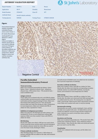
  • 1. Figure: Immunohistochemical analysis of paraffin embedded Human uterus cancer tissue. 1: β I tubulin Monoclonal Antibody(3F7) was diluted at 1:200 (4 degree Celsius,overnight). 2: Sodium citrate pH 6.0 was used for antibody retrieval (>98 degree Celsius,20min). 3: Secondary antibody was diluted at 1:200 (room temperature, 30min). Negative control was used by secondary antibody only. Report Number 96939-a Host Mouse Application IHC-P Clonality Monoclonal Model Number STJ96939 Clone ID 3F7 Antibody Name Anti-β I tubulin antibody Testing Species HUMAN Testing Tissue UTERUS CANCER ANTIBODY VALIDATION REPORT b. (A small amount of distilled water was added into the incubation box to prevent evaporation of antibody). 29. Secondary antibody incubation a. Slides were washed 3 times, with PBS on a shaker for 5min. Shortly after the slides were dried the corresponding secondary antibody solution was added (HRP labelled), covering the tissues, and incubated at room temperature for 30min. b. 30. DAB staining a. Slides were washed 3 times, with PBS on a shaker for 5min. b. Shortly after, the slides were dried and fresh DAB staining buffer was added inside the circles. The staining time was adjusted under a microscope. Yellow-brown colour represented a positive result. Slides were washed with water to stop the staining. c. 31. Haematoxylin staining a. Haematoxylin was used to counter-staining for 1min, and then the slides were washed with water. 1% Hydrochloric acid and alcohol was added for several seconds and then washed with water. Ammonia was used to reveal blue colour, and then flushed with water. b. 32. Desolation and Clearing i. Slides were incubated sequentially into: 75% alcohol 5min, 85% alcohol 5min, Anhydrous ethanol - 5min, Anhydrous ethanol - 5min & Xylene - 5min. Shortly after slides were dried and neutral gum was used to seal the slides. ii. 33. Visualization a. Results were validated with microscope, and the slides were scanned. Paraffin-Embedded Immunohistochemistry Protocol 23. 24. Tissue processing a. Slides were incubated sequentially into Xylene; 15min – Xylene, 15min - Anhydrous ethanol, 5min - Anhydrous ethanol, 5min - 85% alcohol, 5min - 75% alcohol & 5min – wash in distilled water. b. 25. Antigen retrieval a. Tissue slides were incubated with citric acid (PH6.0) antigen retrieval buffer and microwaved for antigen retrieval (heated until boiled and then stopped heating) for 8min. Slides were then heated with medium power for 7min. During this process slides were kept from drying out. After cooling down at room temperature, slides were washed with PBS on shaker for 5min, repeated for 3 times. b. 26. Inhibition of endogenous peroxidase a. Slides were placed in 3% Hydrogen peroxide solution, and incubated for 10 min at room temperature without light exposure. Slides were then washed 3 times with PBS on a shaker for 5mins. b. 27. BSA Blocking a. Shortly after slides were dried, a PAP pen was used to draw circles around the tissue sections (and to prevent draining of the antibody solution). Inside the circles, BSA was used to cover the tissue evenly, blocking for 30min. b. 28. Primary antibody incubation After blocking solution was removed a 1:200 solution of primary antibody/PBS was added on the slide, and incubated overnight at 4°C. St John's Laboratory Ltd. www.stjohnslabs.com
  • 2. Figure: Immunohistochemical analysis of paraffin embedded Human colon tissue. 1: β I tubulin Monoclonal Antibody(3F7) was diluted at 1:200 (4 degree Celsius,overnight). 2: Sodium citrate pH 6.0 was used for antibody retrieval (>98 degree Celsius,20min). 3: Secondary antibody was diluted at 1:200 (room temperature, 30min). Negative control was used by secondary antibody only. Report Number 96939-b Host Mouse Application IHC-P Clonality Monoclonal Model Number STJ96939 Clone ID 3F7 Antibody Name Anti-β I tubulin antibody Testing Species HUMAN Testing Tissue COLON ANTIBODY VALIDATION REPORT b. (A small amount of distilled water was added into the incubation box to prevent evaporation of antibody). 18. Secondary antibody incubation a. Slides were washed 3 times, with PBS on a shaker for 5min. Shortly after the slides were dried the corresponding secondary antibody solution was added (HRP labelled), covering the tissues, and incubated at room temperature for 30min. b. 19. DAB staining a. Slides were washed 3 times, with PBS on a shaker for 5min. b. Shortly after, the slides were dried and fresh DAB staining buffer was added inside the circles. The staining time was adjusted under a microscope. Yellow-brown colour represented a positive result. Slides were washed with water to stop the staining. c. 20. Haematoxylin staining a. Haematoxylin was used to counter-staining for 1min, and then the slides were washed with water. 1% Hydrochloric acid and alcohol was added for several seconds and then washed with water. Ammonia was used to reveal blue colour, and then flushed with water. b. 21. Desolation and Clearing i. Slides were incubated sequentially into: 75% alcohol 5min, 85% alcohol 5min, Anhydrous ethanol - 5min, Anhydrous ethanol - 5min & Xylene - 5min. Shortly after slides were dried and neutral gum was used to seal the slides. ii. 22. Visualization a. Results were validated with microscope, and the slides were scanned. Paraffin-Embedded Immunohistochemistry Protocol 12. 13. Tissue processing a. Slides were incubated sequentially into Xylene; 15min – Xylene, 15min - Anhydrous ethanol, 5min - Anhydrous ethanol, 5min - 85% alcohol, 5min - 75% alcohol & 5min – wash in distilled water. b. 14. Antigen retrieval a. Tissue slides were incubated with citric acid (PH6.0) antigen retrieval buffer and microwaved for antigen retrieval (heated until boiled and then stopped heating) for 8min. Slides were then heated with medium power for 7min. During this process slides were kept from drying out. After cooling down at room temperature, slides were washed with PBS on shaker for 5min, repeated for 3 times. b. 15. Inhibition of endogenous peroxidase a. Slides were placed in 3% Hydrogen peroxide solution, and incubated for 10 min at room temperature without light exposure. Slides were then washed 3 times with PBS on a shaker for 5mins. b. 16. BSA Blocking a. Shortly after slides were dried, a PAP pen was used to draw circles around the tissue sections (and to prevent draining of the antibody solution). Inside the circles, BSA was used to cover the tissue evenly, blocking for 30min. b. 17. Primary antibody incubation After blocking solution was removed a 1:200 solution of primary antibody/PBS was added on the slide, and incubated overnight at 4°C. St John's Laboratory Ltd. www.stjohnslabs.com
  • 3. Figure: Immunohistochemical analysis of paraffin embedded Human appendix tissue. 1: β I tubulin Monoclonal Antibody(3F7) was diluted at 1:200 (4 degree Celsius,overnight). 2: Sodium citrate pH 6.0 was used for antibody retrieval (>98 degree Celsius,20min). 3: Secondary antibody was diluted at 1:200 (room temperature, 30min). Negative control was used by secondary antibody only. Report Number 96939-c Host Mouse Application IHC-P Clonality Monoclonal Model Number STJ96939 Clone ID 3F7 Antibody Name Anti-β I tubulin antibody Testing Species HUMAN Testing Tissue APPENDIX ANTIBODY VALIDATION REPORT b. (A small amount of distilled water was added into the incubation box to prevent evaporation of antibody). 7. Secondary antibody incubation a. Slides were washed 3 times, with PBS on a shaker for 5min. Shortly after the slides were dried the corresponding secondary antibody solution was added (HRP labelled), covering the tissues, and incubated at room temperature for 30min. b. 8. DAB staining a. Slides were washed 3 times, with PBS on a shaker for 5min. b. Shortly after, the slides were dried and fresh DAB staining buffer was added inside the circles. The staining time was adjusted under a microscope. Yellow-brown colour represented a positive result. Slides were washed with water to stop the staining. c. 9. Haematoxylin staining a. Haematoxylin was used to counter-staining for 1min, and then the slides were washed with water. 1% Hydrochloric acid and alcohol was added for several seconds and then washed with water. Ammonia was used to reveal blue colour, and then flushed with water. b. 10. Desolation and Clearing i. Slides were incubated sequentially into: 75% alcohol 5min, 85% alcohol 5min, Anhydrous ethanol - 5min, Anhydrous ethanol - 5min & Xylene - 5min. Shortly after slides were dried and neutral gum was used to seal the slides. ii. 11. Visualization a. Results were validated with microscope, and the slides were scanned. Paraffin-Embedded Immunohistochemistry Protocol 1. 2. Tissue processing a. Slides were incubated sequentially into Xylene; 15min – Xylene, 15min - Anhydrous ethanol, 5min - Anhydrous ethanol, 5min - 85% alcohol, 5min - 75% alcohol & 5min – wash in distilled water. b. 3. Antigen retrieval a. Tissue slides were incubated with citric acid (PH6.0) antigen retrieval buffer and microwaved for antigen retrieval (heated until boiled and then stopped heating) for 8min. Slides were then heated with medium power for 7min. During this process slides were kept from drying out. After cooling down at room temperature, slides were washed with PBS on shaker for 5min, repeated for 3 times. b. 4. Inhibition of endogenous peroxidase a. Slides were placed in 3% Hydrogen peroxide solution, and incubated for 10 min at room temperature without light exposure. Slides were then washed 3 times with PBS on a shaker for 5mins. b. 5. BSA Blocking a. Shortly after slides were dried, a PAP pen was used to draw circles around the tissue sections (and to prevent draining of the antibody solution). Inside the circles, BSA was used to cover the tissue evenly, blocking for 30min. b. 6. Primary antibody incubation After blocking solution was removed a 1:200 solution of primary antibody/PBS was added on the slide, and incubated overnight at 4°C. St John's Laboratory Ltd. www.stjohnslabs.com
  • 4. Figure: Immunohistochemical analysis of paraffin embedded Rat lung tissue. 1: β I tubulin Monoclonal Antibody(3F7) was diluted at 1:200 (4 degree Celsius,overnight). 2: Sodium citrate pH 6.0 was used for antibody retrieval (>98 degree Celsius,20min). 3: Secondary antibody was diluted at 1:200 (room temperature, 30min). Negative control was used by secondary antibody only. Report Number 96939-d Host Mouse Application IHC-P Clonality Monoclonal Model Number STJ96939 Clone ID 3F7 Antibody Name Anti-β I tubulin antibody Testing Species RAT Testing Tissue LUNG ANTIBODY VALIDATION REPORT b. (A small amount of distilled water was added into the incubation box to prevent evaporation of antibody). 34. Secondary antibody incubation a. Slides were washed 3 times, with PBS on a shaker for 5min. Shortly after the slides were dried the corresponding secondary antibody solution was added (HRP labelled), covering the tissues, and incubated at room temperature for 30min. b. 35. DAB staining a. Slides were washed 3 times, with PBS on a shaker for 5min. b. Shortly after, the slides were dried and fresh DAB staining buffer was added inside the circles. The staining time was adjusted under a microscope. Yellow-brown colour represented a positive result. Slides were washed with water to stop the staining. c. 36. Haematoxylin staining a. Haematoxylin was used to counter-staining for 1min, and then the slides were washed with water. 1% Hydrochloric acid and alcohol was added for several seconds and then washed with water. Ammonia was used to reveal blue colour, and then flushed with water. b. 37. Desolation and Clearing i. Slides were incubated sequentially into: 75% alcohol 5min, 85% alcohol 5min, Anhydrous ethanol - 5min, Anhydrous ethanol - 5min & Xylene - 5min. Shortly after slides were dried and neutral gum was used to seal the slides. ii. 38. Visualization a. Results were validated with microscope, and the slides were scanned. Paraffin-Embedded Immunohistochemistry Protocol 39. 40. Tissue processing a. Slides were incubated sequentially into Xylene; 15min – Xylene, 15min - Anhydrous ethanol, 5min - Anhydrous ethanol, 5min - 85% alcohol, 5min - 75% alcohol & 5min – wash in distilled water. b. 41. Antigen retrieval a. Tissue slides were incubated with citric acid (PH6.0) antigen retrieval buffer and microwaved for antigen retrieval (heated until boiled and then stopped heating) for 8min. Slides were then heated with medium power for 7min. During this process slides were kept from drying out. After cooling down at room temperature, slides were washed with PBS on shaker for 5min, repeated for 3 times. b. 42. Inhibition of endogenous peroxidase a. Slides were placed in 3% Hydrogen peroxide solution, and incubated for 10 min at room temperature without light exposure. Slides were then washed 3 times with PBS on a shaker for 5mins. b. 43. BSA Blocking a. Shortly after slides were dried, a PAP pen was used to draw circles around the tissue sections (and to prevent draining of the antibody solution). Inside the circles, BSA was used to cover the tissue evenly, blocking for 30min. b. 44. Primary antibody incubation After blocking solution was removed a 1:200 solution of primary antibody/PBS was added on the slide, and incubated overnight at 4°C. St John's Laboratory Ltd. www.stjohnslabs.com
  • 5. Figure: Immunohistochemical analysis of paraffin embedded Rat kidney tissue. 1: β I tubulin Monoclonal Antibody(3F7) was diluted at 1:200 (4 degree Celsius,overnight). 2: Sodium citrate pH 6.0 was used for antibody retrieval (>98 degree Celsius,20min). 3: Secondary antibody was diluted at 1:200 (room temperature, 30min). Negative control was used by secondary antibody only. Report Number 96939-e Host Mouse Application IHC-P Clonality Monoclonal Model Number STJ96939 Clone ID 3F7 Antibody Name Anti-β I tubulin antibody Testing Species RAT Testing Tissue KIDNEY ANTIBODY VALIDATION REPORT b. (A small amount of distilled water was added into the incubation box to prevent evaporation of antibody). 45. Secondary antibody incubation a. Slides were washed 3 times, with PBS on a shaker for 5min. Shortly after the slides were dried the corresponding secondary antibody solution was added (HRP labelled), covering the tissues, and incubated at room temperature for 30min. b. 46. DAB staining a. Slides were washed 3 times, with PBS on a shaker for 5min. b. Shortly after, the slides were dried and fresh DAB staining buffer was added inside the circles. The staining time was adjusted under a microscope. Yellow-brown colour represented a positive result. Slides were washed with water to stop the staining. c. 47. Haematoxylin staining a. Haematoxylin was used to counter-staining for 1min, and then the slides were washed with water. 1% Hydrochloric acid and alcohol was added for several seconds and then washed with water. Ammonia was used to reveal blue colour, and then flushed with water. b. 48. Desolation and Clearing i. Slides were incubated sequentially into: 75% alcohol 5min, 85% alcohol 5min, Anhydrous ethanol - 5min, Anhydrous ethanol - 5min & Xylene - 5min. Shortly after slides were dried and neutral gum was used to seal the slides. ii. 49. Visualization a. Results were validated with microscope, and the slides were scanned. Paraffin-Embedded Immunohistochemistry Protocol 50. 51. Tissue processing a. Slides were incubated sequentially into Xylene; 15min – Xylene, 15min - Anhydrous ethanol, 5min - Anhydrous ethanol, 5min - 85% alcohol, 5min - 75% alcohol & 5min – wash in distilled water. b. 52. Antigen retrieval a. Tissue slides were incubated with citric acid (PH6.0) antigen retrieval buffer and microwaved for antigen retrieval (heated until boiled and then stopped heating) for 8min. Slides were then heated with medium power for 7min. During this process slides were kept from drying out. After cooling down at room temperature, slides were washed with PBS on shaker for 5min, repeated for 3 times. b. 53. Inhibition of endogenous peroxidase a. Slides were placed in 3% Hydrogen peroxide solution, and incubated for 10 min at room temperature without light exposure. Slides were then washed 3 times with PBS on a shaker for 5mins. b. 54. BSA Blocking a. Shortly after slides were dried, a PAP pen was used to draw circles around the tissue sections (and to prevent draining of the antibody solution). Inside the circles, BSA was used to cover the tissue evenly, blocking for 30min. b. 55. Primary antibody incubation After blocking solution was removed a 1:200 solution of primary antibody/PBS was added on the slide, and incubated overnight at 4°C. St John's Laboratory Ltd. www.stjohnslabs.com
  • 6. Figure: Immunohistochemical analysis of paraffin embedded Mouse testis tissue. 1: β I tubulin Monoclonal Antibody(3F7) was diluted at 1:200 (4 degree Celsius,overnight). 2: Sodium citrate pH 6.0 was used for antibody retrieval (>98 degree Celsius,20min). 3: Secondary antibody was diluted at 1:200 (room temperature, 30min). Negative control was used by secondary antibody only. Report Number 96939-f Host Mouse Application IHC-P Clonality Monoclonal Model Number STJ96939 Clone ID 3F7 Antibody Name Anti-β I tubulin antibody Testing Species MOUSE Testing Tissue TESTIS ANTIBODY VALIDATION REPORT b. (A small amount of distilled water was added into the incubation box to prevent evaporation of antibody). 56. Secondary antibody incubation a. Slides were washed 3 times, with PBS on a shaker for 5min. Shortly after the slides were dried the corresponding secondary antibody solution was added (HRP labelled), covering the tissues, and incubated at room temperature for 30min. b. 57. DAB staining a. Slides were washed 3 times, with PBS on a shaker for 5min. b. Shortly after, the slides were dried and fresh DAB staining buffer was added inside the circles. The staining time was adjusted under a microscope. Yellow-brown colour represented a positive result. Slides were washed with water to stop the staining. c. 58. Haematoxylin staining a. Haematoxylin was used to counter-staining for 1min, and then the slides were washed with water. 1% Hydrochloric acid and alcohol was added for several seconds and then washed with water. Ammonia was used to reveal blue colour, and then flushed with water. b. 59. Desolation and Clearing i. Slides were incubated sequentially into: 75% alcohol 5min, 85% alcohol 5min, Anhydrous ethanol - 5min, Anhydrous ethanol - 5min & Xylene - 5min. Shortly after slides were dried and neutral gum was used to seal the slides. ii. 60. Visualization a. Results were validated with microscope, and the slides were scanned. Paraffin-Embedded Immunohistochemistry Protocol 61. 62. Tissue processing a. Slides were incubated sequentially into Xylene; 15min – Xylene, 15min - Anhydrous ethanol, 5min - Anhydrous ethanol, 5min - 85% alcohol, 5min - 75% alcohol & 5min – wash in distilled water. b. 63. Antigen retrieval a. Tissue slides were incubated with citric acid (PH6.0) antigen retrieval buffer and microwaved for antigen retrieval (heated until boiled and then stopped heating) for 8min. Slides were then heated with medium power for 7min. During this process slides were kept from drying out. After cooling down at room temperature, slides were washed with PBS on shaker for 5min, repeated for 3 times. b. 64. Inhibition of endogenous peroxidase a. Slides were placed in 3% Hydrogen peroxide solution, and incubated for 10 min at room temperature without light exposure. Slides were then washed 3 times with PBS on a shaker for 5mins. b. 65. BSA Blocking a. Shortly after slides were dried, a PAP pen was used to draw circles around the tissue sections (and to prevent draining of the antibody solution). Inside the circles, BSA was used to cover the tissue evenly, blocking for 30min. b. 66. Primary antibody incubation After blocking solution was removed a 1:200 solution of primary antibody/PBS was added on the slide, and incubated overnight at 4°C. St John's Laboratory Ltd. www.stjohnslabs.com
  • 7. Figure: Immunohistochemical analysis of paraffin embedded Mouse lung tissue. 1: β I tubulin Monoclonal Antibody(3F7) was diluted at 1:200 (4 degree Celsius,overnight). 2: Sodium citrate pH 6.0 was used for antibody retrieval (>98 degree Celsius,20min). 3: Secondary antibody was diluted at 1:200 (room temperature, 30min). Negative control was used by secondary antibody only. Report Number 96939-g Host Mouse Application IHC-P Clonality Monoclonal Model Number STJ96939 Clone ID 3F7 Antibody Name Anti-β I tubulin antibody Testing Species MOUSE Testing Tissue LUNG ANTIBODY VALIDATION REPORT b. (A small amount of distilled water was added into the incubation box to prevent evaporation of antibody). 67. Secondary antibody incubation a. Slides were washed 3 times, with PBS on a shaker for 5min. Shortly after the slides were dried the corresponding secondary antibody solution was added (HRP labelled), covering the tissues, and incubated at room temperature for 30min. b. 68. DAB staining a. Slides were washed 3 times, with PBS on a shaker for 5min. b. Shortly after, the slides were dried and fresh DAB staining buffer was added inside the circles. The staining time was adjusted under a microscope. Yellow-brown colour represented a positive result. Slides were washed with water to stop the staining. c. 69. Haematoxylin staining a. Haematoxylin was used to counter-staining for 1min, and then the slides were washed with water. 1% Hydrochloric acid and alcohol was added for several seconds and then washed with water. Ammonia was used to reveal blue colour, and then flushed with water. b. 70. Desolation and Clearing i. Slides were incubated sequentially into: 75% alcohol 5min, 85% alcohol 5min, Anhydrous ethanol - 5min, Anhydrous ethanol - 5min & Xylene - 5min. Shortly after slides were dried and neutral gum was used to seal the slides. ii. 71. Visualization a. Results were validated with microscope, and the slides were scanned. Paraffin-Embedded Immunohistochemistry Protocol 72. 73. Tissue processing a. Slides were incubated sequentially into Xylene; 15min – Xylene, 15min - Anhydrous ethanol, 5min - Anhydrous ethanol, 5min - 85% alcohol, 5min - 75% alcohol & 5min – wash in distilled water. b. 74. Antigen retrieval a. Tissue slides were incubated with citric acid (PH6.0) antigen retrieval buffer and microwaved for antigen retrieval (heated until boiled and then stopped heating) for 8min. Slides were then heated with medium power for 7min. During this process slides were kept from drying out. After cooling down at room temperature, slides were washed with PBS on shaker for 5min, repeated for 3 times. b. 75. Inhibition of endogenous peroxidase a. Slides were placed in 3% Hydrogen peroxide solution, and incubated for 10 min at room temperature without light exposure. Slides were then washed 3 times with PBS on a shaker for 5mins. b. 76. BSA Blocking a. Shortly after slides were dried, a PAP pen was used to draw circles around the tissue sections (and to prevent draining of the antibody solution). Inside the circles, BSA was used to cover the tissue evenly, blocking for 30min. b. 77. Primary antibody incubation After blocking solution was removed a 1:200 solution of primary antibody/PBS was added on the slide, and incubated overnight at 4°C. St John's Laboratory Ltd. www.stjohnslabs.com
  • 8. Figure: Immunohistochemical analysis of paraffin embedded Mouse kidney tissue. 1: β I tubulin Monoclonal Antibody(3F7) was diluted at 1:200 (4 degree Celsius,overnight). 2: Sodium citrate pH 6.0 was used for antibody retrieval (>98 degree Celsius,20min). 3: Secondary antibody was diluted at 1:200 (room temperature, 30min). Negative control was used by secondary antibody only. Report Number 96939-h Host Mouse Application IHC-P Clonality Monoclonal Model Number STJ96939 Clone ID 3F7 Antibody Name Anti-β I tubulin antibody Testing Species MOUSE Testing Tissue KIDNEY ANTIBODY VALIDATION REPORT b. (A small amount of distilled water was added into the incubation box to prevent evaporation of antibody). 78. Secondary antibody incubation a. Slides were washed 3 times, with PBS on a shaker for 5min. Shortly after the slides were dried the corresponding secondary antibody solution was added (HRP labelled), covering the tissues, and incubated at room temperature for 30min. b. 79. DAB staining a. Slides were washed 3 times, with PBS on a shaker for 5min. b. Shortly after, the slides were dried and fresh DAB staining buffer was added inside the circles. The staining time was adjusted under a microscope. Yellow-brown colour represented a positive result. Slides were washed with water to stop the staining. c. 80. Haematoxylin staining a. Haematoxylin was used to counter-staining for 1min, and then the slides were washed with water. 1% Hydrochloric acid and alcohol was added for several seconds and then washed with water. Ammonia was used to reveal blue colour, and then flushed with water. b. 81. Desolation and Clearing i. Slides were incubated sequentially into: 75% alcohol 5min, 85% alcohol 5min, Anhydrous ethanol - 5min, Anhydrous ethanol - 5min & Xylene - 5min. Shortly after slides were dried and neutral gum was used to seal the slides. ii. 82. Visualization a. Results were validated with microscope, and the slides were scanned. Paraffin-Embedded Immunohistochemistry Protocol 83. 84. Tissue processing a. Slides were incubated sequentially into Xylene; 15min – Xylene, 15min - Anhydrous ethanol, 5min - Anhydrous ethanol, 5min - 85% alcohol, 5min - 75% alcohol & 5min – wash in distilled water. b. 85. Antigen retrieval a. Tissue slides were incubated with citric acid (PH6.0) antigen retrieval buffer and microwaved for antigen retrieval (heated until boiled and then stopped heating) for 8min. Slides were then heated with medium power for 7min. During this process slides were kept from drying out. After cooling down at room temperature, slides were washed with PBS on shaker for 5min, repeated for 3 times. b. 86. Inhibition of endogenous peroxidase a. Slides were placed in 3% Hydrogen peroxide solution, and incubated for 10 min at room temperature without light exposure. Slides were then washed 3 times with PBS on a shaker for 5mins. b. 87. BSA Blocking a. Shortly after slides were dried, a PAP pen was used to draw circles around the tissue sections (and to prevent draining of the antibody solution). Inside the circles, BSA was used to cover the tissue evenly, blocking for 30min. b. 88. Primary antibody incubation After blocking solution was removed a 1:200 solution of primary antibody/PBS was added on the slide, and incubated overnight at 4°C. St John's Laboratory Ltd. www.stjohnslabs.com
  • 9. Figure: Immunohistochemical analysis of paraffin embedded Mouse brain tissue. 1: β I tubulin Monoclonal Antibody(3F7) was diluted at 1:200 (4 degree Celsius,overnight). 2: Sodium citrate pH 6.0 was used for antibody retrieval (>98 degree Celsius,20min). 3: Secondary antibody was diluted at 1:200 (room temperature, 30min). Negative control was used by secondary antibody only. Report Number 96939-i Host Mouse Application IHC-P Clonality Monoclonal Model Number STJ96939 Clone ID 3F7 Antibody Name Anti-β I tubulin antibody Testing Species MOUSE Testing Tissue BRAIN ANTIBODY VALIDATION REPORT b. (A small amount of distilled water was added into the incubation box to prevent evaporation of antibody). 89. Secondary antibody incubation a. Slides were washed 3 times, with PBS on a shaker for 5min. Shortly after the slides were dried the corresponding secondary antibody solution was added (HRP labelled), covering the tissues, and incubated at room temperature for 30min. b. 90. DAB staining a. Slides were washed 3 times, with PBS on a shaker for 5min. b. Shortly after, the slides were dried and fresh DAB staining buffer was added inside the circles. The staining time was adjusted under a microscope. Yellow-brown colour represented a positive result. Slides were washed with water to stop the staining. c. 91. Haematoxylin staining a. Haematoxylin was used to counter-staining for 1min, and then the slides were washed with water. 1% Hydrochloric acid and alcohol was added for several seconds and then washed with water. Ammonia was used to reveal blue colour, and then flushed with water. b. 92. Desolation and Clearing i. Slides were incubated sequentially into: 75% alcohol 5min, 85% alcohol 5min, Anhydrous ethanol - 5min, Anhydrous ethanol - 5min & Xylene - 5min. Shortly after slides were dried and neutral gum was used to seal the slides. ii. 93. Visualization a. Results were validated with microscope, and the slides were scanned. Paraffin-Embedded Immunohistochemistry Protocol 94. 95. Tissue processing a. Slides were incubated sequentially into Xylene; 15min – Xylene, 15min - Anhydrous ethanol, 5min - Anhydrous ethanol, 5min - 85% alcohol, 5min - 75% alcohol & 5min – wash in distilled water. b. 96. Antigen retrieval a. Tissue slides were incubated with citric acid (PH6.0) antigen retrieval buffer and microwaved for antigen retrieval (heated until boiled and then stopped heating) for 8min. Slides were then heated with medium power for 7min. During this process slides were kept from drying out. After cooling down at room temperature, slides were washed with PBS on shaker for 5min, repeated for 3 times. b. 97. Inhibition of endogenous peroxidase a. Slides were placed in 3% Hydrogen peroxide solution, and incubated for 10 min at room temperature without light exposure. Slides were then washed 3 times with PBS on a shaker for 5mins. b. 98. BSA Blocking a. Shortly after slides were dried, a PAP pen was used to draw circles around the tissue sections (and to prevent draining of the antibody solution). Inside the circles, BSA was used to cover the tissue evenly, blocking for 30min. b. 99. Primary antibody incubation After blocking solution was removed a 1:200 solution of primary antibody/PBS was added on the slide, and incubated overnight at 4°C. St John's Laboratory Ltd. www.stjohnslabs.com