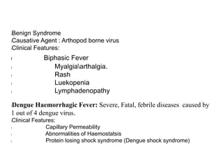
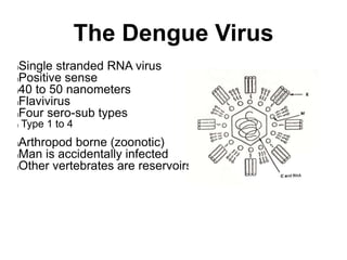
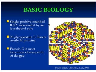
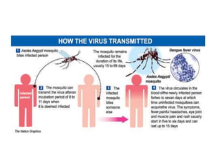
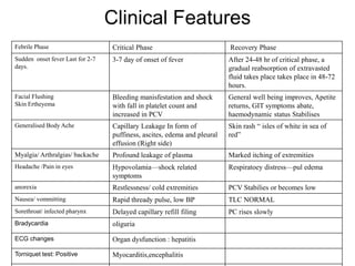
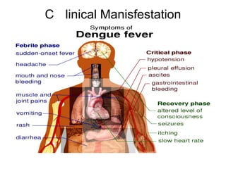
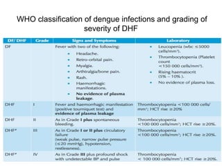
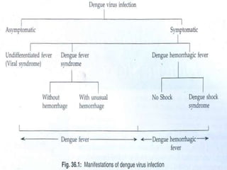
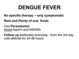
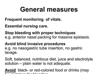
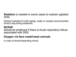
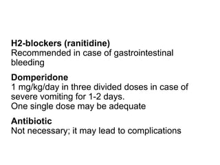
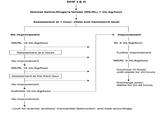
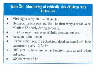
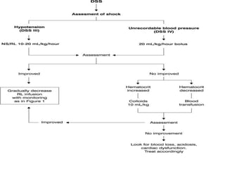
Dengue
By
Dr. Liza Bulsara
lBenign Syndrome
lCausative Agent : Arthopod borne virus
lClinical Features:
l Biphasic Fever
l Myalgiaarthalgia.
l Rash
l Luekopenia
l Lymphadenopathy
lDengue Haemorrhagic Fever: Severe, Fatal, febrile diseases caused by
1 out of 4 dengue virus.
lClinical Features:
l Capillary Permeability
l Abnormalities of Haemostatsis
l Protein losing shock syndrome (Dengue shock syndrome)
The Dengue Virus
lSingle stranded RNA virus
lPositive sense
l40 to 50 nanometers
lFlavivirus
lFour sero-sub types
l Type 1 to 4
lArthropod borne (zoonotic)
lMan is accidentally infected
lOther vertebrates are reservoirs
Etiology
l4 Distinct antigenic types of flavivirus.
lDengue 1
lDengue 2
lDengue 3
lDengue 4
lMembers of Family Flaviviridae.
lDengue like illness are caused by arthropod borne virus
lTogavirus : Chikungunya (Africa, India, Southeast Asia, USA)
lTogavirus : O'nyong-nyong (East Africa)
lFlavivirus : West Nile Fever (Europe, Africa, Middle east, India)
Epidemiology
lDengue virus are transmitted by mosquitoes
“STEGOMYIA” family, AEDES AGEYTI, a day
time biting mosquito, is the principal vector.
lA. Aegyti, is highly urbanised , breeding in water
stored for drinking or bathing and in rainwater
collected in any container .
lMore than 390 million dengue infections occur
annually, 96 million have clincial diseases,
lA. Aegyti has limited flight range, spread of
epidemic occcurs mainly through viremic human
beings and follows the main line of transportation,
nearly simultaneous secondary infection give the
appearance of a contagious disease.
Transmission Cycle
Intrinsic Incubation Period:
3-14 days
Viraemia & Fever: 5-7 days
Vector
Humidity:
Rainfall & Temp.
Susceptible hosts,
(population)
Source patients
Extrinsic
Incubation
Period:
8-10 days
Dengue Haemorrhagic fever
lEndemic in asia and tropical america, wgere
warm temperatures and the practise of water
storage in homes plus outdoor breeding sites
result in large, permanent populations of
A.Aegypti.
lPrimary Infection : First Infection .
lSecondary Infection: It is followed by Primary
infection with different dengue virus.
Pathogenesis
lThe virus is taken up by dendritic cells, and after
antigenic processing, presents it to T- Cell, leading
to immune activation and release cascade of
cytokines that are believed to mdiate systemic
effect of plasma leakeage and circulatory
insufficiency.
lThrombocytopenia develops due to the presence
of cross-reacting antibodies to platelets and is
responsible for bleeding diathesis.
lThe original phenomeon of “Original antigenic
shift” explains the increased severuty of the illness
suring secondary infection, due to presence of
antibodies to the previously infecting serotype.it
Clinical Features
Febrile Phase Critical Phase Recovery Phase
Sudden onset fever Last for 2-7
days.
3-7 day of onset of fever After 24-48 hr of critical phase, a
gradual reabsorption of extravasted
fluid takes place takes place in 48-72
hours.
Facial Flushing
Skin Ertheyema
Bleeding manisfestation and shock
with fall in platelet count and
increased in PCV
General well being improves, Apetite
returns, GIT symptoms abate,
haemodynamic status Stabilises
Generalised Body Ache Capillary Leakage In form of
puffiness, ascites, edema and pleural
effusion (Right side)
Skin rash “ isles of white in sea of
red”
Myalgia/ Arthralgias/ backache Profound leakage of plasma Marked itching of extremities
Headache /Pain in eyes Hypovolamia—shock related
symptoms
Respiratoey distress—pul edema
anorexia Restlessness/ cold extremities PCV Stabilies or becomes low
Nausea/ vommitting Rapid thready pulse, low BP TLC NORMAL
Sorethroat/ infected pharynx Delayed capillary refill filing PC rises slowly
Bradycardia oliguria
ECG changes Organ dysfunction : hepatitis
Torniquet test: Positive Myocarditis,encephalitis
C linical Manisfestation
lIncubation period: 4-10 days
lClinical criteria
lhigh grade fever
lhemorrhgic manifestation (a positive torniquet
test)
lTender hepatomegaly
lEffusion in body cavities and shock.
lLaboratory criteria
lThrombocytopenia
lRising hametocrit
Tourniquet Test
Inflate blood pressure
cuff to a point midway
between systolic and
diastolic pressure for 5
min
Positive test: 20 or more
petechiae / inch2 (6.25
cm2)
In an epidemic situation,
the test is positive in
50% on the 1st day, and in
80% by the end of the febrile
WHO classification of dengue infections and grading of
severity of DHF
Differential Diagnosis
lInfluenza
lMalaria
lEnteric fever
lLeptospirosis
lFlu
lRicketessial fever
lChikungunya fever
Laboratory investigation
lHametological studies:
lGradual increase in PCV
lDecrease in Platelet, leukocyte count.
lIncrease in lymphocyte
lRising hematocrit or single hamatocrit more than
40% S/o capillary hemoconc. Due to capillary
leakage
llow WBC with febrile illness in endemic season is
a pointer towards possible dengue infection
lperipheral smear shows transformed lymphocytes
lshock reveals DIC
lBlood collection in tubes or vials
lThe following are the steps for blood collection in tubes or vials:
lCollect 2 ml–10 ml of venous blood with aseptic precautions.
lUse adhesive tape marked with pencil, indelible ink, or a typewritten/printed self-adhesive label
to identify the container. The name of the patient, identification number and date of collection
must be indicated on the label.
lUse vacuum tubes or sterile vials with screw caps and gasket, if possible, for collection.
lSecure the cap with adhesive tape, wax or other sealing material to prevent leakage during
ltransport.
lIn case of an anticipated delay of more than 24 hours before specimens can be submitted to the
laboratory, separate the serum from the red blood cells and store frozen. Do not freeze whole
blood as haemolysis may interfere with serology test results.
lShip specimens to the laboratory on wet ice (blood) or dry ice (serum) as soon as possible.
lThe shipment should adhere to national/international guidelines on shipment of infectious
material
Biochemistry
lDecrese in total protein & albumin
lTransaminases raised
lIncrease in SGOT than SGPT s/o dengue
infection
lSevere Cases: Hyponateremia & Acidosis.
Increase in BUN and creatinine.
lSkiagram & Ultrasonography: Pleural effusion
(Right side)
lUltrasonography: Ascites & enlarged gall bladder
due to wall edema
lAbnormal ECG & mycordial dysfunctionon ECG
lDecrease in cardiac output
lDecrease preload
lDecrease in left ventricular
lActivation of complement system leads to
profound depression of C3 AND C5 level.
Confirmatory test
lAntibody detection
l Hemagglutination Inhibition (HAI)
l ELISA (IgG/IgM)
l Rapid test (IgG/IgM)
lAntigen detection
l NS1 & E/M antigen
l RNA detection
lPCR
l Viral isolation
lPrimary Infection
l NS1 antigen : Day 1 after onset of fever and up
to day 9
l IgM antibody : Day 5 of infection, sometimes as
early as Day 3
lIgM levels : peak in 2 weeks, followed by a 2
week rapid decay
lUndetectable 2 to 3 months after infection
lLow levels of IgG are detected in the early
convalescent phase, not during the acute phase
lSecondary Infection
lNS1 antigen : day 1 after onset of fever and up to
day 9IgM response is more varied
l Usually preceded by IgG and appears quite late
during the febrile phase
l Minority of patients will show no detectable levels
of IgM
lMay not be produced until 20 days after onset of
infection●
lHigh levels of IgG are detectable during the acute
phase Persist for 30-40 days then decline to levels
found in primary or past infection.
Approximate timeline of primary and secondary dengue virus infections and the
diagnostic methods that can be used to detect infection
Treatment
DENGUE FEVER
No specific therapy – only symptomatic
Rest and Plenty of oral fluids
Use Paracetamol
Avoid Aspirin and NSAIDs
Follow up preferably everyday - from the 3rd day
until afebrile for 24-48 hours
General measures
Frequent monitoring of vitals.
Essential nursing care.
Stop bleeding with proper techniques
e.g. anterior nasal packing for massive epistaxis.
Avoid blind invasive procedures
e.g. no nasogastric tube insertion, no gastric
lavage.
Soft, balanced, nutritious diet, juice and electrolyte
solution – plain water is not adequate.
Avoid black- or red-colored food or drinks (may
Sedation is needed in some cases to restrain agitated
child.
Chloral hydrate(12.5-50 mg/kg), orally or rectally recommended.
Avoid Long-acting sedatives.
NCPAP
should be preferred if there is Acute respiratory failure
associated with DSS.
Oxygen via face mask/nasal cannula
in case of shock/impending shock.
H2-blockers (ranitidine)
Recommended in case of gastrointestinal
bleeding
Domperidone
1 mg/kg/day in three divided doses in case of
severe vomiting for 1-2 days.
One single dose may be adequate
Antibiotic
Not necessary; it may lead to complications
FLUID MANAGEMENT
lIn young infants without shock-
lN/2 saline in 5% dextrose
lIn patients who already have volume overload, i.e., massive
pleural effusion
lcolloid solutions
lHydroxyethyl starch at 6% may be preferred in children
with severe shock; the use of dextran is associated with
various adverse reactions
In case of no response to IV fluids:
May have myocardial dysfunction and decreased LV
performance, which may be easily detected by
echocardiography.
Consider and correct
Massive plasma leakage
Concealed internal bleeding – decrease in Hct
Hypoglycemia – Blood sugar < 60 mg%
Hyponatremia, hypocalcemia – electrolytes
Acidosis – indicates metabolic acidosis in blood gas analysis
Blood and platelet transfusion
Platelet Transfusion: Thrombocytopenia with
significant bleeding.
Platelet count < 10,000/mm3
DOSE: 10-20 mL/kg
Platelets or blood should NOT be transfused based upon
platelet count alone.
Low platelet count may not be predictive of bleeding.
Only 0.4% of DHF pts need platelet
transfusion.
Mild reductions in platelet counts are usually not
associated with significant bleeding.
In children with severe thrombocytopenia in absence of
significant bleeding, platelet infusion does not alter the
outcome.
Platelets return to normal within 7-9 days.
Fresh Whole blood / Packed red cell transfusion
Significant blood loss > 10% (6-8 mL/kg)
Concealed internal bleeding
Hemolysis
DOSE
Fresh whole blood 10 mL/kg/dose
Packed red cells 5 mL/kg/dose
Complications of DF/DSS
DIC
Myocardial dysfunction incl. Cardiomyopathy
Hepatitis
Reye-like syndrome
Encephalitis
ARDS
Glomerulonephritis
Treatment of complications
Fluid overload
AVOID
Early IV fluid therapy- in the febrile phase
Excessive use of hypotonic solutions
Non-reduction in the rate of IV fluid after initial resuscitation
Blood loss replaced with fluids in cases with occult bleeding
Treatment
Judicious fluid removal
colloids with controlled diuresis (furosemide 1 mg/kg infusion over 4 hours) or
dialysis
Electrolyte imbalance
Hyponatremia
Hypocalcemia – 10% Ca gluconate 1 mL/kg/dose, slow IV
push every 6 hour.
Large pleural effusions, ascites
Careful titration of intravenous fluids.
Avoid insertion of intercostal drains and tracheal intubation.
Large pleural effusions during recovery phase after 48 hours
furosemide (0.25-0.5 mg/kg at 6 hours’ interval for 1 to 2
doses).
Disseminated intravascular
coagulation
Frequent Clinical assessment
Regular Coagulation profile
PT, aPTT, fibrinogen, platelet and FDP mandatory, as indicated.
Seriously sick patients with bleeding & DIC have benefited from :
Platelets (4 units/m2 or 10-20 mL/kg) within 1 hr and
Fresh frozen plasma (FFP 10-20 mL/kg).
Prognosis
DF is a very incapacitating disease; however, its prognosis is favorable.
Significant morbidity and mortality can result if early recognition and
monitoring of severe forms are not done.
If left untreated, the mortality of DHF or DSS patients may be as high as
40-50%.
Early recognition of illness, careful monitoring and adequate and
appropriate fluid therapy have decreased mortality to 1%.
If shock is identified when pulse pressure starts to drop and intravenous
fluids are administered, the outcome will be excellent.
Recovery is fast and most patients recover in 24-48 hours without any
sequelae.
The outcome may not be so good if the patient develops cold
extremities.
Most deaths from DHF/DSS are caused by
prolonged shock
massive bleeding
fluid overload and
acute liver failure with encephalopathy.
Severe refractory shock, DIC, ARDS, liver failure and neurological
manifestations singly or in combination were the commonest causes of
death in a recent series.
The case fatality rate is high with shortage of experienced medical
teams..
Criteria for Discharge of patients
lVisible clinical improvement with return of appetite
lStable pulse, blood pressure and respiratory rate
lAfebrile for 24 hours (without anti-fever therapy)
lMinimum of 3 days after recovery from shock
lGood urinary output and stable haematocrit levels
lPlatelet count > 100,000/mm3
lNo respiratory distress /pleural effusion /ascites
lNo evidence of external or internal bleeding
lConvalescent confluent petechial rash
VECTOR CONTROL
The control is primarily dependent on eradicating mosquito.
Public spraying for mosquitoes is the most important aspect of this
approach.
PERSONAL PROTECTION
Avoiding endemic areas
Cover exposed skin
Mosquito nets, repellents
Use of DEET-impregnated bed nets
Immunopathogenic
lCascade of DHF/DSS
lMacrophage – monocyte infection
lPrevious infection with heterologous
Dengue serotype results in
production
of non protective antiviral antibodies
lThese Ab bind to the virion’s surface
Fc receptor and focus the Dengue
virus
on to the target cells –
macro/monocytes
lT cell - cytokines, interferon, TNF alpha
Dengue liza
Dengue liza

Dengue liza

  • 1.
  • 2.
    lBenign Syndrome lCausative Agent: Arthopod borne virus lClinical Features: l Biphasic Fever l Myalgiaarthalgia. l Rash l Luekopenia l Lymphadenopathy lDengue Haemorrhagic Fever: Severe, Fatal, febrile diseases caused by 1 out of 4 dengue virus. lClinical Features: l Capillary Permeability l Abnormalities of Haemostatsis l Protein losing shock syndrome (Dengue shock syndrome)
  • 3.
    The Dengue Virus lSinglestranded RNA virus lPositive sense l40 to 50 nanometers lFlavivirus lFour sero-sub types l Type 1 to 4 lArthropod borne (zoonotic) lMan is accidentally infected lOther vertebrates are reservoirs
  • 5.
    Etiology l4 Distinct antigenictypes of flavivirus. lDengue 1 lDengue 2 lDengue 3 lDengue 4 lMembers of Family Flaviviridae. lDengue like illness are caused by arthropod borne virus lTogavirus : Chikungunya (Africa, India, Southeast Asia, USA) lTogavirus : O'nyong-nyong (East Africa) lFlavivirus : West Nile Fever (Europe, Africa, Middle east, India)
  • 6.
    Epidemiology lDengue virus aretransmitted by mosquitoes “STEGOMYIA” family, AEDES AGEYTI, a day time biting mosquito, is the principal vector. lA. Aegyti, is highly urbanised , breeding in water stored for drinking or bathing and in rainwater collected in any container . lMore than 390 million dengue infections occur annually, 96 million have clincial diseases, lA. Aegyti has limited flight range, spread of epidemic occcurs mainly through viremic human beings and follows the main line of transportation, nearly simultaneous secondary infection give the appearance of a contagious disease.
  • 8.
    Transmission Cycle Intrinsic IncubationPeriod: 3-14 days Viraemia & Fever: 5-7 days Vector Humidity: Rainfall & Temp. Susceptible hosts, (population) Source patients Extrinsic Incubation Period: 8-10 days
  • 10.
    Dengue Haemorrhagic fever lEndemicin asia and tropical america, wgere warm temperatures and the practise of water storage in homes plus outdoor breeding sites result in large, permanent populations of A.Aegypti. lPrimary Infection : First Infection . lSecondary Infection: It is followed by Primary infection with different dengue virus.
  • 11.
    Pathogenesis lThe virus istaken up by dendritic cells, and after antigenic processing, presents it to T- Cell, leading to immune activation and release cascade of cytokines that are believed to mdiate systemic effect of plasma leakeage and circulatory insufficiency. lThrombocytopenia develops due to the presence of cross-reacting antibodies to platelets and is responsible for bleeding diathesis. lThe original phenomeon of “Original antigenic shift” explains the increased severuty of the illness suring secondary infection, due to presence of antibodies to the previously infecting serotype.it
  • 13.
    Clinical Features Febrile PhaseCritical Phase Recovery Phase Sudden onset fever Last for 2-7 days. 3-7 day of onset of fever After 24-48 hr of critical phase, a gradual reabsorption of extravasted fluid takes place takes place in 48-72 hours. Facial Flushing Skin Ertheyema Bleeding manisfestation and shock with fall in platelet count and increased in PCV General well being improves, Apetite returns, GIT symptoms abate, haemodynamic status Stabilises Generalised Body Ache Capillary Leakage In form of puffiness, ascites, edema and pleural effusion (Right side) Skin rash “ isles of white in sea of red” Myalgia/ Arthralgias/ backache Profound leakage of plasma Marked itching of extremities Headache /Pain in eyes Hypovolamia—shock related symptoms Respiratoey distress—pul edema anorexia Restlessness/ cold extremities PCV Stabilies or becomes low Nausea/ vommitting Rapid thready pulse, low BP TLC NORMAL Sorethroat/ infected pharynx Delayed capillary refill filing PC rises slowly Bradycardia oliguria ECG changes Organ dysfunction : hepatitis Torniquet test: Positive Myocarditis,encephalitis
  • 14.
  • 15.
    lIncubation period: 4-10days lClinical criteria lhigh grade fever lhemorrhgic manifestation (a positive torniquet test) lTender hepatomegaly lEffusion in body cavities and shock. lLaboratory criteria lThrombocytopenia lRising hametocrit
  • 16.
    Tourniquet Test Inflate bloodpressure cuff to a point midway between systolic and diastolic pressure for 5 min Positive test: 20 or more petechiae / inch2 (6.25 cm2) In an epidemic situation, the test is positive in 50% on the 1st day, and in 80% by the end of the febrile
  • 17.
    WHO classification ofdengue infections and grading of severity of DHF
  • 19.
  • 20.
    Laboratory investigation lHametological studies: lGradualincrease in PCV lDecrease in Platelet, leukocyte count. lIncrease in lymphocyte lRising hematocrit or single hamatocrit more than 40% S/o capillary hemoconc. Due to capillary leakage llow WBC with febrile illness in endemic season is a pointer towards possible dengue infection lperipheral smear shows transformed lymphocytes lshock reveals DIC
  • 21.
    lBlood collection intubes or vials lThe following are the steps for blood collection in tubes or vials: lCollect 2 ml–10 ml of venous blood with aseptic precautions. lUse adhesive tape marked with pencil, indelible ink, or a typewritten/printed self-adhesive label to identify the container. The name of the patient, identification number and date of collection must be indicated on the label. lUse vacuum tubes or sterile vials with screw caps and gasket, if possible, for collection. lSecure the cap with adhesive tape, wax or other sealing material to prevent leakage during ltransport. lIn case of an anticipated delay of more than 24 hours before specimens can be submitted to the laboratory, separate the serum from the red blood cells and store frozen. Do not freeze whole blood as haemolysis may interfere with serology test results. lShip specimens to the laboratory on wet ice (blood) or dry ice (serum) as soon as possible. lThe shipment should adhere to national/international guidelines on shipment of infectious material
  • 22.
    Biochemistry lDecrese in totalprotein & albumin lTransaminases raised lIncrease in SGOT than SGPT s/o dengue infection lSevere Cases: Hyponateremia & Acidosis. Increase in BUN and creatinine.
  • 23.
    lSkiagram & Ultrasonography:Pleural effusion (Right side) lUltrasonography: Ascites & enlarged gall bladder due to wall edema lAbnormal ECG & mycordial dysfunctionon ECG lDecrease in cardiac output lDecrease preload lDecrease in left ventricular lActivation of complement system leads to profound depression of C3 AND C5 level.
  • 24.
    Confirmatory test lAntibody detection lHemagglutination Inhibition (HAI) l ELISA (IgG/IgM) l Rapid test (IgG/IgM) lAntigen detection l NS1 & E/M antigen l RNA detection lPCR l Viral isolation
  • 25.
    lPrimary Infection l NS1antigen : Day 1 after onset of fever and up to day 9 l IgM antibody : Day 5 of infection, sometimes as early as Day 3 lIgM levels : peak in 2 weeks, followed by a 2 week rapid decay lUndetectable 2 to 3 months after infection lLow levels of IgG are detected in the early convalescent phase, not during the acute phase
  • 26.
    lSecondary Infection lNS1 antigen: day 1 after onset of fever and up to day 9IgM response is more varied l Usually preceded by IgG and appears quite late during the febrile phase l Minority of patients will show no detectable levels of IgM lMay not be produced until 20 days after onset of infection● lHigh levels of IgG are detectable during the acute phase Persist for 30-40 days then decline to levels found in primary or past infection.
  • 28.
    Approximate timeline ofprimary and secondary dengue virus infections and the diagnostic methods that can be used to detect infection
  • 29.
  • 30.
    DENGUE FEVER No specifictherapy – only symptomatic Rest and Plenty of oral fluids Use Paracetamol Avoid Aspirin and NSAIDs Follow up preferably everyday - from the 3rd day until afebrile for 24-48 hours
  • 31.
    General measures Frequent monitoringof vitals. Essential nursing care. Stop bleeding with proper techniques e.g. anterior nasal packing for massive epistaxis. Avoid blind invasive procedures e.g. no nasogastric tube insertion, no gastric lavage. Soft, balanced, nutritious diet, juice and electrolyte solution – plain water is not adequate. Avoid black- or red-colored food or drinks (may
  • 32.
    Sedation is neededin some cases to restrain agitated child. Chloral hydrate(12.5-50 mg/kg), orally or rectally recommended. Avoid Long-acting sedatives. NCPAP should be preferred if there is Acute respiratory failure associated with DSS. Oxygen via face mask/nasal cannula in case of shock/impending shock.
  • 33.
    H2-blockers (ranitidine) Recommended incase of gastrointestinal bleeding Domperidone 1 mg/kg/day in three divided doses in case of severe vomiting for 1-2 days. One single dose may be adequate Antibiotic Not necessary; it may lead to complications
  • 37.
    FLUID MANAGEMENT lIn younginfants without shock- lN/2 saline in 5% dextrose lIn patients who already have volume overload, i.e., massive pleural effusion lcolloid solutions lHydroxyethyl starch at 6% may be preferred in children with severe shock; the use of dextran is associated with various adverse reactions
  • 38.
    In case ofno response to IV fluids: May have myocardial dysfunction and decreased LV performance, which may be easily detected by echocardiography. Consider and correct Massive plasma leakage Concealed internal bleeding – decrease in Hct Hypoglycemia – Blood sugar < 60 mg% Hyponatremia, hypocalcemia – electrolytes Acidosis – indicates metabolic acidosis in blood gas analysis
  • 39.
    Blood and platelettransfusion Platelet Transfusion: Thrombocytopenia with significant bleeding. Platelet count < 10,000/mm3 DOSE: 10-20 mL/kg
  • 40.
    Platelets or bloodshould NOT be transfused based upon platelet count alone. Low platelet count may not be predictive of bleeding. Only 0.4% of DHF pts need platelet transfusion. Mild reductions in platelet counts are usually not associated with significant bleeding. In children with severe thrombocytopenia in absence of significant bleeding, platelet infusion does not alter the outcome. Platelets return to normal within 7-9 days.
  • 41.
    Fresh Whole blood/ Packed red cell transfusion Significant blood loss > 10% (6-8 mL/kg) Concealed internal bleeding Hemolysis DOSE Fresh whole blood 10 mL/kg/dose Packed red cells 5 mL/kg/dose
  • 42.
    Complications of DF/DSS DIC Myocardialdysfunction incl. Cardiomyopathy Hepatitis Reye-like syndrome Encephalitis ARDS Glomerulonephritis
  • 43.
    Treatment of complications Fluidoverload AVOID Early IV fluid therapy- in the febrile phase Excessive use of hypotonic solutions Non-reduction in the rate of IV fluid after initial resuscitation Blood loss replaced with fluids in cases with occult bleeding Treatment Judicious fluid removal colloids with controlled diuresis (furosemide 1 mg/kg infusion over 4 hours) or dialysis
  • 44.
    Electrolyte imbalance Hyponatremia Hypocalcemia –10% Ca gluconate 1 mL/kg/dose, slow IV push every 6 hour. Large pleural effusions, ascites Careful titration of intravenous fluids. Avoid insertion of intercostal drains and tracheal intubation. Large pleural effusions during recovery phase after 48 hours furosemide (0.25-0.5 mg/kg at 6 hours’ interval for 1 to 2 doses).
  • 45.
    Disseminated intravascular coagulation Frequent Clinicalassessment Regular Coagulation profile PT, aPTT, fibrinogen, platelet and FDP mandatory, as indicated. Seriously sick patients with bleeding & DIC have benefited from : Platelets (4 units/m2 or 10-20 mL/kg) within 1 hr and Fresh frozen plasma (FFP 10-20 mL/kg).
  • 46.
    Prognosis DF is avery incapacitating disease; however, its prognosis is favorable. Significant morbidity and mortality can result if early recognition and monitoring of severe forms are not done. If left untreated, the mortality of DHF or DSS patients may be as high as 40-50%. Early recognition of illness, careful monitoring and adequate and appropriate fluid therapy have decreased mortality to 1%. If shock is identified when pulse pressure starts to drop and intravenous fluids are administered, the outcome will be excellent.
  • 47.
    Recovery is fastand most patients recover in 24-48 hours without any sequelae. The outcome may not be so good if the patient develops cold extremities. Most deaths from DHF/DSS are caused by prolonged shock massive bleeding fluid overload and acute liver failure with encephalopathy. Severe refractory shock, DIC, ARDS, liver failure and neurological manifestations singly or in combination were the commonest causes of death in a recent series. The case fatality rate is high with shortage of experienced medical teams..
  • 48.
    Criteria for Dischargeof patients lVisible clinical improvement with return of appetite lStable pulse, blood pressure and respiratory rate lAfebrile for 24 hours (without anti-fever therapy) lMinimum of 3 days after recovery from shock lGood urinary output and stable haematocrit levels lPlatelet count > 100,000/mm3 lNo respiratory distress /pleural effusion /ascites lNo evidence of external or internal bleeding lConvalescent confluent petechial rash
  • 50.
    VECTOR CONTROL The controlis primarily dependent on eradicating mosquito. Public spraying for mosquitoes is the most important aspect of this approach. PERSONAL PROTECTION Avoiding endemic areas Cover exposed skin Mosquito nets, repellents Use of DEET-impregnated bed nets
  • 51.
    Immunopathogenic lCascade of DHF/DSS lMacrophage– monocyte infection lPrevious infection with heterologous Dengue serotype results in production of non protective antiviral antibodies lThese Ab bind to the virion’s surface Fc receptor and focus the Dengue virus on to the target cells – macro/monocytes lT cell - cytokines, interferon, TNF alpha