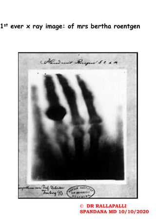
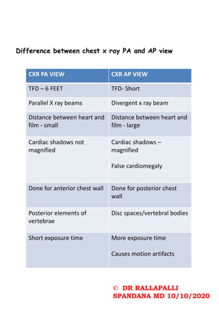
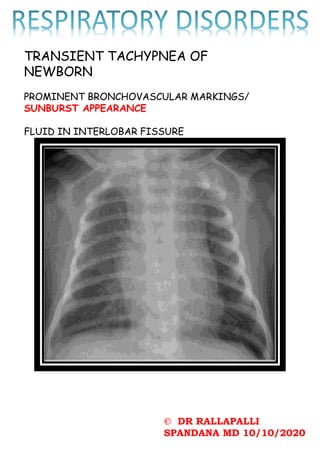
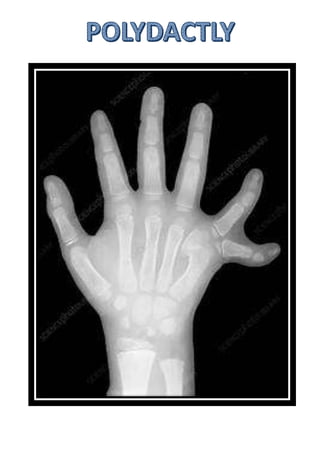
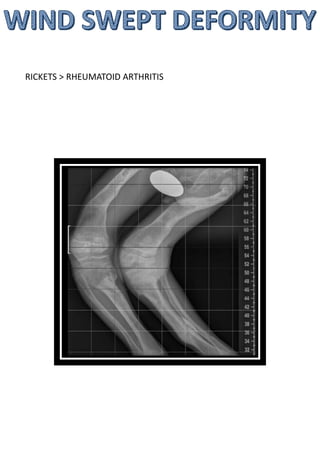
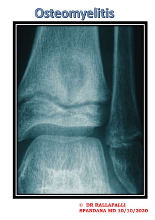
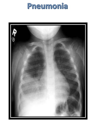
4. •Pediatric patients are more
radiosensitive than adults (i.e., the
cancer risk per unit dose of ionizing
radiation is higher);
•Use of equipment and exposure settings
designed for adults may result in
excessive radiation exposure if used on
smaller patients;
•Pediatric patients have a longer
expected lifetime, putting them at higher
risk of cancer from the effects of
radiation exposure.
5. For a child we don’t talk about chest alone ,
we talk about chest and abdomen.
Baby is small - chest and abdomen fit in one
single film.
Examine from trachea to periphery .
Some prefer to read from periphery to the
center.
Look at heart and lungs last.
Look both domes of diaphragm.
Look at costophrenic and cardiophrenic
angles.
6. In abdomen look for –
stomach shadow
Transverse colon
Rectal gas shadow
Near lung fields:
All ribs
Vertebrae
Soft tissue shadows
Upper part of femur and humerus also
helps in diagnosis.
11. Posterior costophrenic recess.
Fluid collection (pleural effusion)
Normal finding in a lateral chest x
ray
1) Retrosternal lucency
2) Retrocardiac lucency
3) Lucency along vertebral
column must increase from top
to bottom
20. The causes of neonatal
pneumoperitoneum are different from
adult pneumoperitoneum and include:
•perforated hollow viscus
• necrotising enterocolitis (NEC):
most common
• meconium ileus in cystic fibrosis
• Hirschsprung disease
• intestinal atresia or web
• peptic ulcer disease
•iatrogenic
• intubation/mechanical ventilation
• rectal thermometer
• enema
21. Radiographic features
Although these are essentially the
same as in adults, erect chest X-rays
are not obtained, and thus the
diagnosis should be made with supine
films. Additional horizontal cross
table shoot through films or left
decubitus shoot though films may be
performed.
Signs
•football sign
•Rigler's sign
23. RESPIRATORY DISTRESS SYNDROME
WHITE OUT LUNG/GROUND GLASS APPEARANCE OF
LUNG/ reticulonodular appearance
(due to absence of air entry into the lung)
Air bronchogram
(due to air in the bronchus and bronchioles)
27. •indistinct diaphragm with
opacification of part of or all
the hemithorax (typically left
sided)
•scaphoid abdomen
•deviation of lines 3
• endotracheal tube
• nasogastric tube
• umbilical arterial and
venous catheters
28. Congenital diaphragmatic herniation
can be classified into two basic types
on location:
1.Bochdalek hernia
1. most common fetal congenital
diaphragmatic hernia
2.commoner on the left: 75-90%
3.posterolateral
4.large and associated with poorer
outcome
5.presents earlier
6.mnemonic: BBBBB
2.Morgagni hernia
1. less common
2.anterior
3.presents later
32. The thumb sign in epiglottitis is a
manifestation of an oedematous and
enlarged epiglottis which is seen on lateral
soft-tissue radiograph of the neck, and it
suggests a diagnosis of acute
infectious epiglottitis. This is the
radiographic corollary of the omega sign.
Thumb sign is a term also used in other
conditions:
•thumb sign (Marfan disease) (also known
as Steinberg sign) : a clinical test in which
the tip of the thumb is visible medial to
the little finger when it is clasped in the
clenched hand
•thumb sign (chordoma): a radiological sign
showing a clival tumour projection
indenting the pons
35. Frankel line: dense zone of
provisional calcification
Trummerfeld zone: lucent
metaphyseal band underlying
frankel line.
Pelken spur: metaphyseal spurs
which result in cupping of the
metaphysis.
40. It is not surprising that these
features are most prominent at the
bones where growth is greatest:
•knee: distal femur, proximal tibia
•wrist: especially the ulna 1
•anterior rib ends: rachitic rosary
As osteomalacia co-occurs with
rickets, it is important to remember
that even bones that appear
mineralised are weak and result in
bowing, most commonly seen in the
lower limbs once the child is walking.
The legs bow outwards with variable
deformity of the hips (both coxa
vara and coxa valga are seen 1).
41. Other bone deformities are
also noted such as genu valga
and vara as well as protrusio
acetabuli .
The lower ribs may also be
drawn inwards inferiorly by
the attachment of the
diaphragm (Harrison's sulcus).
42. The differential for leg bowing
in children includes :
•developmental or congenital
bowing
•Blount disease
•osteogenesis imperfecta
•many others that are not
usually a consideration (see leg
bowing in children)
43. The differential for flaring of the
metaphysis includes:
•Anaemias
•fibrous dysplasia
•storage diseases
•chronic lead poisoning
•bone dysplasias
44. The differential for widening of
the growth plate includes:
•Schmid-type metaphyseal
chondrodysplasia
•hypovitaminosis C (scurvy)
•delayed maturation due to illness
•endocrine disturbances
• growth hormone excess
• hyperparathyroidism
• hypothyroidism
98. Septic arthritis of hip in infancy.
Results in complete destruction
of cartilaginous femoral head.
Presentation is a child in his
preschool age with painless limp.
Affected limb is shorter.
X ray shows complete absence of
head and neck of femur.
102. While plain radiographs may show joint
effusions and a loss of soft tissue
planes, there may not be an immediate
indication of bone infection.7 It may
take 10 to 14 days to show 30 to 50
percent of bone mineral loss before
osteomyelitic changes are evident.
A differential diagnosis for
radiographic bony lesions should include
osteomyelitis, leukemia, round cell
carcinomas, Ewing’s sarcoma, metastatic
neuroblastomas, eosinophilic granulomas,
histiocytosis X and tuberculosis.
If radiographs are normal and one
suspects osteomyelitis, proceed to
obtain technetium-99m bone scans.
However, keep in mind that while this
imaging is sensitive for certain bony
conditions, it is not specific.
116. Female>male
50% cases bilateral
Defect- distal end of the radius growth
plate fuses early
Defect in growth (but ulna continues to
grow normally)
Deformity:
Ulna is more prominent than the radius.
Volar subluxation of hand – dinner fork
deformity
123. Hyperinflated lungs: increased radiolucency of
lungs
Flattening of domes of diaphragm
Pulmonary infiltrates
Segmental collapse
124.
125. Head , neck and spine
Basilar invagination
Wormian bones
Kyphoscoliosis
Verebral compression fractures
Codfish vertebrae
Platyspondyly
Chest
Pectus excavatumor carinatum
Pelvis
Protrusio acetabuli
Coxa vara
126. General
Severe osteoporosis
Deformed bones
Cortical thining
Popcorn calcification: the
metaphysis and epiphysis exhibit
numerous scalloped radiolucent
areas with sclerotic margins
Zebra stripe sign : cyclic
bisphosphonate treatment produces
sclerotic growth recovery lines in
the long bones.
Formation of pseudoarthrosis at
sites of healing fractures.