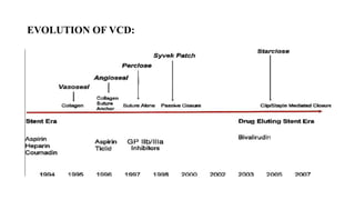
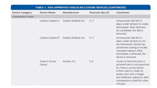
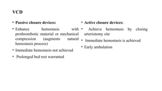
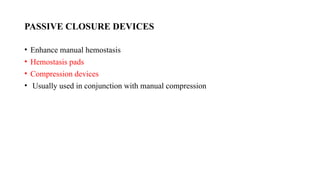
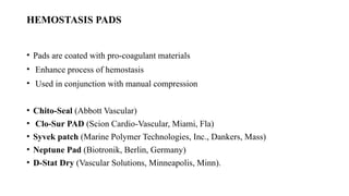
VASCULAR DEVICE CLOSURE
MODERATOR : DR.SENGOTTUVELU/ DR SENTHIL
PRESENTOR: DR PRATHABAN
INTRODUCTION:
• Angiography is currently one of the most common procedures in diagnostic and
interventional cardiology, vascular surgery and interventional radiology.
• Traditionally, hemostasis at the access site has been achieved via manual
compression, the gold standard.
• However, a wide array of vascular access closure devices have been created to
assist the proceduralist in achieving hemostasis.
• These devices can be useful for patients with large body habitus, patients on
anticoagulation and antiplatelet therapy, or when extended bed rest is undesirable
such as in the case of a patient with extensive pressure ulcers.
INTRODUCTION:
• Manual compression needs to be applied for an extended period or with
considerable force.
• Complications that can result from inadequate manual compression include
hematoma and pseudoaneurysm formation.
• Studies have shown that use of these devices is safe even when patients have
received thrombolytic therapy.
• Vascular access closure devices are usually employed whenever a sheath that is a
6F catheter or larger is used for an arterial procedure.
INTRODUCTION:
• Incidence of major complications – 1-17%
• Femoral site complications :- Diagnostic – 1.8%. Intervention – 4%
• Bleeding rates = 2-14%
MANUAL COMPRESSION
Easy, “ golden standard”.
• Have to wait for the ACT to decrease.
• Patient and doctor discomfort
• 6 hours bedrest : back pain and urinary retention
• 15–30 min for a 6 Fr sheath
• 3 min per F for arterial,
• 2 min per F for venous.
Full pressure for 5 min, then
• 75% for 5 min,
• 50% for 5 min,
• 25% for 3-5 min.
WHY DEVICE CLOSURE ?
• Manual compression is dependent on operator technique and expertise.
• Cumbersome
• Requires prolonged bed rest, patient discomfort
• Increase in structural interventions, use of MCS devices – need of large bore
access (>10F)
ADVANTAGE OF VCD:
• Shorten time to hemostasis
• Reduced bleeding and access site complications
• Early patient ambulation and reduced discomfort
• Does not depend on anticoagulation
EVOLUTION OF VCD:
VCD –TYPES
• ACTIVE: physically close the arteriotomy with the use of a suture or a nitinol
clip.
• PASSIVE: deploy a plug, sealant, or gel at the arteriotomy site without actively
closing the arteriotomy.
• EXTERNAL HEMOSTATIC DEVICES :are placed on top of the skin and are
designed to achieve hemostasis by providing mechanical pressure at the
arteriotomy site or by accelerating the clotting cascade.
ACTIVE VCD
PASSIVE VCD
VCD
• Passive closure devices:
• Enhance hemostasis with
prothrombotic material or mechanical
compression (augments natural
hemostasis process)
• Immediate hemostasis not achieved
• Prolonged bed rest warranted
• Active closure devices:
• Achieve hemostasis by closing
arteriotomy site
• Immediate hemostasis is achieved
• Early ambulation
PASSIVE CLOSURE DEVICES
• Enhance manual hemostasis
• Hemostasis pads
• Compression devices
• Usually used in conjunction with manual compression
HEMOSTASIS PADS
• Pads are coated with pro-coagulant materials
• Enhance process of hemostasis
• Used in conjunction with manual compression
• Chito-Seal (Abbott Vascular)
• Clo-Sur PAD (Scion Cardio-Vascular, Miami, Fla)
• Syvek patch (Marine Polymer Technologies, Inc., Dankers, Mass)
• Neptune Pad (Biotronik, Berlin, Germany)
• D-Stat Dry (Vascular Solutions, Minneapolis, Minn).
CHITO SEAL
Placed over puncture site
• Coated with Chitosan gel
• Positively charged
• chitosan molecules which
attract negatively charged
RBC and platelets towards it
• Expedites clot formation and
achieves hemostasis
SYVEK PATCH
• Topical hemostasis pad Contains poly-N-acetylglucosamine fibers in a lyophilised
3-D structure
• Achieves faster hemostasis
• Can be used in patients with ACT upto 300
• Allows almost immediate sheath removal after procedure
CLO SUR PAD.
• Topical hemostasis pad
• Contains polyprolate, known chitosan biopolymer
• Positive charge attracts negatively charged RBC and platelets -enhances
hemostasis
• Also provides an antimicrobial barrier over access sites
D STAT DRY
• Contains thrombin
• Activates the intrinsic coagulation coagulation cascade
• Also contains silver chloride– works as an antimicrobial agent.
• OTHERS:Neptune PAD (calcium alginate), Quick Clot, V+ PAD (D-glucosamine
enriched fibres)
• Comparison of Chito-seal and Clo-sur pad with manual compression
• Time to hemostasis significantly reduced (16.2 +/- 4.9, 16.0 +/- 5.3, 18.3 +/- 5.7
min)
• Major and minor bleeding rates similar
• Overall time to ambulation was similar in all groups
• Removal of sheath at higher ACT levels enabled early ambulation post PCI
COMPRESSION DEVICES
• Used as a substitute to manual compression (hands free manual pressure)
• Can provide longer and sustained compression
• Easy and less cumbersome for the operator
• Femostop system
• Clamp ease
• Compass system
• Safeguard dressing
• X-press device
FEMOSTOP SYSTEM
• Adjustable belt allows placement of device over arterial access site.
• Digital manometer helps to accurately maintain desired pressure
• Transparent dome allows visualization of access site
FEMOSTOP SYSTEM
Ensure proper
compression of
access site.
- Large
hematoma
-Excessive pain
despite
analgesia
FEMOSTOP SYSTEM
• Diagnostic cases- 20-30 mins
• Interventional cases- 30-60 mins
• Interventional cases on OAC- 60-90 mins
CLAMP EASE
• Metal plate is placed under the patient such that C-arm is directly above the access
site
• As sheath is removed, C-arm clamp with pressure pad is lowered down on the
access site
• Transparent pad allows visualization of access site
• Comparison of mechanical clamp compression vs hand pressure
• Primary end point was a composite of ultrasound-defined femoral vascular
complications
• Compared to manual compression, mechanical clamp hemostasis reduced the
primary adverse end point by 63% (p = 0.041)
• Reduces operator burden
X-PRESS DEVICE
• Apply the device and rotate the handle to appropriate pressure.
• Can be visualised.
• Stationary position for 2 hrs and ambulatory position for 2 hrs.
COMPRESSION DEVICES
• Reduces burden on the operator
• Reduces hand fatigue
• Able to take care of more patients
• Patient discomfort ++
ACTIVE CLOSURE DEVICE
• COLLAGEN BASED DEVICES-ANGIOSEAL, VASOSEAL
• POLYGLYCOLIC ACID- MYNX, EXOSEAL, DUETT
• SUTURE BASED DEVICES- PROGLIDE, PROSTAR
• CLIP BASED DEVICES-STARCLIP, EVS
PATIENT SELECTION
• Femoral angiogram - to assess femoral anatomy and site of puncture
Precaution to be exercised if :
• High or low puncture
• Femoral artery calcium
• Significant peripheral arterial disease
• Small femoral arteries
• Access via vascular graft
• Bleeding diathesis, patient on Gp IIb/IIIa inhibitors
BOOMERANG
• Wire based device which causes internal tamponade of arteriotomy site
• Device has nitinol wire and nitinol braided mesh disc
• Site specific compression between arteriotomy and tissue tract – internal
• compression
• Recoil of arteriotomy site – boomerang effect
• Manual compression needed after device removal
• For 4-10 F arteriotomies
STARCLOSE SE
• Delivers an extravascular flexible nitinol clip to the adventitial surface of the
vessel wall to complete a circumferential arteriotomy closure.
• The StarClose is designed for closure of 5- to 6-French sheath sizes.
• The main advantage of the StarClose is that there is no implanted intraluminal
material.
• A theoretical disadvantage is that there is a residual permanent metal implant
• The CLIP trial (RCT of 596 patients) randomized to closure with StarClose or MC
• Reduced times to hemostasis and ambulation (1.5 vs 15.5 and 163 vs 269 min)
SUTURE MEDIATED -PERCLOSE PROGLIDE
• Delivers a single pretied non-biodegradable monofilament polypropylene suture
to close the arteriotomy.
• Licensed in the closure of sheath sizes from 5-French to 21-French.
• Sheath sizes greater then 8-French require at least two devices using the pre-close
technique particularly in the setting of endovascular aneurysm repair.
• Following completion of the procedure, the sheath is removed over a wire, whilst
the predeployed sutures are tensioned.
The advantage of this pre-close technique:
 Large sheath sizes can be closed percutaneously.
 A guide wire can be retained during tensioning of the sutures to allow
insertion of a further VCD or temporary sheath to stop bleeding.
COLLAGEN BASED VCD
ANGIOSEAL:
• Collagen based device which closes arteriotomy site
• Consists of absorbable anchor, absorbable collagen plug and suture
• Used for closure of 6-8F arterial access sites
• Needs to be stored in cool place (10-25ºC)
ANGIOSEAL:
• Insertion sheath
• Angioseal device
• Anchor, collagen plug, suture
• Arteriotomy locator
• Guidewire
ANGIOSEAL
• All components of angioseal device are absorbed within 90 days
• Re puncture can be done at the same site – 1 cm higher than previous one
• No definite contraindications for angioseal use
• Femoral sheath angiogram should be taken before deciding on usage of vascular
closure device.
ANGIOSEAL
• Angioseal vs manual compression (n=435)
• Device success rate = 96%
• Time to hemostasis was significantly shorter in Angioseal group
• Complication rates were lower in Angioseal – bleeding and hematoma
MYNX
• Collagen based vascular device
• Contains Polyethylene glycol, water soluble, bio-inert polymer
• Combination of mechanical closure with an extravascular sealant
• Tamponade balloon on inside, polymer sealant on outside
• Used for closure of 5-7F arterial access sites
• Device success = 91-93%
MYNX
MECHANISM
• Once the sealant enters the tissue tract, the body’s temperature and pH level cause
the Grip Tip to soften and securely adhere to the vessel wall, effectively gripping
the artery and providing active closure.
• The sealant’s porous structure absorbs blood and subcutaneous fluids. The sealant
swells three to four times its original size, filling the tissue tract.
EXOSEAL
• Collagen plug based device
• 5-7F arteriotomies
• 12 cm working length needed
• No anchor left inside the artery
• 2 unique visual indicators for precise positioning
• Device success = 94%
• Step 1 – Insert Exoseal device through sheath until marker band
• Step 2 – Retract sheath upto sheath adaptor on device. Back bleed noted. Indicator
wire uncovered in artery
• Step 3 – Pull back entire assembly until back bleed slows down or stops. Continue
retraction till graphic pattern in indicator window changes from black-white to
solid black.
• Step 4 – Plug deployment. Indicator wire automatically retracts back and plug gets
deployed.
FISH
• Uses bioabsorbable extracellular matrix patch, made from small intestinal
submucosa.
• Used for 5-8F arteriotomies
• Patch straddles arteriotomy site and suture incorporates the patch firmly at site
• Portion of patch remains intravascular
• No documented complications
• Success rates = 98%
DUETT
• Collagen based device which closes both arteriotomy and tissue tract
• Uses a liquid procoagulant for sealing
• Procoagulant – combination of collagen and thrombin
• Consists of low profile balloon and sleeve
• Recommended for 5-9F arteriotomies
• Does not leave any residual material intravascular or extravascular
DEPLOYMENT
Step 1 – Insert device into sheath upto black marker. Inflate balloon with
saline
DEPLOYMENT
Step 2 :
Balloon withdrawn till tip of sheath (1st resistance)
Whole system withdrawn till balloon anchors against arterial wall (2nd resistance)
DEPLOYMENT
Step 3 :
Pull back sheath
slightly With light tension on
device, procoagulant
liquid is injected
through side port of
sheath
DEPLOYMENT
Step 4 :
After ensuring hemostasis,
deflate balloon,
Pull back entire system while
maintaining manual
compression proximally
MANTA
• Collagen based closure device
• Used for large bore arteriotomies
• (12-25F)
• Available in 2 sizes – 14 F and 18 F
• Radiopaque stainless steel lock – acts as marker for future access.
• N = 263
• Manta VCD used in EVAR,TEVAR and TAVR procedures (majority TAVR ~
80%), Success rate = 97.7%
• Majority had single device usage (99.6%)
• Major vascular complications noted in 4.2% (managed with covered stent, balloon
tamponade or surgical repair).
COMPLICATIONS
• Bleeding is the most common vascular complication related to endovascular
procedures comprising ~70% of all complications, followed by pseudoaneurysm
(~20%).
• Trials that reported an intention-to-treat approach, the risk of hematoma was
higher and the risk of pseudoaneurysm was higher with VCDs.
• VCDs increased the risk of groin infection and tended to increase the risk of leg
ischemia and a complication requiring surgical repair
COMPLICATIONS
• The indications for surgery in the MC patients were primarily pseudoaneurysm
• (71%), hemorrhage (32%) and arterial venous fistula (15%), all of which tended
to
• occur more often with MC compared with VCDs.
• Infectious complications (5%) and limb ischemia (7%) were infrequent
indications for surgery following MC but were significantly more common in the
VCD patients that required surgery (infectious in 39%, ischemia in 28%).
COMPLICATIONS
• Femoral angiography should be performed before using an active VCD to confirm
that the arteriotomy is in the common femoral artery, superior to the femoral
artery bifurcation, to confirm the absence of peripheral arterial disease and in
particular vascular calcification at the access site.
• The clinical factors associated with the greatest risk for vascular complications
include female gender, advanced age (≥ 70 years), and low body surface area (<
1.6 m2) .
• Operator Experience/Learning Curve plays a role
• THANK YOU .

VASCULAR_DEVICE_CLOSURE., cardiologypptx

  • 1.
    VASCULAR DEVICE CLOSURE MODERATOR: DR.SENGOTTUVELU/ DR SENTHIL PRESENTOR: DR PRATHABAN
  • 2.
    INTRODUCTION: • Angiography iscurrently one of the most common procedures in diagnostic and interventional cardiology, vascular surgery and interventional radiology. • Traditionally, hemostasis at the access site has been achieved via manual compression, the gold standard. • However, a wide array of vascular access closure devices have been created to assist the proceduralist in achieving hemostasis. • These devices can be useful for patients with large body habitus, patients on anticoagulation and antiplatelet therapy, or when extended bed rest is undesirable such as in the case of a patient with extensive pressure ulcers.
  • 3.
    INTRODUCTION: • Manual compressionneeds to be applied for an extended period or with considerable force. • Complications that can result from inadequate manual compression include hematoma and pseudoaneurysm formation. • Studies have shown that use of these devices is safe even when patients have received thrombolytic therapy. • Vascular access closure devices are usually employed whenever a sheath that is a 6F catheter or larger is used for an arterial procedure.
  • 4.
    INTRODUCTION: • Incidence ofmajor complications – 1-17% • Femoral site complications :- Diagnostic – 1.8%. Intervention – 4% • Bleeding rates = 2-14%
  • 5.
    MANUAL COMPRESSION Easy, “golden standard”. • Have to wait for the ACT to decrease. • Patient and doctor discomfort • 6 hours bedrest : back pain and urinary retention • 15–30 min for a 6 Fr sheath • 3 min per F for arterial, • 2 min per F for venous. Full pressure for 5 min, then • 75% for 5 min, • 50% for 5 min, • 25% for 3-5 min.
  • 6.
    WHY DEVICE CLOSURE? • Manual compression is dependent on operator technique and expertise. • Cumbersome • Requires prolonged bed rest, patient discomfort • Increase in structural interventions, use of MCS devices – need of large bore access (>10F)
  • 7.
    ADVANTAGE OF VCD: •Shorten time to hemostasis • Reduced bleeding and access site complications • Early patient ambulation and reduced discomfort • Does not depend on anticoagulation
  • 8.
  • 9.
    VCD –TYPES • ACTIVE:physically close the arteriotomy with the use of a suture or a nitinol clip. • PASSIVE: deploy a plug, sealant, or gel at the arteriotomy site without actively closing the arteriotomy. • EXTERNAL HEMOSTATIC DEVICES :are placed on top of the skin and are designed to achieve hemostasis by providing mechanical pressure at the arteriotomy site or by accelerating the clotting cascade.
  • 10.
  • 11.
  • 14.
    VCD • Passive closuredevices: • Enhance hemostasis with prothrombotic material or mechanical compression (augments natural hemostasis process) • Immediate hemostasis not achieved • Prolonged bed rest warranted • Active closure devices: • Achieve hemostasis by closing arteriotomy site • Immediate hemostasis is achieved • Early ambulation
  • 15.
    PASSIVE CLOSURE DEVICES •Enhance manual hemostasis • Hemostasis pads • Compression devices • Usually used in conjunction with manual compression
  • 16.
    HEMOSTASIS PADS • Padsare coated with pro-coagulant materials • Enhance process of hemostasis • Used in conjunction with manual compression • Chito-Seal (Abbott Vascular) • Clo-Sur PAD (Scion Cardio-Vascular, Miami, Fla) • Syvek patch (Marine Polymer Technologies, Inc., Dankers, Mass) • Neptune Pad (Biotronik, Berlin, Germany) • D-Stat Dry (Vascular Solutions, Minneapolis, Minn).
  • 17.
    CHITO SEAL Placed overpuncture site • Coated with Chitosan gel • Positively charged • chitosan molecules which attract negatively charged RBC and platelets towards it • Expedites clot formation and achieves hemostasis
  • 18.
    SYVEK PATCH • Topicalhemostasis pad Contains poly-N-acetylglucosamine fibers in a lyophilised 3-D structure • Achieves faster hemostasis • Can be used in patients with ACT upto 300 • Allows almost immediate sheath removal after procedure
  • 20.
    CLO SUR PAD. •Topical hemostasis pad • Contains polyprolate, known chitosan biopolymer • Positive charge attracts negatively charged RBC and platelets -enhances hemostasis • Also provides an antimicrobial barrier over access sites
  • 21.
    D STAT DRY •Contains thrombin • Activates the intrinsic coagulation coagulation cascade • Also contains silver chloride– works as an antimicrobial agent. • OTHERS:Neptune PAD (calcium alginate), Quick Clot, V+ PAD (D-glucosamine enriched fibres)
  • 22.
    • Comparison ofChito-seal and Clo-sur pad with manual compression • Time to hemostasis significantly reduced (16.2 +/- 4.9, 16.0 +/- 5.3, 18.3 +/- 5.7 min) • Major and minor bleeding rates similar • Overall time to ambulation was similar in all groups • Removal of sheath at higher ACT levels enabled early ambulation post PCI
  • 23.
    COMPRESSION DEVICES • Usedas a substitute to manual compression (hands free manual pressure) • Can provide longer and sustained compression • Easy and less cumbersome for the operator • Femostop system • Clamp ease • Compass system • Safeguard dressing • X-press device
  • 24.
    FEMOSTOP SYSTEM • Adjustablebelt allows placement of device over arterial access site. • Digital manometer helps to accurately maintain desired pressure • Transparent dome allows visualization of access site
  • 25.
    FEMOSTOP SYSTEM Ensure proper compressionof access site. - Large hematoma -Excessive pain despite analgesia
  • 26.
    FEMOSTOP SYSTEM • Diagnosticcases- 20-30 mins • Interventional cases- 30-60 mins • Interventional cases on OAC- 60-90 mins
  • 27.
    CLAMP EASE • Metalplate is placed under the patient such that C-arm is directly above the access site • As sheath is removed, C-arm clamp with pressure pad is lowered down on the access site • Transparent pad allows visualization of access site
  • 28.
    • Comparison ofmechanical clamp compression vs hand pressure • Primary end point was a composite of ultrasound-defined femoral vascular complications • Compared to manual compression, mechanical clamp hemostasis reduced the primary adverse end point by 63% (p = 0.041) • Reduces operator burden
  • 29.
    X-PRESS DEVICE • Applythe device and rotate the handle to appropriate pressure. • Can be visualised. • Stationary position for 2 hrs and ambulatory position for 2 hrs.
  • 30.
    COMPRESSION DEVICES • Reducesburden on the operator • Reduces hand fatigue • Able to take care of more patients • Patient discomfort ++
  • 31.
    ACTIVE CLOSURE DEVICE •COLLAGEN BASED DEVICES-ANGIOSEAL, VASOSEAL • POLYGLYCOLIC ACID- MYNX, EXOSEAL, DUETT • SUTURE BASED DEVICES- PROGLIDE, PROSTAR • CLIP BASED DEVICES-STARCLIP, EVS
  • 32.
    PATIENT SELECTION • Femoralangiogram - to assess femoral anatomy and site of puncture Precaution to be exercised if : • High or low puncture • Femoral artery calcium • Significant peripheral arterial disease • Small femoral arteries • Access via vascular graft • Bleeding diathesis, patient on Gp IIb/IIIa inhibitors
  • 33.
    BOOMERANG • Wire baseddevice which causes internal tamponade of arteriotomy site • Device has nitinol wire and nitinol braided mesh disc • Site specific compression between arteriotomy and tissue tract – internal • compression • Recoil of arteriotomy site – boomerang effect • Manual compression needed after device removal • For 4-10 F arteriotomies
  • 34.
    STARCLOSE SE • Deliversan extravascular flexible nitinol clip to the adventitial surface of the vessel wall to complete a circumferential arteriotomy closure. • The StarClose is designed for closure of 5- to 6-French sheath sizes. • The main advantage of the StarClose is that there is no implanted intraluminal material. • A theoretical disadvantage is that there is a residual permanent metal implant • The CLIP trial (RCT of 596 patients) randomized to closure with StarClose or MC • Reduced times to hemostasis and ambulation (1.5 vs 15.5 and 163 vs 269 min)
  • 36.
    SUTURE MEDIATED -PERCLOSEPROGLIDE • Delivers a single pretied non-biodegradable monofilament polypropylene suture to close the arteriotomy. • Licensed in the closure of sheath sizes from 5-French to 21-French. • Sheath sizes greater then 8-French require at least two devices using the pre-close technique particularly in the setting of endovascular aneurysm repair. • Following completion of the procedure, the sheath is removed over a wire, whilst the predeployed sutures are tensioned.
  • 37.
    The advantage ofthis pre-close technique:  Large sheath sizes can be closed percutaneously.  A guide wire can be retained during tensioning of the sutures to allow insertion of a further VCD or temporary sheath to stop bleeding.
  • 38.
    COLLAGEN BASED VCD ANGIOSEAL: •Collagen based device which closes arteriotomy site • Consists of absorbable anchor, absorbable collagen plug and suture • Used for closure of 6-8F arterial access sites • Needs to be stored in cool place (10-25ºC)
  • 39.
    ANGIOSEAL: • Insertion sheath •Angioseal device • Anchor, collagen plug, suture • Arteriotomy locator • Guidewire
  • 40.
    ANGIOSEAL • All componentsof angioseal device are absorbed within 90 days • Re puncture can be done at the same site – 1 cm higher than previous one • No definite contraindications for angioseal use • Femoral sheath angiogram should be taken before deciding on usage of vascular closure device.
  • 41.
    ANGIOSEAL • Angioseal vsmanual compression (n=435) • Device success rate = 96% • Time to hemostasis was significantly shorter in Angioseal group • Complication rates were lower in Angioseal – bleeding and hematoma
  • 42.
    MYNX • Collagen basedvascular device • Contains Polyethylene glycol, water soluble, bio-inert polymer • Combination of mechanical closure with an extravascular sealant • Tamponade balloon on inside, polymer sealant on outside • Used for closure of 5-7F arterial access sites • Device success = 91-93%
  • 43.
  • 44.
    MECHANISM • Once thesealant enters the tissue tract, the body’s temperature and pH level cause the Grip Tip to soften and securely adhere to the vessel wall, effectively gripping the artery and providing active closure. • The sealant’s porous structure absorbs blood and subcutaneous fluids. The sealant swells three to four times its original size, filling the tissue tract.
  • 45.
    EXOSEAL • Collagen plugbased device • 5-7F arteriotomies • 12 cm working length needed • No anchor left inside the artery • 2 unique visual indicators for precise positioning • Device success = 94%
  • 47.
    • Step 1– Insert Exoseal device through sheath until marker band • Step 2 – Retract sheath upto sheath adaptor on device. Back bleed noted. Indicator wire uncovered in artery • Step 3 – Pull back entire assembly until back bleed slows down or stops. Continue retraction till graphic pattern in indicator window changes from black-white to solid black. • Step 4 – Plug deployment. Indicator wire automatically retracts back and plug gets deployed.
  • 50.
    FISH • Uses bioabsorbableextracellular matrix patch, made from small intestinal submucosa. • Used for 5-8F arteriotomies • Patch straddles arteriotomy site and suture incorporates the patch firmly at site • Portion of patch remains intravascular • No documented complications • Success rates = 98%
  • 52.
    DUETT • Collagen baseddevice which closes both arteriotomy and tissue tract • Uses a liquid procoagulant for sealing • Procoagulant – combination of collagen and thrombin • Consists of low profile balloon and sleeve • Recommended for 5-9F arteriotomies • Does not leave any residual material intravascular or extravascular
  • 53.
    DEPLOYMENT Step 1 –Insert device into sheath upto black marker. Inflate balloon with saline
  • 54.
    DEPLOYMENT Step 2 : Balloonwithdrawn till tip of sheath (1st resistance) Whole system withdrawn till balloon anchors against arterial wall (2nd resistance)
  • 55.
    DEPLOYMENT Step 3 : Pullback sheath slightly With light tension on device, procoagulant liquid is injected through side port of sheath
  • 56.
    DEPLOYMENT Step 4 : Afterensuring hemostasis, deflate balloon, Pull back entire system while maintaining manual compression proximally
  • 57.
    MANTA • Collagen basedclosure device • Used for large bore arteriotomies • (12-25F) • Available in 2 sizes – 14 F and 18 F • Radiopaque stainless steel lock – acts as marker for future access.
  • 59.
    • N =263 • Manta VCD used in EVAR,TEVAR and TAVR procedures (majority TAVR ~ 80%), Success rate = 97.7% • Majority had single device usage (99.6%) • Major vascular complications noted in 4.2% (managed with covered stent, balloon tamponade or surgical repair).
  • 61.
    COMPLICATIONS • Bleeding isthe most common vascular complication related to endovascular procedures comprising ~70% of all complications, followed by pseudoaneurysm (~20%). • Trials that reported an intention-to-treat approach, the risk of hematoma was higher and the risk of pseudoaneurysm was higher with VCDs. • VCDs increased the risk of groin infection and tended to increase the risk of leg ischemia and a complication requiring surgical repair
  • 62.
    COMPLICATIONS • The indicationsfor surgery in the MC patients were primarily pseudoaneurysm • (71%), hemorrhage (32%) and arterial venous fistula (15%), all of which tended to • occur more often with MC compared with VCDs. • Infectious complications (5%) and limb ischemia (7%) were infrequent indications for surgery following MC but were significantly more common in the VCD patients that required surgery (infectious in 39%, ischemia in 28%).
  • 63.
    COMPLICATIONS • Femoral angiographyshould be performed before using an active VCD to confirm that the arteriotomy is in the common femoral artery, superior to the femoral artery bifurcation, to confirm the absence of peripheral arterial disease and in particular vascular calcification at the access site. • The clinical factors associated with the greatest risk for vascular complications include female gender, advanced age (≥ 70 years), and low body surface area (< 1.6 m2) . • Operator Experience/Learning Curve plays a role
  • 64.