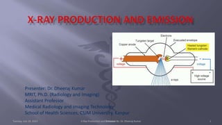
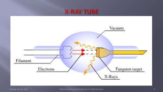
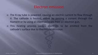
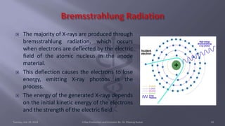
Presenter: Dr. Dheeraj Kumar
MRIT, Ph.D. (Radiology and Imaging)
Assistant Professor
Medical Radiology and Imaging Technology
School of Health Sciences, CSJM University, Kanpur
Tuesday, July 18, 2023 X-Ray Production and Emission By- Dr. Dheeraj Kumar 1
๏‚จ X-ray production is a fundamental process used in medical imaging,
scientific research, industrial applications, and various other fields. X-
rays are a form of electromagnetic radiation with high energy and
short wavelengths, capable of penetrating most substances to varying
degrees.
๏‚จ The production of X-rays involves the interaction between high-energy
electrons and matter, typically within a specialized device called an X-
ray tube.
Tuesday, July 18, 2023 X-Ray Production and Emission By- Dr. Dheeraj Kumar 2
๏‚จ An X-ray tube consists of two primary components: a cathode and an
anode. The cathode is a filament made of tungsten that emits
electrons when heated.
๏‚จ The anode, typically made of tungsten or another high atomic number
material, serves as a target for the electrons and produces X-rays when
struck by them.
๏‚จ The cathode and anode are enclosed in a vacuum-sealed glass or
metal housing to prevent unwanted interactions with air molecules.
Tuesday, July 18, 2023 X-Ray Production and Emission By- Dr. Dheeraj Kumar 3
Tuesday, July 18, 2023 X-Ray Production and Emission By- Dr. Dheeraj Kumar 4
๏‚จ Electron Emission
๏‚จ Electron Acceleration
๏‚จ Electron Deceleration
๏‚จ X-ray Production
๏‚จ X-ray Emission and Collimation
๏‚จ X-ray Detection
Tuesday, July 18, 2023 X-Ray Production and Emission By- Dr. Dheeraj Kumar 5
๏‚จ The X-ray tube is powered, causing an electric current to flow through
it. The cathode is heated, either by passing a current through the
filament or by using an electron beam from an electron gun.
๏‚จ This heating process causes electrons to be emitted from the
cathode's surface due to thermionic emission.
Tuesday, July 18, 2023 X-Ray Production and Emission By- Dr. Dheeraj Kumar 6
๏‚จ A high voltage is applied across the X-ray tube, creating an electric field
between the cathode and the anode.
๏‚จ This field accelerates the emitted electrons towards the anode.
Tuesday, July 18, 2023 X-Ray Production and Emission By- Dr. Dheeraj Kumar 7
๏‚จ As the high-energy electrons approach the anode, they experience a
strong electrostatic force, causing them to rapidly decelerate.
๏‚จ This deceleration leads to the emission of X-rays.
Tuesday, July 18, 2023 X-Ray Production and Emission By- Dr. Dheeraj Kumar 8
๏‚จ When the high-speed electrons strike the anode, two primary
processes contribute to X-ray generation:
๏‚จ Bremsstrahlung radiation and
๏‚จ Characteristic radiation.
Tuesday, July 18, 2023 X-Ray Production and Emission By- Dr. Dheeraj Kumar 9
๏‚จ The majority of X-rays are produced through
bremsstrahlung radiation, which occurs
when electrons are deflected by the electric
field of the atomic nucleus in the anode
material.
๏‚จ This deflection causes the electrons to lose
energy, emitting X-ray photons in the
process.
๏‚จ The energy of the generated X-rays depends
on the initial kinetic energy of the electrons
and the strength of the electric field.
Tuesday, July 18, 2023 X-Ray Production and Emission By- Dr. Dheeraj Kumar 10
๏‚จ At high energies, some electrons dislodge
inner-shell electrons of the anode material.
๏‚จ Electrons from higher energy levels then
transition to fill the resulting vacancies,
emitting X-ray photons with specific energies
characteristic of the target material.
๏‚จ This process produces characteristic X-rays,
which have discrete energy levels
corresponding to the electron transitions
within the atoms of the anode.
Tuesday, July 18, 2023 X-Ray Production and Emission By- Dr. Dheeraj Kumar 11
๏‚จ The generated X-rays are
emitted in all directions from the
X-ray tube. To create a useful X-
ray beam, a collimator, typically
a lead or tungsten aperture, is
used to restrict the X-ray beam
to a desired size and shape.
๏‚จ This helps to focus the X-rays
onto the target area and reduce
scatter radiation.
Tuesday, July 18, 2023 X-Ray Production and Emission By- Dr. Dheeraj Kumar 12
๏‚จ After passing through the
object being imaged, the X-
rays are detected by a device
such as an X-ray film, image
intensifier, or digital X-ray
detector.
๏‚จ The detector converts the X-
ray photons into an image that
can be visualized and
interpreted by medical
professionals or scientists.
Tuesday, July 18, 2023 X-Ray Production and Emission By- Dr. Dheeraj Kumar 13
๏‚จ It's important to note that X-ray production can be potentially harmful
due to the ionizing nature of X-rays. Proper safety precautions, such as
shielding and limited exposure, are essential to protect both patients
and (Radiographer) operators from excessive radiation exposure.
๏‚จ X-ray production involves the acceleration and deceleration of high-
energy electrons in an X-ray tube, resulting in the emission of X-rays
through bremsstrahlung radiation and characteristic radiation. These
X-rays are then controlled, directed, and detected to create valuable
images used for various applications, including medical diagnosis,
material analysis, and industrial inspection.
Tuesday, July 18, 2023 X-Ray Production and Emission By- Dr. Dheeraj Kumar 14
๏‚จ Bushberg, J. T., Seibert, J. A., Leidholdt, E. M., & Boone, J. M. (2011).
The essential physics of medical imaging. Lippincott Williams &
Wilkins.
๏‚จ Faiz, A., & Tavakoli Golpaygani, A. (2013). Medical physics in diagnostic
radiology. Springer Science & Business Media.
๏‚จ Podgorsak, E. B. (2016). Radiation oncology physics: A handbook for
teachers and students. International Atomic Energy Agency.
๏‚จ Seeram, E. (2016). Computed tomography: Physical principles, clinical
applications, and quality control. Saunders.
Tuesday, July 18, 2023 X-Ray Production and Emission By- Dr. Dheeraj Kumar 15
Tuesday, July 18, 2023 X-Ray Production and Emission By- Dr. Dheeraj Kumar 16

X-ray production AND Emission.pptx

  • 1.
    Presenter: Dr. DheerajKumar MRIT, Ph.D. (Radiology and Imaging) Assistant Professor Medical Radiology and Imaging Technology School of Health Sciences, CSJM University, Kanpur Tuesday, July 18, 2023 X-Ray Production and Emission By- Dr. Dheeraj Kumar 1
  • 2.
    ๏‚จ X-ray productionis a fundamental process used in medical imaging, scientific research, industrial applications, and various other fields. X- rays are a form of electromagnetic radiation with high energy and short wavelengths, capable of penetrating most substances to varying degrees. ๏‚จ The production of X-rays involves the interaction between high-energy electrons and matter, typically within a specialized device called an X- ray tube. Tuesday, July 18, 2023 X-Ray Production and Emission By- Dr. Dheeraj Kumar 2
  • 3.
    ๏‚จ An X-raytube consists of two primary components: a cathode and an anode. The cathode is a filament made of tungsten that emits electrons when heated. ๏‚จ The anode, typically made of tungsten or another high atomic number material, serves as a target for the electrons and produces X-rays when struck by them. ๏‚จ The cathode and anode are enclosed in a vacuum-sealed glass or metal housing to prevent unwanted interactions with air molecules. Tuesday, July 18, 2023 X-Ray Production and Emission By- Dr. Dheeraj Kumar 3
  • 4.
    Tuesday, July 18,2023 X-Ray Production and Emission By- Dr. Dheeraj Kumar 4
  • 5.
    ๏‚จ Electron Emission ๏‚จElectron Acceleration ๏‚จ Electron Deceleration ๏‚จ X-ray Production ๏‚จ X-ray Emission and Collimation ๏‚จ X-ray Detection Tuesday, July 18, 2023 X-Ray Production and Emission By- Dr. Dheeraj Kumar 5
  • 6.
    ๏‚จ The X-raytube is powered, causing an electric current to flow through it. The cathode is heated, either by passing a current through the filament or by using an electron beam from an electron gun. ๏‚จ This heating process causes electrons to be emitted from the cathode's surface due to thermionic emission. Tuesday, July 18, 2023 X-Ray Production and Emission By- Dr. Dheeraj Kumar 6
  • 7.
    ๏‚จ A highvoltage is applied across the X-ray tube, creating an electric field between the cathode and the anode. ๏‚จ This field accelerates the emitted electrons towards the anode. Tuesday, July 18, 2023 X-Ray Production and Emission By- Dr. Dheeraj Kumar 7
  • 8.
    ๏‚จ As thehigh-energy electrons approach the anode, they experience a strong electrostatic force, causing them to rapidly decelerate. ๏‚จ This deceleration leads to the emission of X-rays. Tuesday, July 18, 2023 X-Ray Production and Emission By- Dr. Dheeraj Kumar 8
  • 9.
    ๏‚จ When thehigh-speed electrons strike the anode, two primary processes contribute to X-ray generation: ๏‚จ Bremsstrahlung radiation and ๏‚จ Characteristic radiation. Tuesday, July 18, 2023 X-Ray Production and Emission By- Dr. Dheeraj Kumar 9
  • 10.
    ๏‚จ The majorityof X-rays are produced through bremsstrahlung radiation, which occurs when electrons are deflected by the electric field of the atomic nucleus in the anode material. ๏‚จ This deflection causes the electrons to lose energy, emitting X-ray photons in the process. ๏‚จ The energy of the generated X-rays depends on the initial kinetic energy of the electrons and the strength of the electric field. Tuesday, July 18, 2023 X-Ray Production and Emission By- Dr. Dheeraj Kumar 10
  • 11.
    ๏‚จ At highenergies, some electrons dislodge inner-shell electrons of the anode material. ๏‚จ Electrons from higher energy levels then transition to fill the resulting vacancies, emitting X-ray photons with specific energies characteristic of the target material. ๏‚จ This process produces characteristic X-rays, which have discrete energy levels corresponding to the electron transitions within the atoms of the anode. Tuesday, July 18, 2023 X-Ray Production and Emission By- Dr. Dheeraj Kumar 11
  • 12.
    ๏‚จ The generatedX-rays are emitted in all directions from the X-ray tube. To create a useful X- ray beam, a collimator, typically a lead or tungsten aperture, is used to restrict the X-ray beam to a desired size and shape. ๏‚จ This helps to focus the X-rays onto the target area and reduce scatter radiation. Tuesday, July 18, 2023 X-Ray Production and Emission By- Dr. Dheeraj Kumar 12
  • 13.
    ๏‚จ After passingthrough the object being imaged, the X- rays are detected by a device such as an X-ray film, image intensifier, or digital X-ray detector. ๏‚จ The detector converts the X- ray photons into an image that can be visualized and interpreted by medical professionals or scientists. Tuesday, July 18, 2023 X-Ray Production and Emission By- Dr. Dheeraj Kumar 13
  • 14.
    ๏‚จ It's importantto note that X-ray production can be potentially harmful due to the ionizing nature of X-rays. Proper safety precautions, such as shielding and limited exposure, are essential to protect both patients and (Radiographer) operators from excessive radiation exposure. ๏‚จ X-ray production involves the acceleration and deceleration of high- energy electrons in an X-ray tube, resulting in the emission of X-rays through bremsstrahlung radiation and characteristic radiation. These X-rays are then controlled, directed, and detected to create valuable images used for various applications, including medical diagnosis, material analysis, and industrial inspection. Tuesday, July 18, 2023 X-Ray Production and Emission By- Dr. Dheeraj Kumar 14
  • 15.
    ๏‚จ Bushberg, J.T., Seibert, J. A., Leidholdt, E. M., & Boone, J. M. (2011). The essential physics of medical imaging. Lippincott Williams & Wilkins. ๏‚จ Faiz, A., & Tavakoli Golpaygani, A. (2013). Medical physics in diagnostic radiology. Springer Science & Business Media. ๏‚จ Podgorsak, E. B. (2016). Radiation oncology physics: A handbook for teachers and students. International Atomic Energy Agency. ๏‚จ Seeram, E. (2016). Computed tomography: Physical principles, clinical applications, and quality control. Saunders. Tuesday, July 18, 2023 X-Ray Production and Emission By- Dr. Dheeraj Kumar 15
  • 16.
    Tuesday, July 18,2023 X-Ray Production and Emission By- Dr. Dheeraj Kumar 16