Download to read offline
![IOSR Journal of Dental and Medical Sciences (IOSR-JDMS)
e-ISSN: 2279-0853, p-ISSN: 2279-0861.Volume 14, Issue 11 Ver. VII (Nov. 2015), PP 01-07
www.iosrjournals.org
DOI: 10.9790/0853-141160107 www.iosrjournals.org 1 | Page
Visual Evoked Potential in Normal and Amblyopic Children
Kiran Kumar Patnaik1
, Ajay Shankar Kar2
(1)
Assistant Professor, Department of Physiology, Maharajah’s institute of Medical Sciences, Nellimarla,
Vizianagaram, Andhra Pradesh.
(2)
Consultant Ophthalmologist, Seven Hills Hospital, Visakhapatnam.
Abstract:
Background: Amblyopia refers to a decrease in best-corrected visual acuity in an eye having no organic
pathology. Amblyopia is primarily a cortical phenomenon, caused by unequal competitive inputs from the two
eyes into primary visual cortex area 17, although additional structural and functional abnormalities have been
observed in the lateral geniculate nucleus of amblyopic animals and human. It has been estimated to affect 1–
3% of the population Amblyopia usually affects only one eye, but it is possible to be amblyopic in both eyes if
both are similarly deprived of a good, clear visual image. Detecting the condition in early childhood increases
the chance of successful treatment.
I. Objective
1. Recording of VEP in normal individuals.
2. Recording of VEP in Ambylopia individuals.
3. Comparison of the results of the above two groups.
II. Materials and Methods
52 Amblyopic children in the age group of 4-12 years belonging to both sexes were studied. Age and
sex matched control group of 52 normal children were also studied.(Due criteria were adopted for inclusion and
exclusion)Pattern Visual Evoked Potential (PVEP) were recorded on Viking Select neuro diagnostic system.
III. Results
In PVEP , P100 latencies were longer and amplitudes were shorter in amblyopic group compared to
normal group. The data was subjected to various statistical analysis using SPSS-21 software. The difference in
latencies and amplitudes between two groups (amblyopic & normal) was statistically significant when the data
was subjected to the independent samples T test.
IV. Conclusion
P100 latency of PVEP at the time of presentation was significantly related to visual acuity. So PVEP test
may be useful in future to identify amblyopia long before the appearance of symptoms and to follow treatment
progress in pediatric amblyopes.
Keywords- Visual Evoked Potential (VEP), Amblyopia
V. Background
The term amblyopia describes a condition in which there is reduced visual function in one, or
infrequently both, eye(s), despite optimum optical correction and the absence of overt pathology of the visual
system. There is an acquired defect in vision that is due to an abnormal visual experience during a sensitive
period of visual development. The neural basis of amblyopia is the study of the effects of the abnormal
environmental influences on the genetically programmed development of visual processing system. The
prevalence of amblyopia in humans is thought to be around 1% to 3%. Human amblyopes are usually
asymptomatic when viewing with both eyes open, as the vision in the fellow eye is generally normal. The
motivation for research is not necessarily to find a treatment for this generally asymptomatic condition but
rather the realization that amblyopia may provide valuable insight into the role of early experience on the
structure and function of the human brain.[1]
Several studies have been performed with electrophysiological methods used in humans and in animal
models, to investigate the retinal and visual system in amblyopia dysfunction. Reported findings regarding
retinal function are contradictory. The function of the entire visual pathway, from photoreceptors to the visual
cortex, can be evaluated by visual evoked potential (VEP) recordings and the presence of abnormal VEP
responses has been observed in amblyopia.[2]](https://image.slidesharecdn.com/a0141170107-151207085548-lva1-app6892/85/Visual-Evoked-Potential-in-Normal-and-Amblyopic-Children-1-320.jpg)
![IOSR Journal of Dental and Medical Sciences (IOSR-JDMS)
e-ISSN: 2279-0853, p-ISSN: 2279-0861.Volume 14, Issue 11 Ver. VII (Nov. 2015), PP 01-07
www.iosrjournals.org
DOI: 10.9790/0853-141160107 www.iosrjournals.org 1 | Page
Visual Evoked Potential in Normal and Amblyopic Children
Kiran Kumar Patnaik1
, Ajay Shankar Kar2
(1)
Assistant Professor, Department of Physiology, Maharajah’s institute of Medical Sciences, Nellimarla,
Vizianagaram, Andhra Pradesh.
(2)
Consultant Ophthalmologist, Seven Hills Hospital, Visakhapatnam.
Abstract:
Background: Amblyopia refers to a decrease in best-corrected visual acuity in an eye having no organic
pathology. Amblyopia is primarily a cortical phenomenon, caused by unequal competitive inputs from the two
eyes into primary visual cortex area 17, although additional structural and functional abnormalities have been
observed in the lateral geniculate nucleus of amblyopic animals and human. It has been estimated to affect 1–
3% of the population Amblyopia usually affects only one eye, but it is possible to be amblyopic in both eyes if
both are similarly deprived of a good, clear visual image. Detecting the condition in early childhood increases
the chance of successful treatment.
I. Objective
1. Recording of VEP in normal individuals.
2. Recording of VEP in Ambylopia individuals.
3. Comparison of the results of the above two groups.
II. Materials and Methods
52 Amblyopic children in the age group of 4-12 years belonging to both sexes were studied. Age and
sex matched control group of 52 normal children were also studied.(Due criteria were adopted for inclusion and
exclusion)Pattern Visual Evoked Potential (PVEP) were recorded on Viking Select neuro diagnostic system.
III. Results
In PVEP , P100 latencies were longer and amplitudes were shorter in amblyopic group compared to
normal group. The data was subjected to various statistical analysis using SPSS-21 software. The difference in
latencies and amplitudes between two groups (amblyopic & normal) was statistically significant when the data
was subjected to the independent samples T test.
IV. Conclusion
P100 latency of PVEP at the time of presentation was significantly related to visual acuity. So PVEP test
may be useful in future to identify amblyopia long before the appearance of symptoms and to follow treatment
progress in pediatric amblyopes.
Keywords- Visual Evoked Potential (VEP), Amblyopia
V. Background
The term amblyopia describes a condition in which there is reduced visual function in one, or
infrequently both, eye(s), despite optimum optical correction and the absence of overt pathology of the visual
system. There is an acquired defect in vision that is due to an abnormal visual experience during a sensitive
period of visual development. The neural basis of amblyopia is the study of the effects of the abnormal
environmental influences on the genetically programmed development of visual processing system. The
prevalence of amblyopia in humans is thought to be around 1% to 3%. Human amblyopes are usually
asymptomatic when viewing with both eyes open, as the vision in the fellow eye is generally normal. The
motivation for research is not necessarily to find a treatment for this generally asymptomatic condition but
rather the realization that amblyopia may provide valuable insight into the role of early experience on the
structure and function of the human brain.[1]
Several studies have been performed with electrophysiological methods used in humans and in animal
models, to investigate the retinal and visual system in amblyopia dysfunction. Reported findings regarding
retinal function are contradictory. The function of the entire visual pathway, from photoreceptors to the visual
cortex, can be evaluated by visual evoked potential (VEP) recordings and the presence of abnormal VEP
responses has been observed in amblyopia.[2]](https://image.slidesharecdn.com/a0141170107-151207085548-lva1-app6892/75/Visual-Evoked-Potential-in-Normal-and-Amblyopic-Children-1-2048.jpg)
![Visual Evoked Potential in Normal and Amblyopic Children
DOI: 10.9790/0853-141160107 www.iosrjournals.org 2 | Page
As the VEP readings assess the bioelectrical response of the visual cortex, the observations derived from
previous studies do not suggest specific information on whether the reported VEP abnormalities may be
selectively related to a retinal dysfunction, a postretinal dysfunction, or both. That postretinal structures, in
particular the lateral geniculate nucleus (LGN), may be involved in amblyopia dysfunctional processes was first
suggested by Hubel and Wiessel and later documented in several studies in which morphologic and functional
changes of the LGN were detected.[3]
Understanding Evoked Potentials- An evoked potential (or "evoked response") is an electrical potential
recorded from the nervous system of a human or other animal following presentation of a stimulus, as distinct
from spontaneous potentials detected by electroencephalography (EEG) or electromyography (EMG). Evoked
potential amplitudes tend to be low, ranging from less than a microvolt to several micro volts, compared to tens
of micro volts for EEG, mill volts for EMG, and often close to a volt for ECG. To resolve these low-amplitude
potentials against the background of ongoing EEG, ECG, EMG and other biological signals and ambient noise,
signal averaging is usually required. The signal is time-locked to the stimulus and most of the noise occurs
randomly, allowing the noise to be averaged out with averaging of repeated responses.[4]
Signals can be recorded from cerebral cortex, brain stem, spinal cord and peripheral nerves. Usually the term
"evoked potential" is reserved for responses involving either recording from, or stimulation of, central
nervous system structures. Sensory evoked potentials (SEP) are recorded from the central nervous system
following stimulation of sense organs (for example, visual evoked potentials elicited by a flashing light or
changing pattern on a monitor; auditory evoked potentials by a click or tone stimulus presented through
earphones) or by tactile or somatosensory evoked potential (SSEP) elicited by tactile or electrical stimulation of
a sensory or mixed nerve in the periphery. They have been widely used in clinical diagnostic medicine since the
1970s, and also in intraoperative neurophysiology monitoring (IONM), also known as surgical neurophysiology.
Neuroanatomical and Neurophysiological Abnormalities in Amblyopia
Foveal vision in amblyopia resembles peripheral vision in normals. This suggests that inappropriately
large receptor fields (spatial summation) have developed in the foveal visual cortex. This hypothesis would
explain the loss of contrast sensitivity at high spatial frequencies with preservation of low spatial frequencies.
The phenomenon of spatial uncertainty, defects in judging line offset effects (vernier acuity) and the altered
psyhovisual performance when tested with crowded targets.
Amblyogenic Mechanisms - Disuse versus Competition
Two amblyogenic mechanisms have been proposedand that these may be effective, individually or, in
unison, in the various forms of amblyopia. [5,6]
Disuse - A lack of adequate retinal stimulation during infancy,
causing visual deprivation with arrest of development at a stage at which the interference began, or disuse
atrophy of afferent connections that were already present at birth. This is not regarded as being a major factor in
the development of strabismic amblyopia is now being disputed. Since the salient feature of strabismic
amblyopia is not the lack of afference but the incompatibility of visual impressions received by both eyes.
Competition- This is based on the view that stimulation of corresponding retinal points with unequal
images causes rivalry between the two eyes which is decided in favour of the fixating eye, the other eye
becoming amblyopic. Binocular deprivation and strabismus experiments support that competition rather than
disuse is the main cause of the observed changes. The right circumstances must exist, however, for the
competition to occur, since cells in the normal visual cortex tend to be dominated by one eye or the other, and
the dominant eye does not take over the cell completely. It appears that the incompatibility of the visual input
received by the two eyes causes a decrease or even blockage of synaptic transmission of the afferent impulses
originating from the nonfixing eye
VI. Materials And Methods
Subjects- 52 amblyopic patients of different etiologies were selected for the study. The patients who
were not treated earlier for refractive error, amblyopia or ocular disease were considered for study. Clear media
and normal fundus on ophthalmoscopic examination was a prerequisite for selection criteria.
The 52 patients, based on the etiologies are divided as:
Strabismic- 11
Anisometropic- 16
Isometropic- 25
1.Age group: 4-13 years
2.Sex distribution: Out of 52 patients 32 were female and 20 were male children.
3.Type of study: Prospective study
4.Inclusion criteria: All the children with amblyopia above three years of age were included in the study group.
5.Exclusion criteria: Children below three years](https://image.slidesharecdn.com/a0141170107-151207085548-lva1-app6892/85/Visual-Evoked-Potential-in-Normal-and-Amblyopic-Children-2-320.jpg)
![Visual Evoked Potential in Normal and Amblyopic Children
DOI: 10.9790/0853-141160107 www.iosrjournals.org 6 | Page
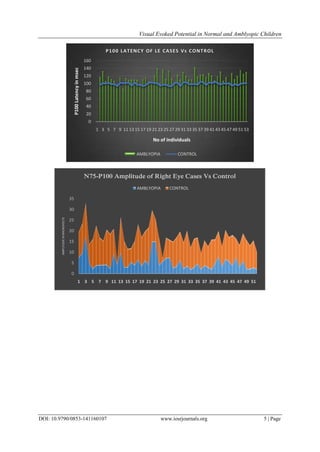
VII. Results
VEPs
1. The mean age of non-amblyopic group is 8.867+/-1.716 (33 males,19 females).The mean age of
amblyopic group is 8.058+/-1.984 (20 males,32 females).
2. The mean value of latencies in milliseconds in right side in non-amblyopic group -N75 is 71.65+/-
6.312, P100 is 98.19+/-3.726 and N145 is 137.62 +/-7.217 where as in amblyopics N75 is 74.23+/-
6.676, P100 is 115.50+/-13.457 and N145 is 151.69 +/-12.298.
3. The mean value of amplitudes(in microvolts)for right side in non-amblyopics include N75-P100 is
11.82+/-3.367 where as in amblyopics N75-P100 is 5.30+/-3.399.
4. The mean value of latencies in milliseconds in left side in non-amblyopic group N75 is 72.60+/-6.669,
P100 is 99.10+/-3.471 and N145 is 138.83+/-5.648,where as in amblyopics N75 is 74.44+/-10.357,
P100 is 115,06+/-11.77 and N145 is 153.88+/-13.88.
5. The mean value of amplitudes(in microvolts)for left side in non-amblyopics include N75-P100 is
11.306+/-3.747,where as in amblyopics N75-P100 is 5.59+/-3.743.
Independent Samples T Test shows statistically significant variations in P100 latencies of right side (p=0.016)
when amblyopes were compared with age matched controls.While similar analysis between left side P100
latencies was not statistically significant. The N75-P100 amplitude (in microvolts) was statistically significant
(p=0.012) in right side when age matched amblyopes were compared with controls. The N75-P100 amplitude in
the left eyes were not statistically significant,
VIII. Discussion
The response of visual cortex to patterned repetitive visual stimuli was tested in normal and amblyopic
children, The VEP latencies and amplitudes were compared between normal and amblyopic children. A
significant correlation was established in VEP P100 latencies and VEP N75-P100 amplitudes between normal
and amblyopia groups. The P100 latency was significantly prolonged (statistically significant,p˂0.05) and there
was a significant decrease in N75-P100 amplitude(p˂0.05) in amblyopic group which is in accordance to
previous studies.[7,8]
There were no differences in other VEP parameters ( N75, N145 ) between the two groups.
In current study, the right eye of the amblyopic group showed more correlation than left. This may provide some
information regarding specialization of functions in cerebral hemispheres. As all the patients examined
happened to be right handed i.e left hemi sphere dominance (motor activities, speech etc.) it may be coupled
with localization of some functions in left hemisphere related to cognitive process.[9]
VEP P100is a long latency
evoked potential. P-100 response of the visual evoked potential to pattern stimulation is a cortically originated
wave either produced exclusively by area 17 or 18 or by a multiplicity of cortical neuronal pools.[10]
Prolongation
of P100 latency in our study, strongly suggest an abnormality at cortical level in amblyopia which is consistent
with previous studies.[11,12]
Also right sided P100 latencies more significant than left side suggesting,
impairment of some cognitive function localized to left hemisphere based on impairment of processing of visual
information
0
5
10
15
20
25
30
35
40
1 3 5 7 9 11 13 15 17 19 21 23 25 27 29 31 33 35 37 39 41 43 45 47 49 51
AMPLITUDEINMICROVOLTS
N75-P100 Amplitude of Left Eye CasesVs Control
AMBLYOPIA CONTROL](https://image.slidesharecdn.com/a0141170107-151207085548-lva1-app6892/85/Visual-Evoked-Potential-in-Normal-and-Amblyopic-Children-6-320.jpg)
![Visual Evoked Potential in Normal and Amblyopic Children
DOI: 10.9790/0853-141160107 www.iosrjournals.org 7 | Page
IX. Conclusion
VEP is a long latency evoked potential and P100 is a more reliable signal for processing information at
cortical level. P100 latencies are prolonged and amplitudes are reduced in amblyopic group. P100 latency of
PVEP at the time of presentation was significantly related to visual acuity. So PVEP test may be useful in future
to identify amblyopia long before the appearance of symptoms and to follow treatment progress in pediatric
amblyopes,[13,14]
Further investigation is needed to determine whether these results are due to physiologic
change(s) of amblyopia itself or to fixation in stability during the test.
Bibliography
[1]. Brenden T. Barrett, Arthur Bradley, Paul V. Mcgraw;Understanding the Neural Basis of Amblyopia, NEUROSCIENTIST; 2004;
10(2):106–117.
[2]. Celesia GG, Bodis-Wollner I, Chatrian GE, Harding GFA, SokolS,Spekreijse H. Recommended standards for
electroretinogramsandvisual evoked potentials: report of an IFCN Committee. ElectroencephClinNeurophysiol. 1993;87:421–436.
[3]. Hubel DH, Wiesel TN. Binocular interaction in striate cortex ofkittens reared with artificial squint. J Neurophysiol. 1965;28:1041–
1059.
[4]. Karl E. Misulis, TouficFakhoury (2001), Spehlmann's Evoked Potential Primer, Butterworth-Heinemann, ISBN 0750673338.
[5]. Wiesel TN. Postnatal development of the visual cortex and the influence of the environment. Nature; 1982; 299:583-591.
[6]. Von Noorden GK, Amblyopia: A multidisciplinary approach. Procter Lecture. Invest Ophthalmol Vis Sci; 1985 ;26:1704-1716.
[7]. Sokol S.Abnormal evoked potential latencies in amblyopia.Br Ophthalmol. 1983 May;67(5):310-4.
[8]. Henc-Petrinović L, Deban N, Gabrić N, Petrinović J. Prognostic value of visual evoked responses in childhood amblyopia.Eur J
Ophthalmol. 1993 Jul-Sep ;3(3):114-20.
[9]. Bankó ÉM, Körtvélyes J, Weiss B, VidnyánszkyZ.How the visual cortex handles stimulus noise: insights from amblyopia.PLoS
One. 2013 Jun 20;8(6):e66583.
[10]. Celesia GG. Anatomy and physiology of visual evoked potentials and electroretinograms.NeurolClin. 1988 Nov;6(4):657-79.
[11]. Xinmei Wang, Dongmei Cui, Ling Zheng, Xiao Yang, Hui Yang, Junwen Zeng. Combination of blood oxygen level–dependent
functional magnetic resonance imaging and visual evoked potential recordings for abnormal visual cortex in two types of
amblyopia.Mol Vis. 2012; 18: 909–919.(http://www.molvis.org/molvis/v18/a95).
[12]. Lisa M. Hamm, Joanna Black, Shuan Dai, and Benjamin Thompson.Global processing in amblyopia: a review. Front Psychol. 2014;
5: 583.
[13]. Friendly DS, Weiss IP, Barnet AB, Saumweber R, Walker JA.Pattern-reversal visual-evoked potentials in the diagnosis of
amblyopia in children. .Am J Ophthalmol. 1986 Sep 15;102(3):329-39.
[14]. McKerral M, Polomeno RC, Leporé F, LachapelleP.Caninterocular pattern reversal visual evoked potential and motor reaction time
differences distinguish anisometropic from strabismic amblyopia?ActaOphthalmol Scand. 1999 Feb;77(1):40-4.](https://image.slidesharecdn.com/a0141170107-151207085548-lva1-app6892/85/Visual-Evoked-Potential-in-Normal-and-Amblyopic-Children-7-320.jpg)

The study examines visual evoked potentials (VEP) in amblyopic children compared to a control group of normal children. It found significant differences in P100 latencies and amplitudes, indicating VEP tests may aid in early detection and monitoring of amblyopia. The research underscores the importance of understanding amblyopia as a cortical phenomenon influenced by visual experience during development.


![PERI-PROSTHETIC FRACTURE NAIL-PLATE CONSTRUCT [NPC].pptx](https://cdn.slidesharecdn.com/ss_thumbnails/drarunkumardrmohamedashrafperiprostheticfrasturenail-plateconstructnpc-260209164459-7e9d15a1-thumbnail.jpg?width=640&height=640&fit=bounds)


![CTEV [ clubfoot] DR ARUN LAL ,DR MOHAMED ASHRAF travancore medical college k...](https://cdn.slidesharecdn.com/ss_thumbnails/ctevclubfootdrarunlaldrmohamedashraftravancoremedicalcollegekollamkeralaindia-260208063247-18fc466c-thumbnail.jpg?width=640&height=640&fit=bounds)
![ONFH[AVN HIP] -TRIPLE REGIME -A NOVAL SURGICAL CONCEPT .pptx](https://cdn.slidesharecdn.com/ss_thumbnails/onfhavnhip2026koaconcalicutdrgokuldevdrmashraf-260210064517-213ec005-thumbnail.jpg?width=640&height=640&fit=bounds)


