Viromer® ONE RED is a preformed and calibrated transfection reagent designed for in vitro delivery of plasmid DNA and mRNA. It comes in single portions of lyophilized material that enable standardized reactions and keep the product fresh until use. Reference data show high-performance transfection over 12 commonly used cell types including cancer and immune cells.
2. 2
Doing transfection is like making coffee. Same procedure, somewhat differ-
ent result each time you do it. Then came Nespresso. An easy, comfortable
and clean way to make your prep.
Regarding coffee we leave it to you. But for standard-
ized transfections, there is Viromer®
ONE RED. Start the
tour and learn how simplicity and control became ONE.
4. 4
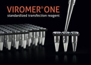
Viromer®
ONE RED is available as a full plate of 96 individual vials, each containing ONE
portion of lyophilized ready-to-use transfection reagent. Simply break a tube or more.
Add DNA or RNA. Get results. Store the rest for next time or share it with your labmates!
What's this?
5. 5
Designed for single to multichannel transfections in 24- to 96-well plate
formats. Just add your diluted DNA (or mRNA) to suspend the reagent and
enable complexation. ONE tube works for ONE well.
ONE step transfection
6. 6
+ Standardized > Preformed, calibrated reagent.
+ Clean > Single portions keep the product fresh until use.
+ ONE step protocol > Just add DNA! Or mRNA …
+ Easy-adjustable > One round of a pre-set optimization.
Easy. Fast. Reliable.
VIROMER®
ONE RED
7. 7
pDNA mRNA
100806040200transfected cells %
Transfection efficiency of Viromer®
ONE RED used for
plasmid DNA and mRNA delivery in a broad spectrum
of cell lines.
Percentages of positive cells (max.) after transfection of a GFP-plas-
mid (3.5 kb) or a GFP encoding mRNA (996 nt).
• 50 -150 ng DNA or mRNA per 96-well depending on optimal re‑
hydration and transfer volumes (for details, see page 11 - 12)
• read-out: 24 hours post-transfection by FACS Calibur
Viromer®
ONE RED
achieves high-performance transfection of easy-to-transfect model
cell lines. Its capacity to deliver mRNA even increases final protein
expression and oversteps some of the limitations relative to plas-
mid transfection.
Viromer®
ONE RED
shows comparable strong efficiency for transfecting plasmid DNA
and mRNA into more specific cell types as cancer cell lines.
Viromer®
ONE RED
in combination with mRNA is THE alternative to properly overex-
press genes in “resistant” cells as macrophages / monocytes.
Benchmark data:
Proven high-performance transfection
CHO
model
cell lines
cancer
cell lines
immune
cells
A549
HEK
H322
HeLa
H358
HepG2
Neuro2A
MDCK
C2C12
J774
THP-1
8. 8
80 µl80 µl
Preparation of pDNA / mRNA
• Dilute your pDNA/mRNA stock solution in water at 10 ng / µl.
• Prepare a volume of 80 µl.
Complexation
• Pierce the foil and rehydrate one vial with 80 µl.
• Mix swiftly by pipetting up and down and incubate for 15 min at
room temperature.
Add the transfection complex on the cells
Per well 96-well 24-well
Transfer volume 10 µl 50 µl
pDNA / mRNA on cells 100 ng 500 ng
Read - out
• Incubate cells as usual. There is no need to change medium
unless high amounts of transfection complex cause toxicity.
• For pDNA monitor effects 24 - 72 hours post-transfection.
• For mRNA, expression can start as early as 6 hours post-transfection.
Basic Transfection Protocol
1
2
1
2
3
4
3
4
10 50
96-well 24-well
µl µl
9. 9
40 µl 50 µl 60 µl 70 µl 80 µl 90 µl
The transfection efficiency is too low or there is toxicity?
Try this 96-well optimization scheme to vary
• the Viromer®
-pDNA (or –mRNA) ratio
• the amount of transfection complexes arriving onto the cells
Preparation of pDNA / mRNA
• Dilute your pDNA/mRNA stock solution in water at 10 ng / µl.
• Prepare a volume of 400 µl.
Complexation
• Rehydrate 6 vials with volumes of 40 - 90 µl.
• Mix swiftly by pipetting up and down and incubate for 15 min
at room temperature.
Add the transfection complex on the cells
Transfer complex onto the cells with 3 different volumes:
5, 10 and 15 µl corresponding to 50, 100 and 150 ng of DNA per well,
respectively.
Read - out
Incubate cells and monitor effects as previously described.
Note: the cell density at seeding time (usually one day prior transfection) and the
duration of incubation between transfection and monitoring of protein expression are
also adjustable parameters. If unknown, the best conditions for your cells should be
determined empirically beforehand.
Optimization Guide
1
2
2
3
3
4
µl
µl
µl
5
10
15
5
10
15
5
10
15
5
10
15
5
10
15
5
10
15
amount of
transfection
complexes
onto the cells
Viromer®
- pDNA
(or - mRNA) ratio
96-well
10. 10
5 µl
5 µl
10 µl
10 µl
15 µl
15 µl
40 µl 40 µl
74,4 75,2
72,0 84,1
63,2 84,4
78,9 75,2
87,3 71,8
80,4 71,8
50 µl 50 µl
78,7 61,1
75,4 75,2
65,7 75,5
83,2 88,5
95,4 88,4
91,1 89,6
60 µl 60 µl
58,9 66,5
76,7 78,8
67,3 79,6
82,9 95,7
95,5 96,1
93,9 92,3
70 µl 70 µl
50,7 56,5
67,8 77,2
69,9 82,1
84,9 97,2
95,5 99,5
96,6 96,3
46,2 58,3
74,2 82,3
72,9 80,4
87,5 99,2
98,7 99,5
97,4 98,4
80 µl 80 µl90 µl 90 µl
40 43,3
67,0 74,7
69,2 83,6
84,5 98,0
96,4 99,9
97,9 100
Optimization - find the best conditions for your
special cells and targets.
pDNA
mRNA
CHO MDCK
We applied the previously detailed op-
timization scheme to identify the best
conditions to transfect 12 different cell
lines.
Example of optimization data for pDNA
and mRNA transfections in CHO and
MDCK cells with Viromer®
ONE RED.
Numbers in circles corresponds to
the percentage of positive cells 24
hours post-transfection of a GFP plas-
mid (3.5 kb) and a GFP encoding mRNA
(996 nt) depending on the rehydra-
tion volume of Viromer®
ONE RED vials
(40 - 90 µl of diluted pDNA or mRNA at
10ng/µl) and the volume of transfec-
tion complex transferred onto plated
cells (5, 10 or 15 µl per 96-well, corre-
sponding to 50, 100 and 150 ng DNA or
mRNA per well, respectively). Read-out:
FACS Calibur.
11. 11
Take a look at our in-house reference data.
pDNA
mRNA
50 µl 80 µl 80 - 90 µl50 - 60 µl 70 µl 40 µl
80µl 80 µl80 - 90 µl 90 µl 90 µl90 µl
CHO
79 % 68 % 99 %90 % 27 % 84 %
99 % 94 % 98 %100 % 75 % 100 %
HEK HELA HepG2 MDCKC2C12
Cell density (cells / 96-well)
MAX % GFP+cells
MAX % GFP+cells
with
with
Viromer®
ONE RED
rehydration
Viromer®
ONE RED
rehydration
DNA/well
RNA/well
transfer
volume/well
transfer
volume/well
1.0 x10⁴
15 x105
7.0 - 103
2.2 x10⁴
3.5 x10⁴
9.0 x10³
modell cell lines
50 ng 100 ng 100 -150 ng
100 -150 ng
50 ng 150 ng 150 ng
100 ng 50 ng 150 ng100 -150 ng 100 ng
5 µl 10 µl 15 µl5 µl 15 µl 15 µl
10 µl 5 µl 15 µl15 µl 10 µl 15 µl
12. 12
pDNA
mRNA
50 µl 70 µl 60 µl90 µl 80 µl 80 µl
80 µl 60 µl90 µl 50 µl 90 µl80 µl
A549
91 % 94 % 8 %52 % 93 % 9 %
100 % 98 % 77 %85 % 100 % 42 %
H322 H358 Neuro2A THP-1J774
Cell density (cells / 96-well)
MAX % GFP+cells
MAX % GFP+cells
with
with
Viromer®
ONE RED
rehydration
Viromer®
ONE RED
rehydration
DNA/well
DNA/well
transfer
volume/well
transfer
volume/well
5.0 x10³
2.0 x10⁴
2.0 x10⁴
2.5 x10⁴
1.2 x10⁴
6.0 x10⁴
cancer cell lines immune cells
50 ng 150 ng 150 ng
100 ng
150 ng 100 ng 50 ng
50 -150 ng 100 ng 100 ng150 ng 150 ng
5 µl 15 µl 15 µl15 µl 10 µl 5 µl
15 µl 10 µl 10 µl15 µl 15 µl 10 µl
And try it on your own!
13. 13
Why and how switching to mRNA transfection?
As shown in the previous set of data, transfecting cells with mRNA
sequences rather than plasmid DNA constructs gives a great chance to
significantly increase protein expression levels. After delivery, mRNA is
directly expressed in the cytosol through a promoter-independent pro-
cess and protein is detectable as early as 6 h post-transfection.
Viromer®
ONE RED has been optimized to work equally strong with DNA
and mRNA. Recent investigations have shown a clear advantage of mRNA
transfection for some specific cells known as “resistant” to plasmid
transfection:
• cells with low division rate, e.g. primary neurons, differentiated skeletal
muscle cells, and
• cells with cytosolic defense mechanisms against foreign DNA (innate
immunity), e.g. AIM2-Interleukin or cGAS-Interferon enzymatic cas‑
cades of macrophages and monocytes.
IMPORTANTE NOTE: To produce stable and high quality mRNA for trans-
fection and subsequent translation, it is recommended to use in vitro
transcription commercial kits enabling 5´ capping and 3´ polyadenylation.
Transcribed mRNA should be then purified.
Use the Viromer®
Start Positive®
Controls!
Positive®
Controls are pre-formulated Viromer®
ONE RED transfection com-
plexes. Use these materials for evaluating transfection of new cell types with
the Viromer®
technology or as reference material, or to compare plasmid DNA
and mRNA transfections.
One kit of Start Positive®
Controls comprises:
• a pCMV-GFP plasmid complexed to the Viromer®
reagent (1 vial)
• a GFP encoding mRNA complexed to the Viromer®
reagent (1 vial)
14. 14
Applications
Viromer®
ONE RED is optimized for in vitro transfection of pDNA and mRNA.
Content and formats
Viromer®
ONE RED 96 transfections VR-01PF-01
Viromer®
ONE RED
+ pDNA- / mRNA-GFP controls
96 transfections
150 µl each
VR-PFBUNDLE-01
ONE plate is sufficient for 96 individual transfections in a 96-well or 24-
well format. Control kits consist of lyophilized Viromer®
RED already
complexed with pCMV-GFP plasmid (1 vial) and GFP mRNA (1 vial).
Storage and use
Viromer®
ONE RED should be stored at +2- 8°C in the provided aluminum
bag. The sealed package is stable for 6 months. Use within 3 months
when opened.
Quality control
Each batch of Viromer®
is tested for transfection using a luciferase re-
porter. MSDS are available at www.viromer-transfection.com.
Product use limitations
This product is intended for research use only; it must not be used
for therapeutic, veterinary or diagnostic applications. The purchase
of Viromer®
reagents implies a limited, non-transferable right to the
purchaser to use these products, or parts from these products, only for
its internal research. All further commercial applications of Viromer®
products require a license from Lipocalyx GmbH.
Product information
15. 15
General information
Technology
Viromer®
ONE RED is a lyophilized polymer-based transfection reagent featuring a viral
mechanism of membrane fusion. It forms transfection complexes with plasmid DNA
(pDNA) or messenger RNA (mRNA), which are taken up by endocytosis, a process that
involves the formation of an acidic compartment. The low pH in late endosomes acts as a
chemical switch that renders the Viromer®
surface hydrophobic and facilitates membrane
crossing. This “Active Endosome Escape” technology is safe and maximizes transfection
efficiency as it is using a natural uptake pathway.
Key Benefits
+ Active Escape Technology > Efficacy and safety during uptake.
+ Zero Charge > Fully compatible with serum or antibiotics.
Fully compatible with suspension cells.
+ Stable Particles > Reproducible results.
+ Lipid free > Works in adipocytes.
+ Reverse Transfection > Ready for High-Throughput Screening.
It is highly effective on a wide range of standard and hard-to-transfect cells including
suspension cells, stem cells and primary cells.
A list of cell types and transfection results is available at:
https://viromer-transfection.com/data-by-cell-type/cell-transfection-a-z
If the cell type of your interest is not listed, talk to our customer service or distributors.
Dr. Sandra Lagauzère (Biologist)
+49 345 55 59 663
sandra.lagauzere@lipocalyx.de
Bettina Weber (Biologist)
+49 345 55 59 625
bettina.weber@lipocalyx.de
Contact