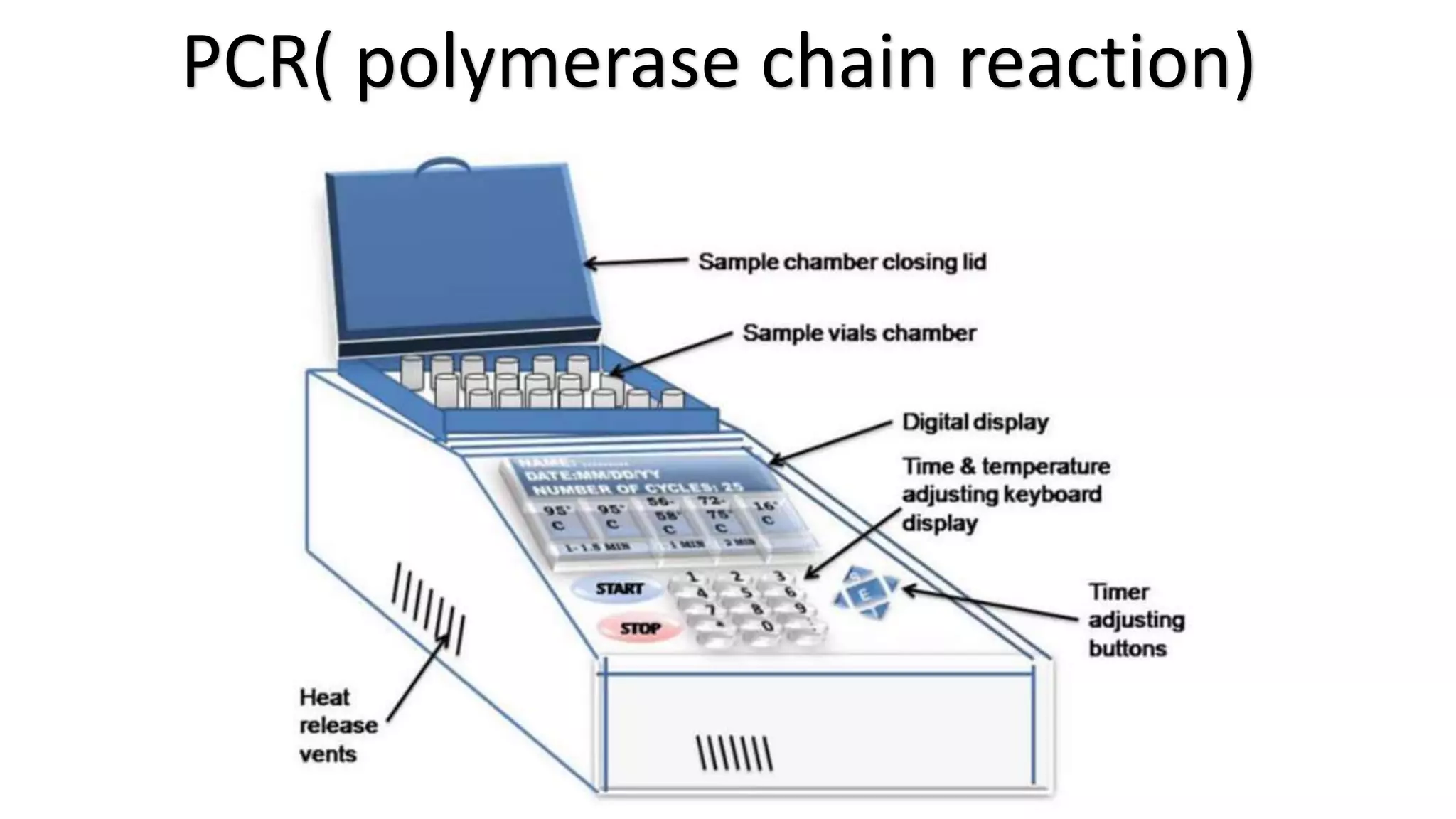
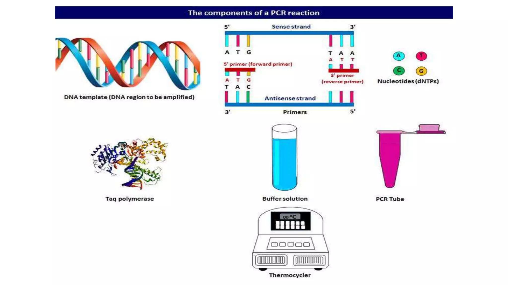
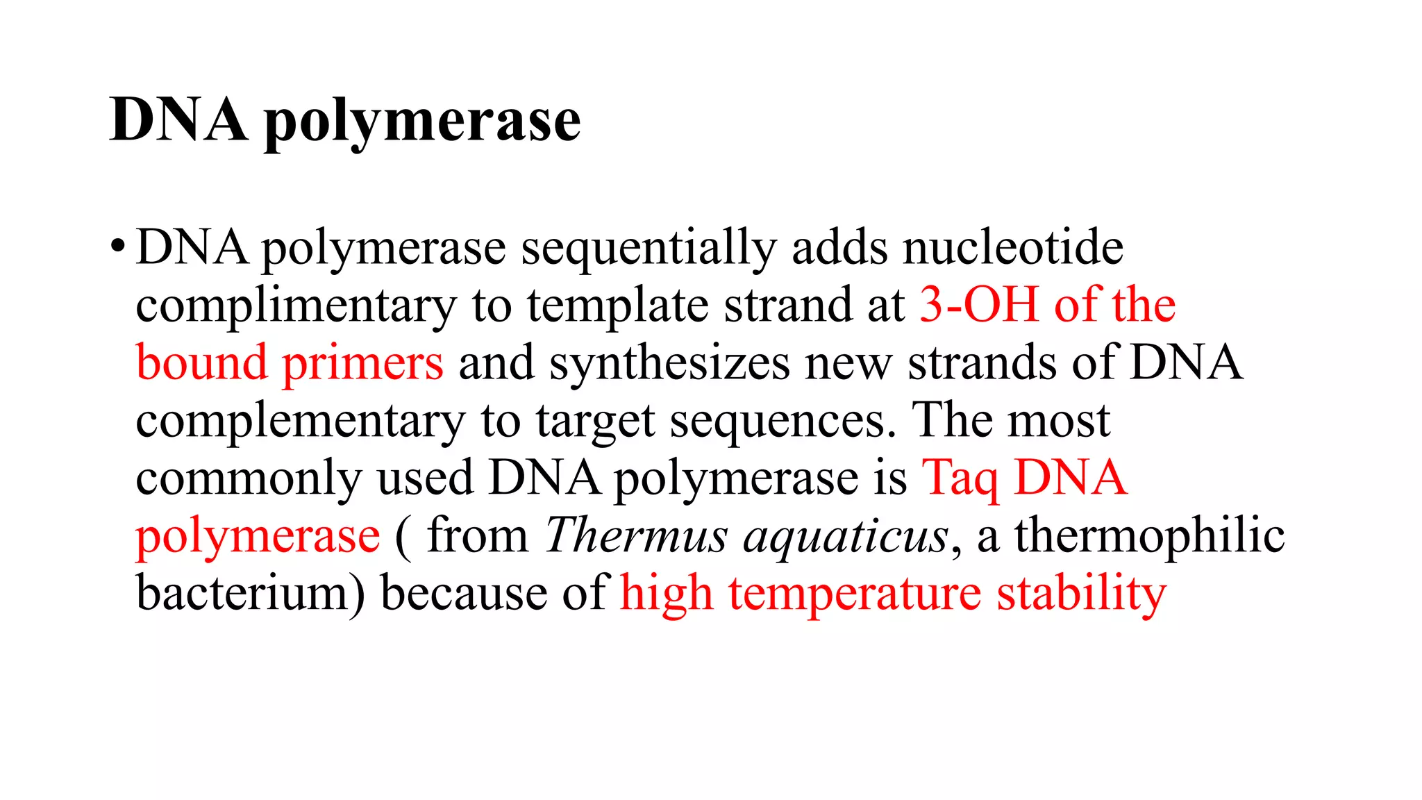
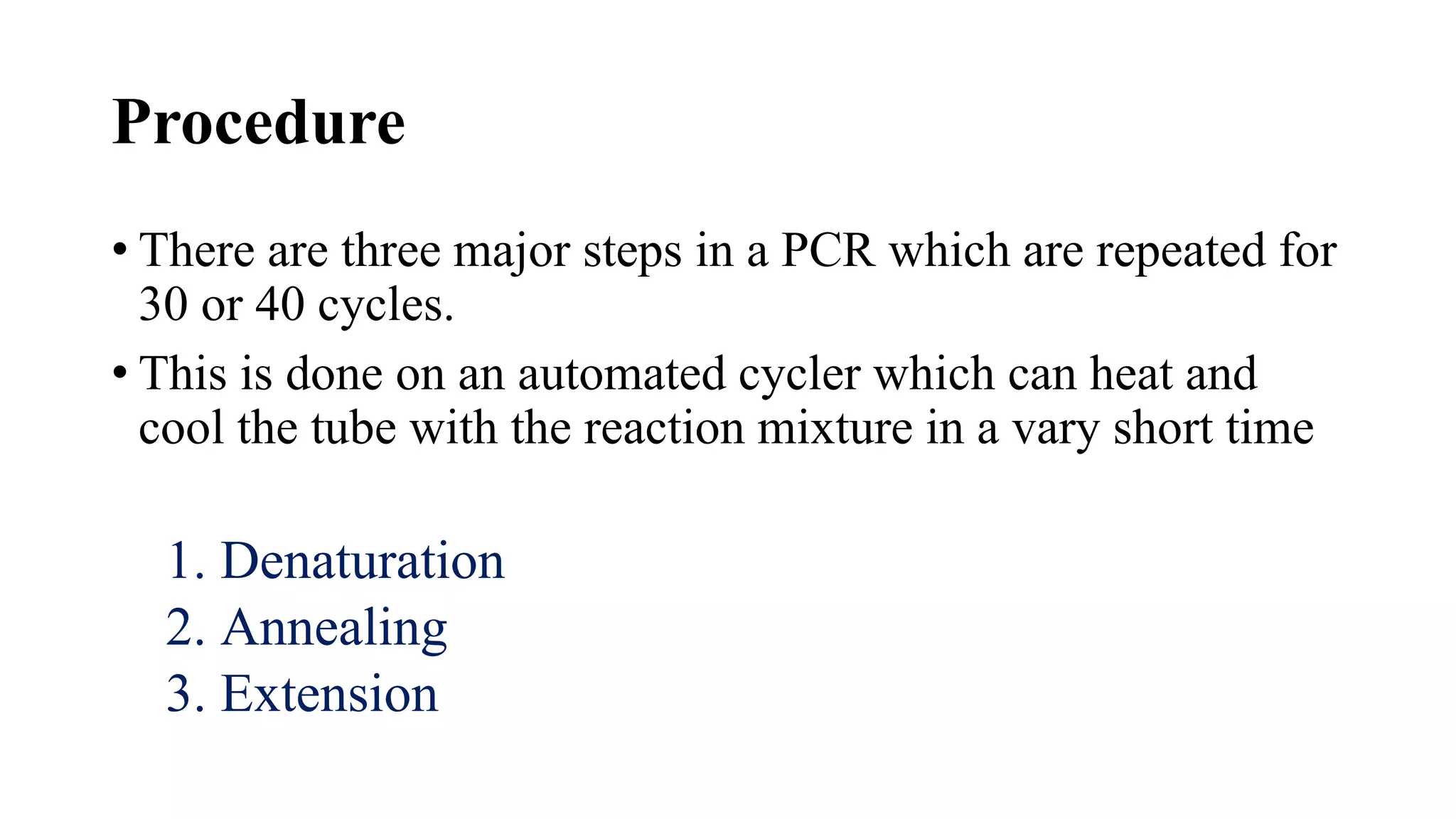
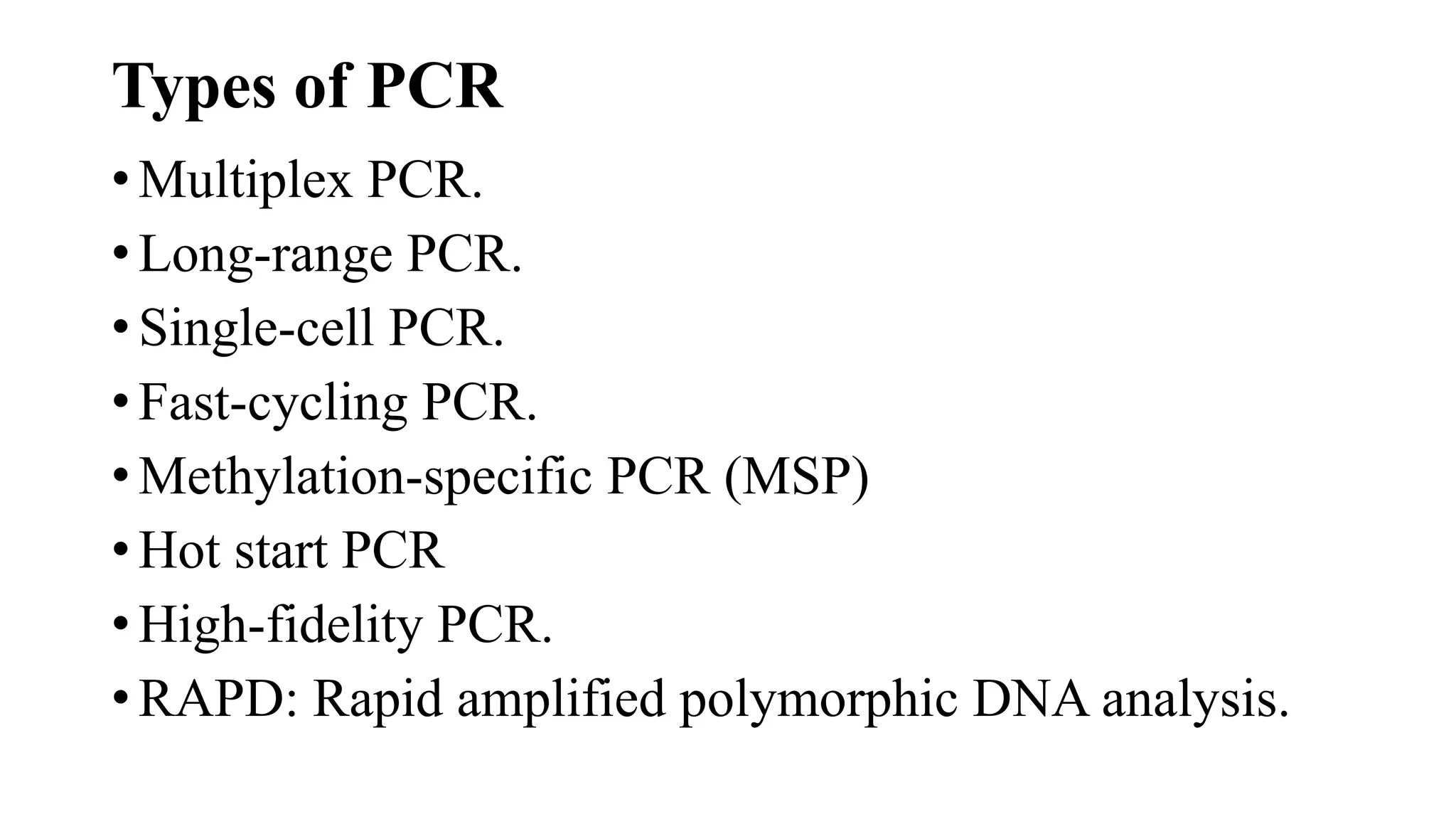
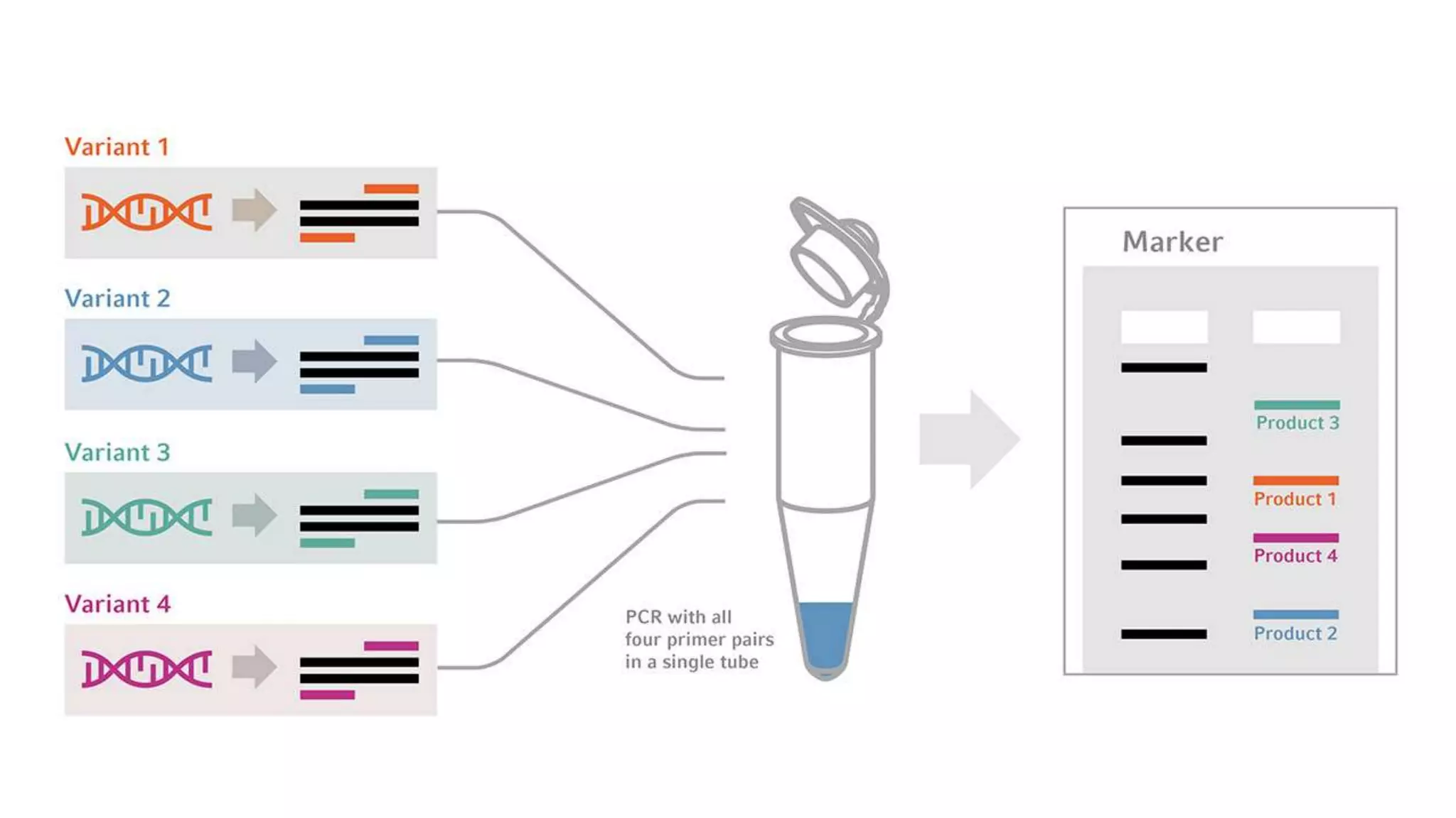
ANNAMALAI UNIVERSITY
FACULTY OF AGRICULTURE
DEPARTMENT OF PLANT PATHOLOGY
PAT-507 TECHNIQUES FOR DETECTION AND
DIAGNOSIS OF PLANT DISEASES
TOPIC
PCR (POLYMERASE CHAIN REACTION)
BY
V.AJAYDESOUZA
1ST YEAR M.sc (Ag) Plant pathology
What is PCR. ?
• It is a molecular technology aim to amplify a single or few
copies of the DNA to thousand or millions of copies
• Developed in 1983 by Kary Mullis PCR is now a common
and often indispensable technique used in medical and
biological research labs for a variety of applications. These
include diagnosis of infectious diseases, DNA sequencing
and DNA-based phylogeny
PCR( polymerase chain reaction)
Components of PCR
Template DNA
DNA template is DNA target sequence. DNA
template is the DNA molecule that contains the
DNA region (segment) to be amplified, the segment
we are concerned which is the target sequence.
DNA polymerase
• DNA polymerase sequentially adds nucleotide
complimentary to template strand at 3-OH of the
bound primers and synthesizes new strands of DNA
complementary to target sequences. The most
commonly used DNA polymerase is Taq DNA
polymerase ( from Thermus aquaticus, a thermophilic
bacterium) because of high temperature stability
• Pfu DNA polymerase (from Pyrococcus furiosus ) is
also used widely because of its higher fidelity
(accuracy of adding complementary nucleotide ).
Mg2+ ions in the buffer act as co- factor for DNA
polymerase enzyme and hence are required for the
reaction
Primers
•Primers are synthetic DNA strands of about 18 to
25 nucleotides complementary to 3’end of the
template strand. DNA polymerase starts
synthesizing new DNA from the 3’ end primers
•Two primers must be designed for PCR the forward
primer and the reverse primer. The forward primer
is complementary to the 3’ end of antisense strand
(3’-5’) and the reverse primer is complementary to
the 3’ end of sense strand (5’-3’) of a gene, for
designing primers, then forward primer is the
beginning of the gene and the reverse primer is the
reverse compliment of the 3’ end of the gene
Nucleotides (DNTPs or deoxynucleotide
triphosphates )
• All types of nucleotides are “ building blocks ” for
new DNA strands and essential for reaction. It includes
Adenine (A), Guanine (G), Cytosine (C), Thymine (T)
or Uracil(U)
Procedure
• There are three major steps in a PCR which are repeated for
30 or 40 cycles.
• This is done on an automated cycler which can heat and
cool the tube with the reaction mixture in a vary short time
1. Denaturation
2. Annealing
3. Extension
Denaturation
•During the denaturation the reaction mixture is
heated to 94⁰ C for 1 min, which causes
separation of DNA double stranded now each
strand act as template for synthesis of
complimentary strand
Annealing
• The reaction temperature is lowered to 54-60⁰ C for
around 20-40 seconds. Here the primers binds to their
complementary sequence on the template DNA
• Primers are single strand sequences of DNA or RNA
around 20 to 30 bases in length
• They serve as the starting point for the synthesis of
DNA
•The two separated strands run in opposite
direction and consequently there are two primers
a forward primers and a reverse primer
Extension
• At this step the temperature is raised to 72-80⁰ C the bases are
added to the 3’ end of the primer by the Taq polymerase
enzyme
• This elongates the DNA in the 5’ to 3’ direction. The DNA
polymerase adds about 1000bp/ minute under optimum
condition
• Taq polymerase can tolerate very high temperature. It
attaches to the primers and adds DNA bases to the single
strand. As a result a double stranded DNA molecule is
obtained
Types of PCR
• Multiplex PCR.
• Long-range PCR.
• Single-cell PCR.
• Fast-cycling PCR.
• Methylation-specific PCR (MSP)
• Hot start PCR
• High-fidelity PCR.
• RAPD: Rapid amplified polymorphic DNA analysis.
Types of PCR ( Plant Pathology)
•Multiplex PCR
•Nested PCR
•Q PCR
•Immune capture PCR
•RT- PCR
Multiplex -PCR
•It is a special type of the PCR used for detection
of multiple pathogens by using multiple primers
sets each one targets a particular pathogen
Uses
This permits the simultaneous analysis of multiple
targets in a single sample
Nested-PCR
• Used to increase the specificity of DNA amplification
• Two sets of primers are used in two successive
reactions
• In the first PCR one pair of primers is used to generate
DNA products which will be the target for second
reaction
• Using one (‘hemi nesting’) or two different primers
whose binding sites are located (nested) within the
first set, thus increasing specificity
Uses
Detection of pathogen that occurs with very few amount
Quantitative - PCR
•Used to measure the specific amount of target
DNA (or RNA) in a sample
•By measuring amplification only within the phase
of true exponential increase, the amount of
measured product more accurately reflects the
initial amount of target,
•Special thermal cyclers are used that monitor the
amount of product during the amplification
Immune capture PCR
• Immunocapture-polymerase chain reaction (IC-PCR)
is a synthesis of two commonly used diagnostic tools.
This method exploits the high-affinity binding of
antibodies to provide a facile method of purification,
usually from a complex matrix, supplying the substrate
for PCR detection.
• Immune capture PCR (IPCR) is a technique capable of
detecting the pathogens with high specificity and
sensitivity
RT-PCR (Reverse Transcription PCR, Real
time- PCR)
• Used to reverse transcribe and amplify RNA to cDNA
• PCR is preceded by a reaction using reverse
transcriptase, an enzyme that converts RNA into
cDNA
• The two reaction may be combined in a tube
Uses
1) Detection of RNA virus
2) Detection of other M.O through targeting of their
Ribosomal RNA
Detection of fungal plant pathogen using PCR
Extraction of DNA of fungal pathogen
(i) Grow the fungal pathogen (Magnaporthe oryzae) in appropriate medium
(V 8 juice agar, Campbell Soup Co. USA) and maintain the culture conditions
that favor optimal growth.
(ii) Grind the mycelium in liquid nitrogen; suspend in 0.4 ml of phenol and
0.8 ml of fungal genomic DNA extraction buffer (100 mM LiCl, 10 mM
EDTA, 10 mM Tris, pH 8.0, 0.5% SDS and 0.1% ß-mercaptoethanol) in
tubes and incubate for 5 min at 60°C.
(iii) Agitate the tubes gently; allow them to cool; add 0.4 ml of chloroform/isoamyl
alcohol (24:1 v/v) and centrifuge at 16,000 g for 10 min.
(iv) Extract 0.7 ml of the upper phase with an equal volume
of chloroform/isoamyl alcohol (24:1, v/v); separate the upper
phase and precipitate DNA with 1.0 ml of cold ethanol
containing 150 mM sodium acetate.
(v) Allow the pellet to dry for 5 min and dissolve in 0.5 ml of
TE buffer ( 1 mM EDTA,10 mM Tris, pH 8.0).
(vi) Treat with RNase (50 mg); extract with
phenol/chloroform and precipitate with ethanol; dissolve the
pellet in 0.1 ml of TE buffer, pH 8.0; determine the DNA
contents spectrophotometrically and adjust the final
concentration to 50 ng/ml
Extraction of DNA from plant tissues
(i) Place the pieces of infected leaf blades in 1.5 ml Eppendorf tubes; add
sufficient extract solution (100 ml) (from the Extract-N-Amp Kit, Sigma
Chemical Co. USA) to cover the leaf tissues and incubate for 10 min at
95°C.
(ii) Add equal volume of dilution solution (from the kit); homogenize the
sample in the tube using a polypropylene pestle; place in ice and dilute 5
ml aliquot tenfold in sterile distilled water.
PCR amplification and detection of
diagnostic amplicon
(i) Use primers pfh2a and pfh2b capable of amplifying the
687-bp region of the Pot2 transposon.
(ii) Perform PCR in a 50 ml rection mixture with DNA Taq
polymerase and purify genomic DNA from pathogen
isolates.
(iii) Perform PCR for plant samples in a 20 ml reaction
mixture from the kit in a DNA thermal cycler
(iv) PCR program consists of initial denaturation of 2
min at 94°C; 30 cycles of 45 s denaturation at 94°C; 45
s of annealing at 55°C; 45 s of extension at 72°C and
final extension at 72°C for 10 min.
(v) Resolve the amplicon after electrophoresis in a 1%
agarose gel; stain for 10 min in an ethidium bromide
solution (10 mg/ml) and visualize the bands with UV
light.
(vi) Use photoimaging system (Stratagene, CA, USA)
for getting gel images.
PCR (polymerase chain reaction) and Extraction of DNA from fungal plant pathogen.pptx

PCR (polymerase chain reaction) and Extraction of DNA from fungal plant pathogen.pptx

  • 1.
    ANNAMALAI UNIVERSITY FACULTY OFAGRICULTURE DEPARTMENT OF PLANT PATHOLOGY PAT-507 TECHNIQUES FOR DETECTION AND DIAGNOSIS OF PLANT DISEASES TOPIC PCR (POLYMERASE CHAIN REACTION) BY V.AJAYDESOUZA 1ST YEAR M.sc (Ag) Plant pathology
  • 2.
    What is PCR.? • It is a molecular technology aim to amplify a single or few copies of the DNA to thousand or millions of copies • Developed in 1983 by Kary Mullis PCR is now a common and often indispensable technique used in medical and biological research labs for a variety of applications. These include diagnosis of infectious diseases, DNA sequencing and DNA-based phylogeny
  • 3.
  • 5.
    Components of PCR TemplateDNA DNA template is DNA target sequence. DNA template is the DNA molecule that contains the DNA region (segment) to be amplified, the segment we are concerned which is the target sequence.
  • 6.
    DNA polymerase • DNApolymerase sequentially adds nucleotide complimentary to template strand at 3-OH of the bound primers and synthesizes new strands of DNA complementary to target sequences. The most commonly used DNA polymerase is Taq DNA polymerase ( from Thermus aquaticus, a thermophilic bacterium) because of high temperature stability
  • 7.
    • Pfu DNApolymerase (from Pyrococcus furiosus ) is also used widely because of its higher fidelity (accuracy of adding complementary nucleotide ). Mg2+ ions in the buffer act as co- factor for DNA polymerase enzyme and hence are required for the reaction
  • 8.
    Primers •Primers are syntheticDNA strands of about 18 to 25 nucleotides complementary to 3’end of the template strand. DNA polymerase starts synthesizing new DNA from the 3’ end primers
  • 9.
    •Two primers mustbe designed for PCR the forward primer and the reverse primer. The forward primer is complementary to the 3’ end of antisense strand (3’-5’) and the reverse primer is complementary to the 3’ end of sense strand (5’-3’) of a gene, for designing primers, then forward primer is the beginning of the gene and the reverse primer is the reverse compliment of the 3’ end of the gene
  • 10.
    Nucleotides (DNTPs ordeoxynucleotide triphosphates ) • All types of nucleotides are “ building blocks ” for new DNA strands and essential for reaction. It includes Adenine (A), Guanine (G), Cytosine (C), Thymine (T) or Uracil(U)
  • 12.
    Procedure • There arethree major steps in a PCR which are repeated for 30 or 40 cycles. • This is done on an automated cycler which can heat and cool the tube with the reaction mixture in a vary short time 1. Denaturation 2. Annealing 3. Extension
  • 13.
    Denaturation •During the denaturationthe reaction mixture is heated to 94⁰ C for 1 min, which causes separation of DNA double stranded now each strand act as template for synthesis of complimentary strand
  • 14.
    Annealing • The reactiontemperature is lowered to 54-60⁰ C for around 20-40 seconds. Here the primers binds to their complementary sequence on the template DNA • Primers are single strand sequences of DNA or RNA around 20 to 30 bases in length • They serve as the starting point for the synthesis of DNA
  • 15.
    •The two separatedstrands run in opposite direction and consequently there are two primers a forward primers and a reverse primer
  • 16.
    Extension • At thisstep the temperature is raised to 72-80⁰ C the bases are added to the 3’ end of the primer by the Taq polymerase enzyme • This elongates the DNA in the 5’ to 3’ direction. The DNA polymerase adds about 1000bp/ minute under optimum condition • Taq polymerase can tolerate very high temperature. It attaches to the primers and adds DNA bases to the single strand. As a result a double stranded DNA molecule is obtained
  • 19.
    Types of PCR •Multiplex PCR. • Long-range PCR. • Single-cell PCR. • Fast-cycling PCR. • Methylation-specific PCR (MSP) • Hot start PCR • High-fidelity PCR. • RAPD: Rapid amplified polymorphic DNA analysis.
  • 20.
    Types of PCR( Plant Pathology) •Multiplex PCR •Nested PCR •Q PCR •Immune capture PCR •RT- PCR
  • 21.
    Multiplex -PCR •It isa special type of the PCR used for detection of multiple pathogens by using multiple primers sets each one targets a particular pathogen Uses This permits the simultaneous analysis of multiple targets in a single sample
  • 23.
    Nested-PCR • Used toincrease the specificity of DNA amplification • Two sets of primers are used in two successive reactions • In the first PCR one pair of primers is used to generate DNA products which will be the target for second reaction
  • 24.
    • Using one(‘hemi nesting’) or two different primers whose binding sites are located (nested) within the first set, thus increasing specificity Uses Detection of pathogen that occurs with very few amount
  • 26.
    Quantitative - PCR •Usedto measure the specific amount of target DNA (or RNA) in a sample •By measuring amplification only within the phase of true exponential increase, the amount of measured product more accurately reflects the initial amount of target, •Special thermal cyclers are used that monitor the amount of product during the amplification
  • 28.
    Immune capture PCR •Immunocapture-polymerase chain reaction (IC-PCR) is a synthesis of two commonly used diagnostic tools. This method exploits the high-affinity binding of antibodies to provide a facile method of purification, usually from a complex matrix, supplying the substrate for PCR detection. • Immune capture PCR (IPCR) is a technique capable of detecting the pathogens with high specificity and sensitivity
  • 31.
    RT-PCR (Reverse TranscriptionPCR, Real time- PCR) • Used to reverse transcribe and amplify RNA to cDNA • PCR is preceded by a reaction using reverse transcriptase, an enzyme that converts RNA into cDNA • The two reaction may be combined in a tube Uses 1) Detection of RNA virus 2) Detection of other M.O through targeting of their Ribosomal RNA
  • 34.
    Detection of fungalplant pathogen using PCR Extraction of DNA of fungal pathogen (i) Grow the fungal pathogen (Magnaporthe oryzae) in appropriate medium (V 8 juice agar, Campbell Soup Co. USA) and maintain the culture conditions that favor optimal growth. (ii) Grind the mycelium in liquid nitrogen; suspend in 0.4 ml of phenol and 0.8 ml of fungal genomic DNA extraction buffer (100 mM LiCl, 10 mM EDTA, 10 mM Tris, pH 8.0, 0.5% SDS and 0.1% ß-mercaptoethanol) in tubes and incubate for 5 min at 60°C. (iii) Agitate the tubes gently; allow them to cool; add 0.4 ml of chloroform/isoamyl alcohol (24:1 v/v) and centrifuge at 16,000 g for 10 min.
  • 35.
    (iv) Extract 0.7ml of the upper phase with an equal volume of chloroform/isoamyl alcohol (24:1, v/v); separate the upper phase and precipitate DNA with 1.0 ml of cold ethanol containing 150 mM sodium acetate. (v) Allow the pellet to dry for 5 min and dissolve in 0.5 ml of TE buffer ( 1 mM EDTA,10 mM Tris, pH 8.0). (vi) Treat with RNase (50 mg); extract with phenol/chloroform and precipitate with ethanol; dissolve the pellet in 0.1 ml of TE buffer, pH 8.0; determine the DNA contents spectrophotometrically and adjust the final concentration to 50 ng/ml
  • 36.
    Extraction of DNAfrom plant tissues (i) Place the pieces of infected leaf blades in 1.5 ml Eppendorf tubes; add sufficient extract solution (100 ml) (from the Extract-N-Amp Kit, Sigma Chemical Co. USA) to cover the leaf tissues and incubate for 10 min at 95°C. (ii) Add equal volume of dilution solution (from the kit); homogenize the sample in the tube using a polypropylene pestle; place in ice and dilute 5 ml aliquot tenfold in sterile distilled water.
  • 37.
    PCR amplification anddetection of diagnostic amplicon (i) Use primers pfh2a and pfh2b capable of amplifying the 687-bp region of the Pot2 transposon. (ii) Perform PCR in a 50 ml rection mixture with DNA Taq polymerase and purify genomic DNA from pathogen isolates. (iii) Perform PCR for plant samples in a 20 ml reaction mixture from the kit in a DNA thermal cycler
  • 38.
    (iv) PCR programconsists of initial denaturation of 2 min at 94°C; 30 cycles of 45 s denaturation at 94°C; 45 s of annealing at 55°C; 45 s of extension at 72°C and final extension at 72°C for 10 min. (v) Resolve the amplicon after electrophoresis in a 1% agarose gel; stain for 10 min in an ethidium bromide solution (10 mg/ml) and visualize the bands with UV light. (vi) Use photoimaging system (Stratagene, CA, USA) for getting gel images.