This document summarizes an experiment that tested the effects of amygdalin (B17) on cervical cancer (HeLa) cells at various concentrations and incubation times. HeLa cells were cultured with B17 at concentrations of 1.25, 2.5, 5, 10, and 20 mg/mL for 24, 48, and 120 hours. An MTT assay was then used to analyze cell viability. The results showed that 10 and 20 mg/mL B17 concentrations caused significant HeLa cell apoptosis after 120 hours of incubation. Lower concentrations had little effect even after longer incubations. Initial issues with low cell density required troubleshooting to obtain clear results from the MTT assay.
Cell culture is the process by which prokaryotic, eukaryotic or plant cells are grown under controlled conditions. Mammalian cell culture technology has become a major field in modern biotechnology; mammalian cell culture refers to the cells of a mammalian, isolated from specific tissues (i.e. skin, liver, glands, etc.) and further cultivated and reproduced in an artificial medium. Cell culture technology is currently playing a major role in toxicity testing, cancer research, virology, genetic engineering, and gene therapy.
OBJECTIVE:
To observe the transfection of CHO and HEK cells with GFP
To observe the recombinant GFP using Western Blotting
To purify the transfected HEK and CHO cells using AKTA Pure Purification
VIROMER® ONE RED - a standardized transfection reagent for plasmid DNA and mRNASandra Lagauzère
Viromer® ONE RED is a preformed and calibrated transfection reagent designed for in vitro delivery of plasmid DNA and mRNA. It comes in single portions of lyophilized material that enable standardized reactions and keep the product fresh until use. Reference data show high-performance transfection over 12 commonly used cell types including cancer and immune cells.
German Scientist “Carl Vogt” was first to describe the principle of apoptosis in 1842. In 1885, Anatomist “Walther Flemming” gave more precise description of Programmed Cell Death. Apoptosis is a form of Programmed Cell Death that occurs in multicellular organisms. It is a Greek word which means falling off. It leads to breakdown and disposal of cells. Macrophages and other Phagocytic Cells remove them by Phagocytosis, without developing any type of inflammation. It is a biochemical event that leads to morphological changes and death. The average adult human looses 50-70 billion cells each day due to apoptosis.
Feiyuebio as manufacurer Supply ELISA kits,Antibody with High Quality and Safe Ship.
For sale:+8618071549908
SEMA3G(Semaphorin-3G) Basic information
Semaphorins are a class of secreted and membrane proteins that were originally identified as axonal growth cone guidance molecules. They primarily act as short-range inhibitory signals and signal through multimeric receptor complexes. Semaphorins are usually cues to deflect axons from inappropriate regions, especially important in the neural system development. The major class of proteins that act as their receptors are called plexins, with neuropilins as their co-receptors in many cases. The main receptors for semaphorins are plexins, which have established roles in regulating Rho-family GTPases. Recent work shows that plexins can also influence R-Ras, which, in turn, can regulate integrins. Such regulation is probably a common feature of semaphorin signalling and contributes substantially to our understanding of semaphorin biology.
Human SEMA3G(Semaphorin-3G) ELISA Kit test method
Feiyue’s Human SEMA3G (Semaphorin-3G) Elisa kit is an ELISA reagent for detection of Neutrophil elastase in serum, plasma, tissue homogenates and other biological fluids.
This kit uses sandwich ELISA to detect the concentration of Semaphorin-3G . SEMA3G (Semaphorin-3G) -specific monoclonal antibody has been pre-coated in the wells of the supplied microplate. Standards samples and controls are added to interact with the immobilized antibody. A sandwich complex is formed by additional anti- SEMA3G (Semaphorin-3G) antibody with HRP-Streptavidin. TMB solution is added to react with the sandwich for ming optical signal measured by microplate reader. The concentration of SEMA3G (Semaphorin-3G) in the sample can be calculated by comparing the absorbance of the sample with the standard curve.
DOI:10.21276/ijlssr.2016.2.4.2
neoplastic progression through the action of viral oncoproteins, mainly E6 and E7.Cervical cancer remains the second
most common cancer in women worldwide with India as a major contributor to global burden with an annual incidence of
132,000 new cases and mortality rate of 74,000 deaths annually. In this study turmeric, neem, tulasi and ginger were
selected as natural anticancer drugs. The objective of the study was to analyze the anticancer property of turmeric
(Curcuma longa), neem (Azadirachta indica), tulasi (Occimum sanctum) and ginger (Zingiber officinale) on HeLa cells.
Turmeric, neem, tulasi and ginger capsules (Himalaya’s Company) were used and aqueous and methanolic extracts of the
turmeric, neem, tulasi and ginger were obtained using a soxhlet extraction. To check the efficacy of these drug MTT assay
was performed, that determines % viability and/or cytotoxicity. IC50 of aqueous turmeric, neem, tulasi and ginger extracts
in case of HeLa cells were 17.8, 22, 79.4, 27.86 respectively and in case of methanolic turmeric, neem, tulasi and ginger
extracts 17, 7.35, 75.24 and 16.1 respectively. To confirm apoptosis as the sole reason behind cell death
immunofluorescence based apoptosis assay was performed using TALI image based cytometer. The study has led to
postulate hypothesis that natural drugs e.g. turmeric, neem, tulasi and ginger are potent anti-cancer compound that are
capable of inhibiting the growth of immortal cells by apoptosis. Key-words- Cervical cancer, Human papillomavirus (HPV), Oncoproteins E6 and E7, Natural compounds, HeLa cell
line (adherent), Cell viability and MTT assay, Apoptosis assay
In vitro methods for the assessment of general cellular toxicity,
End-points for the assessment of general cellular toxicity
Specialized cells commonly used in toxicology
Bacillus thuringiensis, an aerobic, Gram positive, spore forming bacterium produces unique proteinaceous crystalline parasporal inclusions during sporulation which have insecticidal properties. Besides being widely used as an insecticide in agriculture, Bt has been found to be useful in several fields like medicine, endoparasite control, bacteriocin production as well as enzyme production. Parasporin, a new category of bacterial parasporal protein capable of discriminately killing the cancer cells have been discovered. There are six classes of parasporins having different mode of action and cell specificities against cancer and tumor cells (Ohba et al., 2009).Bt proteins have also been used successfully to suppress the population levels of medically important Dipteran pests like mosquitoes by use of mosquitocidal strains that produce Cry proteins (Zhang et al., 2012) as well as potential therapeutic agent against protozoan disease Leishmaniases (El-Sadawy et al., 2008). Crystal proteins, like Cry5B from Bacillus thuringiensis are found to be safe to vertebrates and have been shown to have efficacy against intestinal hookworm parasites (Capello et al., 2006). Thus the multifarious applications of Bacillus thuringiensis have made it a microbe to reckon with and further study its genome for future developments.
In vitro methods of screening of anticancer agentsNikitaSavita
Cancer- It is the leading cause of mortality. Cancer is the disease which is categorized by abnormal cell growth with the dormant to spread to other parts of body.
Expression Purification and Immunodetection of a fusion protein Glutathione S...iosrjce
Glutathione S Transferase(GST) is an enzyme of a multi gene family which is involved in reducing
oxidative damage to cells and detoxification of Xenobiotic compounds and plays critical role in life processes.
The entire work was completely qualitative and the objective of my work was to deal with the induction,
extraction and purification of the GST fusion protein from pGEX 3X vector.In order to achieve high degree of
transformed cells,the E.Coli BL21 host strain was made competent using 0.1M CaCl2 and adding of pGEX 3X
vector into host made it transformed.With the induction of GST protein by 0.1mM IPTG,the desired protein was
purified through glutathione Cl agarose column and was detected by immunoblotting method with the use of
anti GST HRP conjugate Ab which expressed the desired protein.
Expression Purification and Immunodetection of a fusion protein Glutathione S...iosrjce
IOSR Journal of Biotechnology and Biochemistry (IOSR-JBB) covers studies of the chemical processes in living organisms, structure and function of cellular components such as proteins, carbohydrates, lipids, nucleic acids and other biomolecules, chemical properties of important biological molecules, like proteins, in particular the chemistry of enzyme-catalyzed reactions, genetic code (DNA, RNA), protein synthesis, cell membrane transport, and signal transduction. IOSR-JBB is privileged to focus on a wide range of biotechnology as well as high quality articles on genetic engineering, cell and tissue culture technologies, genetics, microbiology, molecular biology, biochemistry, embryology, cell biology, chemical engineering, bioprocess engineering, information technology, biorobotics.
Principles of cell viability assays by surendra.pptxSurendra Chowdary
1.DYE EXCLUSION ASSAYS:
Dye exclusion assays are the simplest methods that are based on utilization of different dyes such as trypan blue, eosin, congo red, and erythrosine B, which are excluded by the living cells, but not by dead cells.
For these assays, although staining procedure is quite straightforward, experimental procedure may be time-consuming in case of large sample sizes.
a. Trypan blue stain assay:
Trypan blue stain assay has initially been developed in 1975 to measure viable cell count and is still used as a confirmatory test for measuring changes in viable cell number caused by a drug or toxin.
Trypan blue stain, a large negatively charged molecule, is one of the simplest assays that are used to determine the number of viable cells in a cell suspension.
Principle:
The principle of this assay is that living cells have intact cell membranes that exclude the trypan blue stain, whereas dead cells do not.
Cell suspension is mixed with the trypan blue stain and examined visually under light microscopy to determine whether cells include or exclude the stain.
A viable cell will have a clear cytoplasm, whereas a nonviable cell will have a blue cytoplasm.
Reagent preparation:
To perform the trypan blue stain assay, 0.4% trypan blue stain and phosphate- buffered saline (PBS) or serum-free medium are obtained.
Trypan blue stain should be stored in dark and filtered after prolonged storage.
As trypan blue stain binds to serum proteins and causing misleading results, serum-free medium should be used to obtain reliable results.
Assay Protocol:
The cell suspension to be tested is centrifuged at 100 g for 5 min.
The supernatant is discarded and the pellet is resuspended in 1-ml PBS solution or serum-free medium.
Then, one portion of this cell suspension is mixed with one portion of trypan blue stain.
The mixture is allowed to stay at room temperature for 3 min. It is important to note that the cells should be counted within 3–5 min of mixing with trypan blue, as longer incubation periods will lead to cell death and hence reduced viability counts.
Following the incubation, a drop of the mixture is applied to a hemocytometer, which is placed on the stage of a binocular microscope.
Viable cells will remain unstained, and nonviable cells will stain, in the hemocytometer and these cells are counted separately.
.
Calculation:
After counting viable and nonviable cells, the total number of viable cells per milliliter of aliquot is determined by multiplying the total number of viable cells by 2, which is the dilution factor for trypan blue.
Similarly, total number of cells per milliliter of aliquot is determined by addition of number of viable and nonviable cells and multiplying it by 2.
Then, the percentage of viable cells is calculated using the following equation.
% Viable cells = Total number of viable cells per milliliter of aliquot × 100.
Total number of cells per milliliter of aliquot
2.COLORIMETRIC ASSAYS:
Colorimetric assays
Similar to HeLaProliferation_b17_AmygdalinStimulated_JasonMorris_05Apr2015 (20)
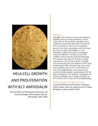
1. HELA CELL GROWTH
AND PROLIFERATION
WITH B17-AMYGDALIN
By Jason Morris of Minneapolis Community and
Technical College, 1501 Hennepin Avenue,
Minneapolis, MN, 55403
ABSTRACT
Amygdalin (B17) is known to have some anticancer
properties and even to target and destroy cervical
cancer cells (1). The aim of this experiment was to
confirm the extent of the above claim. Not only that
B17 was cytotoxic to cells or not was of primary
interest, but at what concentrations and for how long
the cells were under such conditions as well.
Mammalian cell-line HeLa cells were monolayer
cultured in EMEM in T-25 flasks to above 90%
confluency before removing media and undergoing
B17 treatment with media for 24 hours of varying
concentrations. After 24 hours, the B17 and media
were removed and cells were analyzed via MTT assay
method using D-PBS. The above steps were carried
out at incubation times of 48, 72, and 120 hours as
well. After troubleshooting various obstacles, the
results showed that optimal concentrations of B17 for
HeLa cell apoptosis were 10mg/mL and 20mg/mL for
120 hours incubation time. Use high cell density for
B17 on HeLa cells or results will need troubleshooting.
Conducted April 7th-April 27th, 2015
In partial fulfillment of Cell Culture Techniques
Course Project under the supervision of Dr. Rekha
Ganaganur, spring semester, 2015
2. 28th
April, 2015 HeLa cell-line with amygdalin B17 Page 1 of 9
Introduction:
MTT assay is a method often used by scientists interested in culturing cell lines. It is a means of
quickly determining cell viability, proliferation, and activation of cells without having to harvest and
count them individually.2
2-(4,5-dimethylthiazol-2-yl)-2,5-diphenyl tetrazolium bromide (MTT) is
reduced by mitochondrial dehydrogenase enzyme under normal cell conditions. The reduced product is
purple/blue and called formazan. Under normal conditions, when not reduced,it is a yellow, soluble
substrate. It can be used to differentiate between proliferation and cell activation and can be used on
monolayer and suspension culture. In the case of this experiment, monolayer HeLa cells were used with
MTT assay.
Amygdalin, or B17, is a naturally occurring substance derived from seeds of apricots, almonds,
peaches,apples, and several other members of the prunasin family.(1,3)
It is a cyanide former and has been
known to cause apoptosis (programmed cell death) of cervical cancer cell-line HeLa cells.1
Amygdalin is
of growing interest to cancer biologists and researchers of multiple other disciplines. The mode of action
of amygdalin on cervical cancer cells are unknown still.3
It is possible to gain further insight into this
looming question by subjecting HeLa cells with B17 in vitro.
HeLa cell-line was used for this experiment because they proliferate very well for mammalian
cells, especially a human cell line. They are derived from cancer cells of the cervix of one Henrietta
Lacks. This cell line is so resilient, it has been used to develop the polio vaccine, exposed to radiation,
and even shot into space.4
These among other reasons the HeLa cell-line was chosen for this experiment.
3. 28th
April, 2015 HeLa cell-line with amygdalin B17 Page 2 of 9
Materials and Methods:
Passage number 7 of HeLa cell-line was harvested for this experiment at approximately 95%
confluency in the T-25 flasks used. Using a 96 well plate, 1.5*104
cells in 150 uL were plated per well.
B17 was prepared by grounding 100mg tablets with mortar and pestle to a fine powder. Starch in the
tablets were removed from the stock solution by centrifuging and removing the supernatant that contained
soluble B17. Working concentrations of B17 were made for 1.25mg/mL, 2.50mg/mL, 5.0mg/mL,
10mg/mL, and 20mg/mL. It was solubilized in sterile EMEM media and filtered with a 0.22um pore size
membrane filter. The 96 well plating format utilized triplicates of all wells. The controls lacked B17
entirely, but included all other ingredients. The remaining 15 wells in each plate had media, cells, aimed
concentration of B17, and eventually MTT to analyze cytotoxicity and other aspects. The plating plan is
as follows:
B17
control
B17
control
B17
control
1.25mg/mL1.25mg/mL1.25mg/
mL
2.50mg/
mL
2.50mg/
mL
2.50mg/m
L
5.0mg/mL5.0mg
/mL 5.0mg/mL
10.0mg/
mL
10.0mg/
mL
10.0mg/
mL 20.0mg/mL20.0mg/mL20.0mg/mL
The above plating plan was for each plate used. Total of three plates used were 24, 48, and 120
hour exposures to B17. The plates were plated and allowed to adhere and the B17 was added. MTT was
added at time of desired exposure after removing the old media. 15mg of MTT was dissolved in 3mL of
PBS. 50uL MTT was added and let incubate for up to two hours. When formazan forms purple color a
microplate could be read at 570nm wavelength.
4. 28th
April, 2015 HeLa cell-line with amygdalin B17 Page 3 of 9
Results:
24 hour incubation before addition of DMSO 24 hour incubation after addition of DMSO
48 hour incubation before addition of DMSO 48 hour incubation after addition of DMSO
120 hour incubation before addition of DMSO 120 hour incubation after addition of DMSO
7. 28th
April, 2015 HeLa cell-line with amygdalin B17 Page 6 of 9
Discussions and Conclusions:
Since amygdalin is soluble in media and water,additional solvent was unnecessary. Solvent
controls were therefore not used. Differences between drug control wells and wells containing B17 were
not observed in 24 hour incubation plate. The same results were observed for the 48 hour incubation with
the exception of the 20mg/mL concentration of B17. It appeared that there was slight cell apoptosis in that
range. Similar results observed on 120 hour incubation plate with the exception of 10 and 20mg/mL B17
concentrations where cell apoptosis was abundant. This was indicated by the observation of much lighter
color of wells on 10 and 20 mg/mL versus the deep purple on the controls and other wells, indicating
those cells are proliferating well. The results matched expectations in the sense that as incubation and
concentration increased,the cyanide formed should cause cell increased apoptosis. Overall there were not
unusual results.
There was much troubleshooting needed to gain the proper results, however. For the first two
days of incubation, there was no color whatsoever when MTT was added. So when 120 hours came, Dr.
Rekha, technician Shequaya along with other technicians attempted severalmethods of determination of
why MTT was not producing color change. After changing from D-PBS to PBS,using DMSO to open
cells up, and using confirmatory sf-9 cells it was determined that the MTT itself was not an issue. A
young, bright technician by the name of Cody examined them under a hemacytometer and compound
microscope and found that purple color indeed existed. It was determined by Dr. Rekha after 122 hours of
incubation that the problem lied not in reagent,method, or technician, but that the number of cells plated
(cell density) was far too low, not allowing the naked eye to detect a visible color change. Therefore for
future experiments with cell-line HeLa technicians must use at least 1.5*105
or 1.5*106
cells per well.
8. 28th
April, 2015 HeLa cell-line with amygdalin B17 Page 7 of 9
Acknowledgements:
I would like to thank Dr.Rekha Ganaganur for her hard work and dedication to the academic
affairsof her classes and the success of her students. I would also like to thank Shequaya Broadus for her
contributionsto the bettering of this laboratory throughout the semester.
-Jason M Morris
9. 28th
April, 2015 HeLa cell-line with amygdalin B17 Page 8 of 9
Sources:
(1) Chen, Yu, Jinshu Ma, Fang Wang, Jie Hu, Ai Cui, Chengguo Wei, Qing Yang, and Fan Li.
"Amygdalin Induces Apoptosis in Human Cervical Cancer Cell Line HeLa
Cells." Immunopharmacology and Immunotoxicology 35.1 (2013): 43-51. Web. 21 Apr. 2015.
(2) Vega-Avila E, Pugsley MK (2011) An overview of colorimetric assay methods used to assess
survival or proliferation of mammalian cells. Proc West PharmacolSoc 54: 10–14.
(3) Chang, Hyun-Kyung, Mal-Soon Shin, Hye-Young Yang, Jin-Woo Lee, Young-Sick Kim,
Myoung-Hwa Lee,Jullia Kim, Khae-Hawn Kim, and Chang-Ju Kim. "Amygdalin Induces
Apoptosis through Regulation of Bax and Bcl-2 Expressions in Human DU145 and LNCaP
Prostate Cancer Cells." Biological & Pharmaceutical Bulletin 29.8 (2006): 1597-602. Web. 21
Apr. 2015.
(4) Ciaccia L. The Immortal Life of Henrietta Lacks. The Yale Journal of Biology and Medicine.
2010;83(3):165.
(5) Li L-Z, Deng H-X, Lou W-Z, et al. Growth inhibitory effect of 4-phenyl butyric acid on human
gastric cancer cells is associated with cell cycle arrest. World Journal of Gastroenterology : WJG.
2012;18(1):79-83. doi:10.3748/wjg.v18.i1.79.
(6) LaPensee EW,Tuttle TR, Fox SR, Ben-Jonathan N. Bisphenol A at Low Nanomolar Doses
Confers Chemoresistance in Estrogen Receptor-α–Positive and –Negative Breast Cancer
Cells. Environmental Health Perspectives. 2009;117(2):175-180. doi:10.1289/ehp.11788.
(7) Liu J-J, Zhang Y, Guang W-B,Yang H-Z, Lin D-J,Xiao R-Z. Ponicidin Inhibits Monocytic
Leukemia Cell Growth by Induction of Apoptosis. International Journal of Molecular Sciences.
2008;9(11):2265-2277. doi:10.3390/ijms9112265.