SlideShare a Scribd company logo
VENEERS
-Dr. Dhanashree Gunjal (BDS)
Dr.Dhanashree
Gunjal
CONTENT
 Definition
 Indications
 Contraindications
 Types
1. Direct veneer technique
2. Indirect veneer technique
 Repairs of veneers
 Conclusion
Dr.Dhanashree
Gunjal
DEFINITION
 A veneer is a layer of tooth-colored material
that is applied to a tooth to restore localized
or generalized defects and intrinsic
discolorations.
(Sturdevant’s Art and Science of Operative Dentistry)
 Typically, veneers are made of directly applied
composite, processed composite, porcelain, or
pressed ceramic materials.
Dr.Dhanashree
Gunjal
INDICATIONS
 Stained or darkened teeth
 Hypocalcification
 Closure of diastemas
 Peg laterals
 Chipped teeth
 Lingual positioned teeth
 Malposed teeth not requiring
orthodontics
 Fractured teeth
 Esthetically compromised
anterior teeth
Dr.Dhanashree
Gunjal
CONTRAINDICATIONS
 Insufficient tooth substrate (enamel for bonding)
 Labial version
 Excessive interdental spacing
 Poor oral hygiene or caries
 Parafunctional habits (clenching, bruxism)
 Moderate to severe malposition or crowding
Dr.Dhanashree
Gunjal
TYPES
 Based on extent of tooth involved:
1) Partial veneers
2) Full veneers
 Partial veneers are indicated for the restoration of
localized defects or areas of intrinsic discoloration.
 Full veneers are indicated for the restoration of
generalized defects or areas of intrinsic staining
involving most of the facial surface of the tooth.
Dr.Dhanashree
Gunjal
 Full veneers can be accomplished by
the direct or indirect technique.
 Indirect veneers require two appointments but typically
offer three advantages over directly placed full veneers:
1.Indirectly fabricated veneers are much less sensitive to
operator technique. Considerable artistic expertise and
attention to detail are required to consistently achieve
esthetically pleasing and physiologically sound direct
veneers. Indirect veneers are made by a laboratory
technician and are typically more esthetic.
2. If multiple teeth are to be veneered, indirect veneers
usually can be placed much more expeditiously.
Dr.Dhanashree
Gunjal
3. Indirect veneers typically last much longer than do direct
veneers, especially if they are made of porcelain or
pressed ceramic.
Dr.Dhanashree
Gunjal
DIRECT VENEER TECHNIQUE
I. Direct Partial Veneers
 Small localized intrinsic discolorations or defects
that are surrounded by healthy enamel are ideally
treated with direct partial veneers.
 These defects can be restored in one appointment
with a light-cured composite.
 Preliminary steps include cleaning, shade selection,
and isolation with cotton rolls or rubber dam.
 Anesthesia usually is not required unless the defect
is deep, extending into dentin.
Dr.Dhanashree
Gunjal
 The outline form is dictated solely by the extent of the
defect and should include all discolored areas.
 The clinician should use a coarse, elliptical or round
diamond instrument with air-water coolant to remove the
defect. The use of water-air spray is also imperative so
that the tooth can be maintained in a hydrated state.
 After preparation, etching, and restoration of the
defective areas.
A, Patient with overcontoured direct full veneers.
B, After removal ofold veneer, localized white spots are
evident.
C, Models illustrate fault (x) and cavity preparation .
The chamfered margins are irregular in outline.
Dr.Dhanashree
Gunjal
 If the entire defect or stain is removed, a microilled
composite is recommended for restoring the preparation.
 Microfills are excellent “enamel replacement” materials
because of their optical properties. If the tooth has been
maintained in a hydrated state, the micro filled composite
can be positioned on a trial basis to assess the accuracy
of the shade prior to final restoration.
 Nanofilled composites also are excellent material choices
for this technique.
D, Intraenamel preparations
for partial veneer
restorations.
E, Conservative esthetic
result of completed
partial veneers.
Dr.Dhanashree
Gunjal
 If a residual lightly stained area or white spot remains in
enamel, however, an intrinsically less translucent
composite can be used rather than extending the
preparation into dentin to eliminate the defect.
 Most composites filled primarily with radiopaque fillers
(e.g., barium glass) are more optically opaque with
intrinsic masking qualities.
 Use of these types of composites for the restoration of
preparations with light, residual stains is most effective
and conserves the tooth structure.
 In this example, all restorations are of a light-cured micro
filled composite.
Dr.Dhanashree
Gunjal
DIRECT FULL VENEERS
 Extensive enamel hypoplasia involving all maxillary
anterior teeth is treated by placing direct full
veneers.
 A direct technique is used with a light-cured
microilled composite.
 Placing direct full composite veneers is very time
consuming.
 Many dentists find that the preparation, placement,
and finishing of several direct veneers at one time
is too difficult, fatiguing, and time consuming. Some
patients become uncomfortable and restless during
long appointments..
Dr.Dhanashree
Gunjal
Dr.Dhanashree
Gunjal
INDIRECT VENEER TECHNIQUE VENEER
TECHNIQUE
 Indirect veneers are primarily made of
(1) Processed composite,
(2) Feldspathic porcelain, and
(3) Cast or pressed ceramic.
 Feldspathic porcelain has superior strength, durability,
and conservation of the tooth structure, bonded to
intraenamel preparations has historically been the
preferred approach for indirect veneering techniques.
 Some pressed ceramic veneering materials offer
comparable esthetic qualities but may require a deeper
tooth preparation that is often located in dentin.
 Indirect veneers are attached to the enamel by acid
etching and bonding with light-cured resin cement.
Dr.Dhanashree
Gunjal
ETCHED PORCELAIN VENEERS
 The preferred type of indirect veneer is
the etched porcelain (i.e.,feldspathic)
veneer. Porcelain veneers etched with
hydroluoric acid are capable of achieving
high bond strengths to the etched enamel
via a resin-bonding medium.
 In addition to high bond strengths, etched
porcelain veneers are highly esthetic,
stain resistant, and
periodontally compatible.
 The incidence of cohesive fracture for
etched porcelain veneers
is also very low.
Fig-Scanning electron
micrograph (×31,000)
of feldspathic
porcelain
etched with
hydroluoric acid.
(Courtesy Dr. Steven
Bayne.)
Dr.Dhanashree
Gunjal
First appointment
Shade selection
Tooth preparation
Impression
Temporary veneers
Second appointment
Removal temporary
Clinical try-in
Cementation
Dr.Dhanashree
Gunjal
TOOTH PREPARATION
 The veneer preparation is made with a tapered,
rounded-end diamond instrument. It is critical that
the tip diameter of the diamond be measured
because the diamond will serve as the measuring
tool in gauging proper reduction depth.
 A diamond with a tip diameter of 1 to 1.2 mm is
recommended. The tip diameter of the diamond
used in this series is 1.2 mm.
Dr.Dhanashree
Gunjal
Fig- Intraenamel preparation
for an etched porcelain veneer
with a butt-joint incisal edge
design.
A and B, The peripheral outline
form is first established using a
rounded-end diamond
instrument.
C–H, Facial reduction is
achieved by first identifying
and then reducing three
separate facial zones: the
incisal third, the middle third,
and the gingival third (C), in
that order.
Dr.Dhanashree
Gunjal
TOOTH PREPARATION
 Labial reduction
 interproximal reduction
 Incisal modification
 Cervical definition
 The first step in the veneer preparation is establishing
the peripheral outline form.
 Position the diamond to half its depth just facial to the
proximal contact on either proximal surface, and then
extend the bur, while maintaining its occlusogingival
orientation, around the gingival area and then back up
the opposite proximal area, again keeping the diamond
positioned just facial to the proximal contact area.
Dr.Dhanashree
Gunjal
 Facial reduction is achieved by first identifying and then
reducing three separate facial zones: the incisal third, the
middle third, and the gingival in that order.
 Again, the tip of the diamond is used to gauge this
reduction. Reduction depth can be verified by viewing the
tip of the diamond in proximity to the unprepared tooth
structure gingival to this reduced area when viewed from
the proximal, facial, and incisal aspects.
 Reduction of the gingival one third is straightforward and
simply involves removal of the remaining “island” of
unprepared tooth structure to a level consistent with the
surrounding previously prepared tooth structure.
 Incisal reduction is made by orienting the diamond
perpendicular to the incisal edge and then reducing the
incisal edge to attain a minimum reduction of 1 mm or,
more desirably, 1.5 mm. Clinically, this reduction in depth
will be gauged using an incisal reduction index.
Dr.Dhanashree
Gunjal
 Finally, round the facioincisal line angle with the side of
the diamond to reduce internal stresses in the porcelain
veneer.
 The final intraenamel preparation for an etched porcelain
veneer using a butt-joint incisal edge design.
 Frequently, an incisal-lapping preparation is indicated if
the patient has worn or defective areas on the lingual
aspect of the incisal edge.
 The preparation steps for the incisal-lapping preparation
are identical to those for the butt-joint design, including
the steps for incisal reduction; however, additional steps
are required to attain the incisal-lapping feature.
Dr.Dhanashree
Gunjal
I, Incisal reduction is attained. J, The completed
intraenamel preparation for an etched porcelain
veneer with a butt-joint incisal edge.
Dr.Dhanashree
Gunjal
 The first step in achieving this preparation design is to
notch the mesial and distal incisal angles.
 The tip of the same diamond instrument used for the
earlier steps of the veneer preparation is used to
establish these notches.
 Using the diamond, extend the notches completely
through the incisal angles faciolingually to a depth
incisogingivally consistent with the desired amount of
lapping of the lingual surface.
 Once the incisal notches have been generated
incisogingivally to a depth consistent with the desired
amount of lingual lapping, the preparation of the lingual
lap is made.
 Position the diamond into the tooth to a depth of
approximately 0.6 mm and extend the preparation across
the lingual surface from notch to notch.
Dr.Dhanashree
Gunjal
 The resulting sharp incisal angles must then be rounded
to finish the incisal-lapping portion of the preparation.
Care must be taken to include any desired lingual defect.
 The gingival extent of the incisal lap is determined by the
extent of any lingual defect.
 The final lapping portion of the preparation is done. The
facial view of the completed incisal-lapping preparation
with a lingual lap of 0.5 mm is seen.
Dr.Dhanashree
Gunjal
Fig- Intraenamel preparation steps for an etched porcelain veneer with
an incisal-lapping design.
A and B, Incisal notches. C, Preparing the lingual-lapping portion of the
prep. D and E, The completed intraenamel preparation viewed from the
lingual and facial aspects for an etched porcelain veneer with an incisal-
lapping design.
Dr.Dhanashree
Gunjal
Dr.Dhanashree
Gunjal
Fig- Etched porcelain veneers
using an intraenamel
preparation.
A, A patient with severe dental
fluorosis.
B, An incisal reduction index is
made intraorally, since no
significant change in incisal
edge position is desired.
C, Retraction cord is placed.
D, The outline form is first
established.
E–G, Facial reduction is attained
by using three zones of facial
reduction.
H, Incisal reduction is verified
using the incisal reduction index.
Dr.Dhanashree
Gunjal
THE LABORATORY PROCEDURES
Dr.Dhanashree
Gunjal
SECOND APPOINTMENT
Dr.Dhanashree
Gunjal
CEMENTATION
Dr.Dhanashree
Gunjal
I, Finished preparations for
intraenamel preparations.
Note the window preparations
on canines and premolars. J,
Retraction cord is placed for
isolation. K, The it of the
veneer is assessed.
L and M, Etching of the
prepared maxillary central
incisors. N and O, Adhesive is
applied to the etched
enamel and the tooth side of
the porcelain veneer. P, The
veneer is loaded with resin
cement and seated
on the tooth.
Dr.Dhanashree
Gunjal
Fig- Excess cement is
removed with a microbrush.
R, Excess cement is removed
interproximally through
removal of polyester strip. S,
Resin cement cured with
intense curing light. T, No.
12 surgical blade in a Bard-
Parker handle is used for
removing excess cured resin
cement. U and V,
Diamond instruments used to
“dress” marginal areas. W and
X, 30-luted carbide burs and
diamond
impregnated polishing
instruments used to inish and
polish veneer margins
Dr.Dhanashree
Gunjal
Fig- Y and Z, Finished
etched porcelain veneers
as viewed from the
lingual and facial
aspects.
Incisal reduction index
made from a diagnostic
model. A and B, A fast-
set elastomeric
material is used to
record the lingual and
incisal contours of the
diagnostic model. C,
Incisal reduction
index is used to verify
proper incisal
preparation of teeth. D,
Finished etched
porcelain veneers.
Dr.Dhanashree
Gunjal
PRESSED CERAMIC VENEERS
 Pressed ceramic veneers are cast using a lost wax
technique.
 Excellent esthetics is possible using pressed ceramic
materials for most cases involving mild to moderate
discoloration. Because of the more translucent nature
of pressed ceramic veneers, however, dark
discolorations are best treated with etched porcelain
veneers.
 The procedures for tooth preparation, try-in, and
bonding of pressed veneers are the same as for
etched porcelain veneers except that the marginal it is
superior. For that reason, often little marginal
finishing is necessary. Only the excess bonding
medium needs to be removed.
Dr.Dhanashree
Gunjal
VENEER
FOR METAL RE
TORATION
 Esthetic inserts (i.e., partial or full veneers) of a tooth-
colored material can be placed on the facial surface of
a tooth previously restored with a metal restoration.
 For new castings, plans are made at the time of tooth
preparation and during laboratory development of
the wax pattern to incorporate a veneer into the cast
restoration.
 After such a casting has been cemented, the veneer
can be inserted, as described in the next section,
except that the portion of mechanical retention of the
veneer into the casting has been provided in the wax
pattern stage.
Dr.Dhanashree
Gunjal
REPAIR
OF VENEER
 Failures of esthetic veneers occur because of
breakage, discoloration, or wear.
 Small chipped areas on veneers often can be
corrected by recontouring and polishing.
 When a sizable area is broken, it usually can be
repaired if the remaining portion is sound.
Dr.Dhanashree
Gunjal
For direct composite veneers,
 repairs ideally should be made with the same
material that was used originally.
 After cleaning the area and selecting the shade, the
operator should roughen the damaged surface of the
veneer or tooth (or both) with a coarse, tapered,
rounded-end diamond instrument to form a
chamfered cavosurface margin.
 Roughening with microetching (i.e., sandblasting)
also is efective. For more positive retention,
mechanical locks may be placed in the remaining
composite material with a small, round bur.
 Acid etchant is applied to clean the prepared area
and to etch any exposed enamel, which is then
rinsed and dried.
Dr.Dhanashree
Gunjal
 Next, an adhesive is applied to the preparation (i.e.,
existing composite and enamel) and polymerized.
Composite is added, cured, and finished in the
usual manner
A, Fractured veneer on the maxillary canine.
B, Preparation
with rounded-end diamond instrument.
C, Undercuts placed in existing veneer with a No. 1/4 bur.
D, Completed preparation is shown isolated and etched.
E, Veneer restored to original color and contour.
Dr.Dhanashree
Gunjal
For porcelain veneers,
 Repair is done by a hydroluoric acid gel, suitable for
intraoral use (but only with a rubber dam in place),
must be used to etch the fractured porcelain.
 Hydroluoric acid gels are available in approximately
10% bufered concentrations that can be used for
intraoral porcelain repairs if proper isolation with a
rubber dam is used.
 Although caution still must be taken when using
hydroluoric acid gels intraorally, the lower acid
concentration allows for relatively safe intraoral use.
 Full-strength hydroluoric acid should never be used
intraorally for etching porcelain. Isolation of the
porcelain veneer to be repaired should always be
accomplished with a rubber dam to protect gingival
tissue from the irritating efects of the hydroluoric
acid.
Dr.Dhanashree
Gunjal
 A lightly frosted appearance, similar to that of etched
enamel, should be seen if the porcelain has been
properly etched.
 A silane coupling agent may be applied to the etched
porcelain surface before the adhesive is applied.
 Composite material is added, cured, and inished in
the usual manner.
 Large fractures are best treated by replacing the
entire porcelain veneer.
Dr.Dhanashree
Gunjal
CONCLUSION
Dr.Dhanashree
Gunjal

More Related Content

What's hot

ABUTMENT EVALUATION
ABUTMENT EVALUATION ABUTMENT EVALUATION
ABUTMENT EVALUATION
VIGNESH R
 
All ceramic crown preparation seminar
All ceramic crown preparation seminarAll ceramic crown preparation seminar
All ceramic crown preparation seminar
Moataz AboDief
 
Porcelain Laminate Veneer
Porcelain Laminate VeneerPorcelain Laminate Veneer
Porcelain Laminate Veneer
Rajana Raghunath
 
Abutment selection in FPD
Abutment selection in FPDAbutment selection in FPD
Abutment selection in FPD
Dr. Anshul Sahu
 
Resin Bonded Bridges
Resin Bonded BridgesResin Bonded Bridges
Resin Bonded Bridges
Dr. Almas A
 
Laminates & Veneers
Laminates & Veneers Laminates & Veneers
Laminates & Veneers
Self employed
 
Endodontic Retreatment
Endodontic RetreatmentEndodontic Retreatment
Endodontic Retreatment
Dr. Nithin Mathew
 
Fluid control and Soft tissue management in Prosthodontics
Fluid control and Soft tissue management in ProsthodonticsFluid control and Soft tissue management in Prosthodontics
Fluid control and Soft tissue management in Prosthodontics
Vinay Kadavakolanu
 
Class I , II Composites Cavity preparations
 Class I , II Composites Cavity preparations Class I , II Composites Cavity preparations
Class I , II Composites Cavity preparations
Palaniselvi Kamaraj
 
Single visit endodontics
Single visit endodonticsSingle visit endodontics
Single visit endodontics
Rajana Raghunath
 
introduction to removable partial denture
introduction to removable partial dentureintroduction to removable partial denture
introduction to removable partial denture
Said Mosa Fada
 
Internal anatomy of tooth
Internal  anatomy of  toothInternal  anatomy of  tooth
Internal anatomy of tooth
SNEHA RATNANI
 
Gingival finish lines in fixed prosthodontics
Gingival finish lines in fixed prosthodonticsGingival finish lines in fixed prosthodontics
Gingival finish lines in fixed prosthodontics
NAMITHA ANAND
 
rotary instruments in endodontics
rotary instruments in endodonticsrotary instruments in endodontics
rotary instruments in endodontics
Gurmeen Kaur
 
Dental Veneers #2
Dental Veneers #2Dental Veneers #2
Dental Veneers #2
KarolinaSczkowska2
 
Post insertion complaints in complete dentures
Post insertion complaints in complete dentures Post insertion complaints in complete dentures
Post insertion complaints in complete dentures
Vinay Kadavakolanu
 
Root canal anatomy
Root canal anatomyRoot canal anatomy
Root canal anatomy
Lama K Banna
 

What's hot (20)

ABUTMENT EVALUATION
ABUTMENT EVALUATION ABUTMENT EVALUATION
ABUTMENT EVALUATION
 
All ceramic crown preparation seminar
All ceramic crown preparation seminarAll ceramic crown preparation seminar
All ceramic crown preparation seminar
 
Porcelain Laminate Veneer
Porcelain Laminate VeneerPorcelain Laminate Veneer
Porcelain Laminate Veneer
 
Abutment selection in FPD
Abutment selection in FPDAbutment selection in FPD
Abutment selection in FPD
 
Resin Bonded Bridges
Resin Bonded BridgesResin Bonded Bridges
Resin Bonded Bridges
 
Laminates & Veneers
Laminates & Veneers Laminates & Veneers
Laminates & Veneers
 
Endodontic Retreatment
Endodontic RetreatmentEndodontic Retreatment
Endodontic Retreatment
 
Fluid control and Soft tissue management in Prosthodontics
Fluid control and Soft tissue management in ProsthodonticsFluid control and Soft tissue management in Prosthodontics
Fluid control and Soft tissue management in Prosthodontics
 
Complete Denture
Complete DentureComplete Denture
Complete Denture
 
Class I , II Composites Cavity preparations
 Class I , II Composites Cavity preparations Class I , II Composites Cavity preparations
Class I , II Composites Cavity preparations
 
Single visit endodontics
Single visit endodonticsSingle visit endodontics
Single visit endodontics
 
introduction to removable partial denture
introduction to removable partial dentureintroduction to removable partial denture
introduction to removable partial denture
 
Internal anatomy of tooth
Internal  anatomy of  toothInternal  anatomy of  tooth
Internal anatomy of tooth
 
Gingival finish lines in fixed prosthodontics
Gingival finish lines in fixed prosthodonticsGingival finish lines in fixed prosthodontics
Gingival finish lines in fixed prosthodontics
 
Pontics
PonticsPontics
Pontics
 
rotary instruments in endodontics
rotary instruments in endodonticsrotary instruments in endodontics
rotary instruments in endodontics
 
Dental Veneers #2
Dental Veneers #2Dental Veneers #2
Dental Veneers #2
 
Facebow
FacebowFacebow
Facebow
 
Post insertion complaints in complete dentures
Post insertion complaints in complete dentures Post insertion complaints in complete dentures
Post insertion complaints in complete dentures
 
Root canal anatomy
Root canal anatomyRoot canal anatomy
Root canal anatomy
 

Similar to Veneers Dr. Dhanashree Gunjal

the Veneer step by step
 the Veneer step by step the Veneer step by step
the Veneer step by step
Ahmed Alrashedi
 
Minimal Invasive Endodontics
Minimal Invasive EndodonticsMinimal Invasive Endodontics
Minimal Invasive Endodontics
REVATHY M NAIR
 
LAMINATES- CURRENT STATE OF THE ART: A CLINICAL REVIEW
LAMINATES- CURRENT STATE OF THE ART: A CLINICAL REVIEWLAMINATES- CURRENT STATE OF THE ART: A CLINICAL REVIEW
LAMINATES- CURRENT STATE OF THE ART: A CLINICAL REVIEWAbu-Hussein Muhamad
 
Cac buoc veneer
Cac buoc veneerCac buoc veneer
Cac buoc veneer
hieusach-kimnhung
 
IMMEDIATE DENTURES.pptx
IMMEDIATE  DENTURES.pptxIMMEDIATE  DENTURES.pptx
IMMEDIATE DENTURES.pptx
malti19
 
Interproximal Enamel Reduction (stripping)
Interproximal Enamel Reduction (stripping)Interproximal Enamel Reduction (stripping)
Interproximal Enamel Reduction (stripping)
Maher Fouda
 
AESTHETIC REPLACEMENT OF CONGENITALLY MISSING TOOTH USING FIBER-REINFORCED C...
AESTHETIC REPLACEMENT OF CONGENITALLY MISSING TOOTH USING  FIBER-REINFORCED C...AESTHETIC REPLACEMENT OF CONGENITALLY MISSING TOOTH USING  FIBER-REINFORCED C...
AESTHETIC REPLACEMENT OF CONGENITALLY MISSING TOOTH USING FIBER-REINFORCED C...Abu-Hussein Muhamad
 
DIRECT BONDING IN DIASTEMA CLOSURE HIGH DRAMA, IMMEDIATE RESOLUTION: A CASE R...
DIRECT BONDING IN DIASTEMA CLOSURE HIGH DRAMA, IMMEDIATE RESOLUTION: A CASE R...DIRECT BONDING IN DIASTEMA CLOSURE HIGH DRAMA, IMMEDIATE RESOLUTION: A CASE R...
DIRECT BONDING IN DIASTEMA CLOSURE HIGH DRAMA, IMMEDIATE RESOLUTION: A CASE R...Abu-Hussein Muhamad
 
Deep Bite| Braces Treatment| Certification Courses in Fixed Orthodontics in D...
Deep Bite| Braces Treatment| Certification Courses in Fixed Orthodontics in D...Deep Bite| Braces Treatment| Certification Courses in Fixed Orthodontics in D...
Deep Bite| Braces Treatment| Certification Courses in Fixed Orthodontics in D...
Dr. Rajat Sachdeva
 
traumatic injuries management.ppt
traumatic injuries management.ppttraumatic injuries management.ppt
traumatic injuries management.ppt
AsheeshSawhny1
 
Diastema Closure with Direct Composite: Architectural Gingival Contouring
Diastema Closure with Direct Composite: Architectural Gingival ContouringDiastema Closure with Direct Composite: Architectural Gingival Contouring
Diastema Closure with Direct Composite: Architectural Gingival ContouringAbu-Hussein Muhamad
 
Conservative esthetic procedures
Conservative esthetic proceduresConservative esthetic procedures
Conservative esthetic proceduresdukeheart
 
RESTORATION OF ESTHETICS USING CERAMICS LAMINATE VENEER; CLINICAL REVIEW: A CASE
RESTORATION OF ESTHETICS USING CERAMICS LAMINATE VENEER; CLINICAL REVIEW: A CASERESTORATION OF ESTHETICS USING CERAMICS LAMINATE VENEER; CLINICAL REVIEW: A CASE
RESTORATION OF ESTHETICS USING CERAMICS LAMINATE VENEER; CLINICAL REVIEW: A CASEAbu-Hussein Muhamad
 
Single Complete Denture
Single Complete DentureSingle Complete Denture
Single Complete Denture
Dr. Anshul Sahu
 
[Pedo] restorative dentistry
[Pedo] restorative dentistry[Pedo] restorative dentistry
[Pedo] restorative dentistry
dentpress
 
Minimal Invasive Endodontics (1).pptx seminar
Minimal Invasive Endodontics (1).pptx seminarMinimal Invasive Endodontics (1).pptx seminar
Minimal Invasive Endodontics (1).pptx seminar
SiddheshKokitkar
 
Stripping.prof.dr.maher fouda
Stripping.prof.dr.maher foudaStripping.prof.dr.maher fouda
Stripping.prof.dr.maher fouda
SamehYoussef20
 
Mizrahi
MizrahiMizrahi
Mizrahi
ionomer
 
Classvcavitypreparationforamalgamrestorations-gate01
Classvcavitypreparationforamalgamrestorations-gate01Classvcavitypreparationforamalgamrestorations-gate01
Classvcavitypreparationforamalgamrestorations-gate01
Maryam Arbab
 
Inlays & onlays (crown and bridge )
Inlays & onlays (crown and bridge )Inlays & onlays (crown and bridge )
Inlays & onlays (crown and bridge )
dentalcare3
 

Similar to Veneers Dr. Dhanashree Gunjal (20)

the Veneer step by step
 the Veneer step by step the Veneer step by step
the Veneer step by step
 
Minimal Invasive Endodontics
Minimal Invasive EndodonticsMinimal Invasive Endodontics
Minimal Invasive Endodontics
 
LAMINATES- CURRENT STATE OF THE ART: A CLINICAL REVIEW
LAMINATES- CURRENT STATE OF THE ART: A CLINICAL REVIEWLAMINATES- CURRENT STATE OF THE ART: A CLINICAL REVIEW
LAMINATES- CURRENT STATE OF THE ART: A CLINICAL REVIEW
 
Cac buoc veneer
Cac buoc veneerCac buoc veneer
Cac buoc veneer
 
IMMEDIATE DENTURES.pptx
IMMEDIATE  DENTURES.pptxIMMEDIATE  DENTURES.pptx
IMMEDIATE DENTURES.pptx
 
Interproximal Enamel Reduction (stripping)
Interproximal Enamel Reduction (stripping)Interproximal Enamel Reduction (stripping)
Interproximal Enamel Reduction (stripping)
 
AESTHETIC REPLACEMENT OF CONGENITALLY MISSING TOOTH USING FIBER-REINFORCED C...
AESTHETIC REPLACEMENT OF CONGENITALLY MISSING TOOTH USING  FIBER-REINFORCED C...AESTHETIC REPLACEMENT OF CONGENITALLY MISSING TOOTH USING  FIBER-REINFORCED C...
AESTHETIC REPLACEMENT OF CONGENITALLY MISSING TOOTH USING FIBER-REINFORCED C...
 
DIRECT BONDING IN DIASTEMA CLOSURE HIGH DRAMA, IMMEDIATE RESOLUTION: A CASE R...
DIRECT BONDING IN DIASTEMA CLOSURE HIGH DRAMA, IMMEDIATE RESOLUTION: A CASE R...DIRECT BONDING IN DIASTEMA CLOSURE HIGH DRAMA, IMMEDIATE RESOLUTION: A CASE R...
DIRECT BONDING IN DIASTEMA CLOSURE HIGH DRAMA, IMMEDIATE RESOLUTION: A CASE R...
 
Deep Bite| Braces Treatment| Certification Courses in Fixed Orthodontics in D...
Deep Bite| Braces Treatment| Certification Courses in Fixed Orthodontics in D...Deep Bite| Braces Treatment| Certification Courses in Fixed Orthodontics in D...
Deep Bite| Braces Treatment| Certification Courses in Fixed Orthodontics in D...
 
traumatic injuries management.ppt
traumatic injuries management.ppttraumatic injuries management.ppt
traumatic injuries management.ppt
 
Diastema Closure with Direct Composite: Architectural Gingival Contouring
Diastema Closure with Direct Composite: Architectural Gingival ContouringDiastema Closure with Direct Composite: Architectural Gingival Contouring
Diastema Closure with Direct Composite: Architectural Gingival Contouring
 
Conservative esthetic procedures
Conservative esthetic proceduresConservative esthetic procedures
Conservative esthetic procedures
 
RESTORATION OF ESTHETICS USING CERAMICS LAMINATE VENEER; CLINICAL REVIEW: A CASE
RESTORATION OF ESTHETICS USING CERAMICS LAMINATE VENEER; CLINICAL REVIEW: A CASERESTORATION OF ESTHETICS USING CERAMICS LAMINATE VENEER; CLINICAL REVIEW: A CASE
RESTORATION OF ESTHETICS USING CERAMICS LAMINATE VENEER; CLINICAL REVIEW: A CASE
 
Single Complete Denture
Single Complete DentureSingle Complete Denture
Single Complete Denture
 
[Pedo] restorative dentistry
[Pedo] restorative dentistry[Pedo] restorative dentistry
[Pedo] restorative dentistry
 
Minimal Invasive Endodontics (1).pptx seminar
Minimal Invasive Endodontics (1).pptx seminarMinimal Invasive Endodontics (1).pptx seminar
Minimal Invasive Endodontics (1).pptx seminar
 
Stripping.prof.dr.maher fouda
Stripping.prof.dr.maher foudaStripping.prof.dr.maher fouda
Stripping.prof.dr.maher fouda
 
Mizrahi
MizrahiMizrahi
Mizrahi
 
Classvcavitypreparationforamalgamrestorations-gate01
Classvcavitypreparationforamalgamrestorations-gate01Classvcavitypreparationforamalgamrestorations-gate01
Classvcavitypreparationforamalgamrestorations-gate01
 
Inlays & onlays (crown and bridge )
Inlays & onlays (crown and bridge )Inlays & onlays (crown and bridge )
Inlays & onlays (crown and bridge )
 

Recently uploaded

Report Back from SGO 2024: What’s the Latest in Cervical Cancer?
Report Back from SGO 2024: What’s the Latest in Cervical Cancer?Report Back from SGO 2024: What’s the Latest in Cervical Cancer?
Report Back from SGO 2024: What’s the Latest in Cervical Cancer?
bkling
 
Pulmonary Thromboembolism - etilogy, types, medical- Surgical and nursing man...
Pulmonary Thromboembolism - etilogy, types, medical- Surgical and nursing man...Pulmonary Thromboembolism - etilogy, types, medical- Surgical and nursing man...
Pulmonary Thromboembolism - etilogy, types, medical- Surgical and nursing man...
VarunMahajani
 
ACUTE SCROTUM.....pdf. ACUTE SCROTAL CONDITIOND
ACUTE SCROTUM.....pdf. ACUTE SCROTAL CONDITIONDACUTE SCROTUM.....pdf. ACUTE SCROTAL CONDITIOND
ACUTE SCROTUM.....pdf. ACUTE SCROTAL CONDITIOND
DR SETH JOTHAM
 
KDIGO 2024 guidelines for diabetologists
KDIGO 2024 guidelines for diabetologistsKDIGO 2024 guidelines for diabetologists
KDIGO 2024 guidelines for diabetologists
د.محمود نجيب
 
263778731218 Abortion Clinic /Pills In Harare ,
263778731218 Abortion Clinic /Pills In Harare ,263778731218 Abortion Clinic /Pills In Harare ,
263778731218 Abortion Clinic /Pills In Harare ,
sisternakatoto
 
Ophthalmology Clinical Tests for OSCE exam
Ophthalmology Clinical Tests for OSCE examOphthalmology Clinical Tests for OSCE exam
Ophthalmology Clinical Tests for OSCE exam
KafrELShiekh University
 
Physiology of Special Chemical Sensation of Taste
Physiology of Special Chemical Sensation of TastePhysiology of Special Chemical Sensation of Taste
Physiology of Special Chemical Sensation of Taste
MedicoseAcademics
 
Couples presenting to the infertility clinic- Do they really have infertility...
Couples presenting to the infertility clinic- Do they really have infertility...Couples presenting to the infertility clinic- Do they really have infertility...
Couples presenting to the infertility clinic- Do they really have infertility...
Sujoy Dasgupta
 
Evaluation of antidepressant activity of clitoris ternatea in animals
Evaluation of antidepressant activity of clitoris ternatea in animalsEvaluation of antidepressant activity of clitoris ternatea in animals
Evaluation of antidepressant activity of clitoris ternatea in animals
Shweta
 
Tom Selleck Health: A Comprehensive Look at the Iconic Actor’s Wellness Journey
Tom Selleck Health: A Comprehensive Look at the Iconic Actor’s Wellness JourneyTom Selleck Health: A Comprehensive Look at the Iconic Actor’s Wellness Journey
Tom Selleck Health: A Comprehensive Look at the Iconic Actor’s Wellness Journey
greendigital
 
ARTIFICIAL INTELLIGENCE IN HEALTHCARE.pdf
ARTIFICIAL INTELLIGENCE IN  HEALTHCARE.pdfARTIFICIAL INTELLIGENCE IN  HEALTHCARE.pdf
ARTIFICIAL INTELLIGENCE IN HEALTHCARE.pdf
Anujkumaranit
 
Hemodialysis: Chapter 3, Dialysis Water Unit - Dr.Gawad
Hemodialysis: Chapter 3, Dialysis Water Unit - Dr.GawadHemodialysis: Chapter 3, Dialysis Water Unit - Dr.Gawad
Hemodialysis: Chapter 3, Dialysis Water Unit - Dr.Gawad
NephroTube - Dr.Gawad
 
ARTHROLOGY PPT NCISM SYLLABUS AYURVEDA STUDENTS
ARTHROLOGY PPT NCISM SYLLABUS AYURVEDA STUDENTSARTHROLOGY PPT NCISM SYLLABUS AYURVEDA STUDENTS
ARTHROLOGY PPT NCISM SYLLABUS AYURVEDA STUDENTS
Dr. Vinay Pareek
 
Charaka Samhita Sutra sthana Chapter 15 Upakalpaniyaadhyaya
Charaka Samhita Sutra sthana Chapter 15 UpakalpaniyaadhyayaCharaka Samhita Sutra sthana Chapter 15 Upakalpaniyaadhyaya
Charaka Samhita Sutra sthana Chapter 15 Upakalpaniyaadhyaya
Dr KHALID B.M
 
ANATOMY AND PHYSIOLOGY OF URINARY SYSTEM.pptx
ANATOMY AND PHYSIOLOGY OF URINARY SYSTEM.pptxANATOMY AND PHYSIOLOGY OF URINARY SYSTEM.pptx
ANATOMY AND PHYSIOLOGY OF URINARY SYSTEM.pptx
Swetaba Besh
 
Alcohol_Dr. Jeenal Mistry MD Pharmacology.pdf
Alcohol_Dr. Jeenal Mistry MD Pharmacology.pdfAlcohol_Dr. Jeenal Mistry MD Pharmacology.pdf
Alcohol_Dr. Jeenal Mistry MD Pharmacology.pdf
Dr Jeenal Mistry
 
POST OPERATIVE OLIGURIA and its management
POST OPERATIVE OLIGURIA and its managementPOST OPERATIVE OLIGURIA and its management
POST OPERATIVE OLIGURIA and its management
touseefaziz1
 
Flu Vaccine Alert in Bangalore Karnataka
Flu Vaccine Alert in Bangalore KarnatakaFlu Vaccine Alert in Bangalore Karnataka
Flu Vaccine Alert in Bangalore Karnataka
addon Scans
 
Non-respiratory Functions of the Lungs.pdf
Non-respiratory Functions of the Lungs.pdfNon-respiratory Functions of the Lungs.pdf
Non-respiratory Functions of the Lungs.pdf
MedicoseAcademics
 
Superficial & Deep Fascia of the NECK.pptx
Superficial & Deep Fascia of the NECK.pptxSuperficial & Deep Fascia of the NECK.pptx
Superficial & Deep Fascia of the NECK.pptx
Dr. Rabia Inam Gandapore
 

Recently uploaded (20)

Report Back from SGO 2024: What’s the Latest in Cervical Cancer?
Report Back from SGO 2024: What’s the Latest in Cervical Cancer?Report Back from SGO 2024: What’s the Latest in Cervical Cancer?
Report Back from SGO 2024: What’s the Latest in Cervical Cancer?
 
Pulmonary Thromboembolism - etilogy, types, medical- Surgical and nursing man...
Pulmonary Thromboembolism - etilogy, types, medical- Surgical and nursing man...Pulmonary Thromboembolism - etilogy, types, medical- Surgical and nursing man...
Pulmonary Thromboembolism - etilogy, types, medical- Surgical and nursing man...
 
ACUTE SCROTUM.....pdf. ACUTE SCROTAL CONDITIOND
ACUTE SCROTUM.....pdf. ACUTE SCROTAL CONDITIONDACUTE SCROTUM.....pdf. ACUTE SCROTAL CONDITIOND
ACUTE SCROTUM.....pdf. ACUTE SCROTAL CONDITIOND
 
KDIGO 2024 guidelines for diabetologists
KDIGO 2024 guidelines for diabetologistsKDIGO 2024 guidelines for diabetologists
KDIGO 2024 guidelines for diabetologists
 
263778731218 Abortion Clinic /Pills In Harare ,
263778731218 Abortion Clinic /Pills In Harare ,263778731218 Abortion Clinic /Pills In Harare ,
263778731218 Abortion Clinic /Pills In Harare ,
 
Ophthalmology Clinical Tests for OSCE exam
Ophthalmology Clinical Tests for OSCE examOphthalmology Clinical Tests for OSCE exam
Ophthalmology Clinical Tests for OSCE exam
 
Physiology of Special Chemical Sensation of Taste
Physiology of Special Chemical Sensation of TastePhysiology of Special Chemical Sensation of Taste
Physiology of Special Chemical Sensation of Taste
 
Couples presenting to the infertility clinic- Do they really have infertility...
Couples presenting to the infertility clinic- Do they really have infertility...Couples presenting to the infertility clinic- Do they really have infertility...
Couples presenting to the infertility clinic- Do they really have infertility...
 
Evaluation of antidepressant activity of clitoris ternatea in animals
Evaluation of antidepressant activity of clitoris ternatea in animalsEvaluation of antidepressant activity of clitoris ternatea in animals
Evaluation of antidepressant activity of clitoris ternatea in animals
 
Tom Selleck Health: A Comprehensive Look at the Iconic Actor’s Wellness Journey
Tom Selleck Health: A Comprehensive Look at the Iconic Actor’s Wellness JourneyTom Selleck Health: A Comprehensive Look at the Iconic Actor’s Wellness Journey
Tom Selleck Health: A Comprehensive Look at the Iconic Actor’s Wellness Journey
 
ARTIFICIAL INTELLIGENCE IN HEALTHCARE.pdf
ARTIFICIAL INTELLIGENCE IN  HEALTHCARE.pdfARTIFICIAL INTELLIGENCE IN  HEALTHCARE.pdf
ARTIFICIAL INTELLIGENCE IN HEALTHCARE.pdf
 
Hemodialysis: Chapter 3, Dialysis Water Unit - Dr.Gawad
Hemodialysis: Chapter 3, Dialysis Water Unit - Dr.GawadHemodialysis: Chapter 3, Dialysis Water Unit - Dr.Gawad
Hemodialysis: Chapter 3, Dialysis Water Unit - Dr.Gawad
 
ARTHROLOGY PPT NCISM SYLLABUS AYURVEDA STUDENTS
ARTHROLOGY PPT NCISM SYLLABUS AYURVEDA STUDENTSARTHROLOGY PPT NCISM SYLLABUS AYURVEDA STUDENTS
ARTHROLOGY PPT NCISM SYLLABUS AYURVEDA STUDENTS
 
Charaka Samhita Sutra sthana Chapter 15 Upakalpaniyaadhyaya
Charaka Samhita Sutra sthana Chapter 15 UpakalpaniyaadhyayaCharaka Samhita Sutra sthana Chapter 15 Upakalpaniyaadhyaya
Charaka Samhita Sutra sthana Chapter 15 Upakalpaniyaadhyaya
 
ANATOMY AND PHYSIOLOGY OF URINARY SYSTEM.pptx
ANATOMY AND PHYSIOLOGY OF URINARY SYSTEM.pptxANATOMY AND PHYSIOLOGY OF URINARY SYSTEM.pptx
ANATOMY AND PHYSIOLOGY OF URINARY SYSTEM.pptx
 
Alcohol_Dr. Jeenal Mistry MD Pharmacology.pdf
Alcohol_Dr. Jeenal Mistry MD Pharmacology.pdfAlcohol_Dr. Jeenal Mistry MD Pharmacology.pdf
Alcohol_Dr. Jeenal Mistry MD Pharmacology.pdf
 
POST OPERATIVE OLIGURIA and its management
POST OPERATIVE OLIGURIA and its managementPOST OPERATIVE OLIGURIA and its management
POST OPERATIVE OLIGURIA and its management
 
Flu Vaccine Alert in Bangalore Karnataka
Flu Vaccine Alert in Bangalore KarnatakaFlu Vaccine Alert in Bangalore Karnataka
Flu Vaccine Alert in Bangalore Karnataka
 
Non-respiratory Functions of the Lungs.pdf
Non-respiratory Functions of the Lungs.pdfNon-respiratory Functions of the Lungs.pdf
Non-respiratory Functions of the Lungs.pdf
 
Superficial & Deep Fascia of the NECK.pptx
Superficial & Deep Fascia of the NECK.pptxSuperficial & Deep Fascia of the NECK.pptx
Superficial & Deep Fascia of the NECK.pptx
 

Veneers Dr. Dhanashree Gunjal

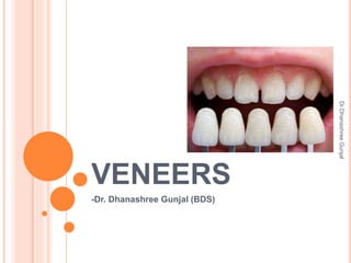
  • 1. VENEERS -Dr. Dhanashree Gunjal (BDS) Dr.Dhanashree Gunjal
  • 2. CONTENT  Definition  Indications  Contraindications  Types 1. Direct veneer technique 2. Indirect veneer technique  Repairs of veneers  Conclusion Dr.Dhanashree Gunjal
  • 3. DEFINITION  A veneer is a layer of tooth-colored material that is applied to a tooth to restore localized or generalized defects and intrinsic discolorations. (Sturdevant’s Art and Science of Operative Dentistry)  Typically, veneers are made of directly applied composite, processed composite, porcelain, or pressed ceramic materials. Dr.Dhanashree Gunjal
  • 4. INDICATIONS  Stained or darkened teeth  Hypocalcification  Closure of diastemas  Peg laterals  Chipped teeth  Lingual positioned teeth  Malposed teeth not requiring orthodontics  Fractured teeth  Esthetically compromised anterior teeth Dr.Dhanashree Gunjal
  • 5. CONTRAINDICATIONS  Insufficient tooth substrate (enamel for bonding)  Labial version  Excessive interdental spacing  Poor oral hygiene or caries  Parafunctional habits (clenching, bruxism)  Moderate to severe malposition or crowding Dr.Dhanashree Gunjal
  • 6. TYPES  Based on extent of tooth involved: 1) Partial veneers 2) Full veneers  Partial veneers are indicated for the restoration of localized defects or areas of intrinsic discoloration.  Full veneers are indicated for the restoration of generalized defects or areas of intrinsic staining involving most of the facial surface of the tooth. Dr.Dhanashree Gunjal
  • 7.  Full veneers can be accomplished by the direct or indirect technique.  Indirect veneers require two appointments but typically offer three advantages over directly placed full veneers: 1.Indirectly fabricated veneers are much less sensitive to operator technique. Considerable artistic expertise and attention to detail are required to consistently achieve esthetically pleasing and physiologically sound direct veneers. Indirect veneers are made by a laboratory technician and are typically more esthetic. 2. If multiple teeth are to be veneered, indirect veneers usually can be placed much more expeditiously. Dr.Dhanashree Gunjal
  • 8. 3. Indirect veneers typically last much longer than do direct veneers, especially if they are made of porcelain or pressed ceramic. Dr.Dhanashree Gunjal
  • 9. DIRECT VENEER TECHNIQUE I. Direct Partial Veneers  Small localized intrinsic discolorations or defects that are surrounded by healthy enamel are ideally treated with direct partial veneers.  These defects can be restored in one appointment with a light-cured composite.  Preliminary steps include cleaning, shade selection, and isolation with cotton rolls or rubber dam.  Anesthesia usually is not required unless the defect is deep, extending into dentin. Dr.Dhanashree Gunjal
  • 10.  The outline form is dictated solely by the extent of the defect and should include all discolored areas.  The clinician should use a coarse, elliptical or round diamond instrument with air-water coolant to remove the defect. The use of water-air spray is also imperative so that the tooth can be maintained in a hydrated state.  After preparation, etching, and restoration of the defective areas. A, Patient with overcontoured direct full veneers. B, After removal ofold veneer, localized white spots are evident. C, Models illustrate fault (x) and cavity preparation . The chamfered margins are irregular in outline. Dr.Dhanashree Gunjal
  • 11.  If the entire defect or stain is removed, a microilled composite is recommended for restoring the preparation.  Microfills are excellent “enamel replacement” materials because of their optical properties. If the tooth has been maintained in a hydrated state, the micro filled composite can be positioned on a trial basis to assess the accuracy of the shade prior to final restoration.  Nanofilled composites also are excellent material choices for this technique. D, Intraenamel preparations for partial veneer restorations. E, Conservative esthetic result of completed partial veneers. Dr.Dhanashree Gunjal
  • 12.  If a residual lightly stained area or white spot remains in enamel, however, an intrinsically less translucent composite can be used rather than extending the preparation into dentin to eliminate the defect.  Most composites filled primarily with radiopaque fillers (e.g., barium glass) are more optically opaque with intrinsic masking qualities.  Use of these types of composites for the restoration of preparations with light, residual stains is most effective and conserves the tooth structure.  In this example, all restorations are of a light-cured micro filled composite. Dr.Dhanashree Gunjal
  • 13. DIRECT FULL VENEERS  Extensive enamel hypoplasia involving all maxillary anterior teeth is treated by placing direct full veneers.  A direct technique is used with a light-cured microilled composite.  Placing direct full composite veneers is very time consuming.  Many dentists find that the preparation, placement, and finishing of several direct veneers at one time is too difficult, fatiguing, and time consuming. Some patients become uncomfortable and restless during long appointments.. Dr.Dhanashree Gunjal
  • 15. INDIRECT VENEER TECHNIQUE VENEER TECHNIQUE  Indirect veneers are primarily made of (1) Processed composite, (2) Feldspathic porcelain, and (3) Cast or pressed ceramic.  Feldspathic porcelain has superior strength, durability, and conservation of the tooth structure, bonded to intraenamel preparations has historically been the preferred approach for indirect veneering techniques.  Some pressed ceramic veneering materials offer comparable esthetic qualities but may require a deeper tooth preparation that is often located in dentin.  Indirect veneers are attached to the enamel by acid etching and bonding with light-cured resin cement. Dr.Dhanashree Gunjal
  • 16. ETCHED PORCELAIN VENEERS  The preferred type of indirect veneer is the etched porcelain (i.e.,feldspathic) veneer. Porcelain veneers etched with hydroluoric acid are capable of achieving high bond strengths to the etched enamel via a resin-bonding medium.  In addition to high bond strengths, etched porcelain veneers are highly esthetic, stain resistant, and periodontally compatible.  The incidence of cohesive fracture for etched porcelain veneers is also very low. Fig-Scanning electron micrograph (×31,000) of feldspathic porcelain etched with hydroluoric acid. (Courtesy Dr. Steven Bayne.) Dr.Dhanashree Gunjal
  • 17. First appointment Shade selection Tooth preparation Impression Temporary veneers Second appointment Removal temporary Clinical try-in Cementation Dr.Dhanashree Gunjal
  • 18. TOOTH PREPARATION  The veneer preparation is made with a tapered, rounded-end diamond instrument. It is critical that the tip diameter of the diamond be measured because the diamond will serve as the measuring tool in gauging proper reduction depth.  A diamond with a tip diameter of 1 to 1.2 mm is recommended. The tip diameter of the diamond used in this series is 1.2 mm. Dr.Dhanashree Gunjal
  • 19. Fig- Intraenamel preparation for an etched porcelain veneer with a butt-joint incisal edge design. A and B, The peripheral outline form is first established using a rounded-end diamond instrument. C–H, Facial reduction is achieved by first identifying and then reducing three separate facial zones: the incisal third, the middle third, and the gingival third (C), in that order. Dr.Dhanashree Gunjal
  • 20. TOOTH PREPARATION  Labial reduction  interproximal reduction  Incisal modification  Cervical definition  The first step in the veneer preparation is establishing the peripheral outline form.  Position the diamond to half its depth just facial to the proximal contact on either proximal surface, and then extend the bur, while maintaining its occlusogingival orientation, around the gingival area and then back up the opposite proximal area, again keeping the diamond positioned just facial to the proximal contact area. Dr.Dhanashree Gunjal
  • 21.  Facial reduction is achieved by first identifying and then reducing three separate facial zones: the incisal third, the middle third, and the gingival in that order.  Again, the tip of the diamond is used to gauge this reduction. Reduction depth can be verified by viewing the tip of the diamond in proximity to the unprepared tooth structure gingival to this reduced area when viewed from the proximal, facial, and incisal aspects.  Reduction of the gingival one third is straightforward and simply involves removal of the remaining “island” of unprepared tooth structure to a level consistent with the surrounding previously prepared tooth structure.  Incisal reduction is made by orienting the diamond perpendicular to the incisal edge and then reducing the incisal edge to attain a minimum reduction of 1 mm or, more desirably, 1.5 mm. Clinically, this reduction in depth will be gauged using an incisal reduction index. Dr.Dhanashree Gunjal
  • 22.  Finally, round the facioincisal line angle with the side of the diamond to reduce internal stresses in the porcelain veneer.  The final intraenamel preparation for an etched porcelain veneer using a butt-joint incisal edge design.  Frequently, an incisal-lapping preparation is indicated if the patient has worn or defective areas on the lingual aspect of the incisal edge.  The preparation steps for the incisal-lapping preparation are identical to those for the butt-joint design, including the steps for incisal reduction; however, additional steps are required to attain the incisal-lapping feature. Dr.Dhanashree Gunjal
  • 23. I, Incisal reduction is attained. J, The completed intraenamel preparation for an etched porcelain veneer with a butt-joint incisal edge. Dr.Dhanashree Gunjal
  • 24.  The first step in achieving this preparation design is to notch the mesial and distal incisal angles.  The tip of the same diamond instrument used for the earlier steps of the veneer preparation is used to establish these notches.  Using the diamond, extend the notches completely through the incisal angles faciolingually to a depth incisogingivally consistent with the desired amount of lapping of the lingual surface.  Once the incisal notches have been generated incisogingivally to a depth consistent with the desired amount of lingual lapping, the preparation of the lingual lap is made.  Position the diamond into the tooth to a depth of approximately 0.6 mm and extend the preparation across the lingual surface from notch to notch. Dr.Dhanashree Gunjal
  • 25.  The resulting sharp incisal angles must then be rounded to finish the incisal-lapping portion of the preparation. Care must be taken to include any desired lingual defect.  The gingival extent of the incisal lap is determined by the extent of any lingual defect.  The final lapping portion of the preparation is done. The facial view of the completed incisal-lapping preparation with a lingual lap of 0.5 mm is seen. Dr.Dhanashree Gunjal
  • 26. Fig- Intraenamel preparation steps for an etched porcelain veneer with an incisal-lapping design. A and B, Incisal notches. C, Preparing the lingual-lapping portion of the prep. D and E, The completed intraenamel preparation viewed from the lingual and facial aspects for an etched porcelain veneer with an incisal- lapping design. Dr.Dhanashree Gunjal
  • 28. Fig- Etched porcelain veneers using an intraenamel preparation. A, A patient with severe dental fluorosis. B, An incisal reduction index is made intraorally, since no significant change in incisal edge position is desired. C, Retraction cord is placed. D, The outline form is first established. E–G, Facial reduction is attained by using three zones of facial reduction. H, Incisal reduction is verified using the incisal reduction index. Dr.Dhanashree Gunjal
  • 32. I, Finished preparations for intraenamel preparations. Note the window preparations on canines and premolars. J, Retraction cord is placed for isolation. K, The it of the veneer is assessed. L and M, Etching of the prepared maxillary central incisors. N and O, Adhesive is applied to the etched enamel and the tooth side of the porcelain veneer. P, The veneer is loaded with resin cement and seated on the tooth. Dr.Dhanashree Gunjal
  • 33. Fig- Excess cement is removed with a microbrush. R, Excess cement is removed interproximally through removal of polyester strip. S, Resin cement cured with intense curing light. T, No. 12 surgical blade in a Bard- Parker handle is used for removing excess cured resin cement. U and V, Diamond instruments used to “dress” marginal areas. W and X, 30-luted carbide burs and diamond impregnated polishing instruments used to inish and polish veneer margins Dr.Dhanashree Gunjal
  • 34. Fig- Y and Z, Finished etched porcelain veneers as viewed from the lingual and facial aspects. Incisal reduction index made from a diagnostic model. A and B, A fast- set elastomeric material is used to record the lingual and incisal contours of the diagnostic model. C, Incisal reduction index is used to verify proper incisal preparation of teeth. D, Finished etched porcelain veneers. Dr.Dhanashree Gunjal
  • 35. PRESSED CERAMIC VENEERS  Pressed ceramic veneers are cast using a lost wax technique.  Excellent esthetics is possible using pressed ceramic materials for most cases involving mild to moderate discoloration. Because of the more translucent nature of pressed ceramic veneers, however, dark discolorations are best treated with etched porcelain veneers.  The procedures for tooth preparation, try-in, and bonding of pressed veneers are the same as for etched porcelain veneers except that the marginal it is superior. For that reason, often little marginal finishing is necessary. Only the excess bonding medium needs to be removed. Dr.Dhanashree Gunjal
  • 36. VENEER FOR METAL RE TORATION  Esthetic inserts (i.e., partial or full veneers) of a tooth- colored material can be placed on the facial surface of a tooth previously restored with a metal restoration.  For new castings, plans are made at the time of tooth preparation and during laboratory development of the wax pattern to incorporate a veneer into the cast restoration.  After such a casting has been cemented, the veneer can be inserted, as described in the next section, except that the portion of mechanical retention of the veneer into the casting has been provided in the wax pattern stage. Dr.Dhanashree Gunjal
  • 37. REPAIR OF VENEER  Failures of esthetic veneers occur because of breakage, discoloration, or wear.  Small chipped areas on veneers often can be corrected by recontouring and polishing.  When a sizable area is broken, it usually can be repaired if the remaining portion is sound. Dr.Dhanashree Gunjal
  • 38. For direct composite veneers,  repairs ideally should be made with the same material that was used originally.  After cleaning the area and selecting the shade, the operator should roughen the damaged surface of the veneer or tooth (or both) with a coarse, tapered, rounded-end diamond instrument to form a chamfered cavosurface margin.  Roughening with microetching (i.e., sandblasting) also is efective. For more positive retention, mechanical locks may be placed in the remaining composite material with a small, round bur.  Acid etchant is applied to clean the prepared area and to etch any exposed enamel, which is then rinsed and dried. Dr.Dhanashree Gunjal
  • 39.  Next, an adhesive is applied to the preparation (i.e., existing composite and enamel) and polymerized. Composite is added, cured, and finished in the usual manner A, Fractured veneer on the maxillary canine. B, Preparation with rounded-end diamond instrument. C, Undercuts placed in existing veneer with a No. 1/4 bur. D, Completed preparation is shown isolated and etched. E, Veneer restored to original color and contour. Dr.Dhanashree Gunjal
  • 40. For porcelain veneers,  Repair is done by a hydroluoric acid gel, suitable for intraoral use (but only with a rubber dam in place), must be used to etch the fractured porcelain.  Hydroluoric acid gels are available in approximately 10% bufered concentrations that can be used for intraoral porcelain repairs if proper isolation with a rubber dam is used.  Although caution still must be taken when using hydroluoric acid gels intraorally, the lower acid concentration allows for relatively safe intraoral use.  Full-strength hydroluoric acid should never be used intraorally for etching porcelain. Isolation of the porcelain veneer to be repaired should always be accomplished with a rubber dam to protect gingival tissue from the irritating efects of the hydroluoric acid. Dr.Dhanashree Gunjal
  • 41.  A lightly frosted appearance, similar to that of etched enamel, should be seen if the porcelain has been properly etched.  A silane coupling agent may be applied to the etched porcelain surface before the adhesive is applied.  Composite material is added, cured, and inished in the usual manner.  Large fractures are best treated by replacing the entire porcelain veneer. Dr.Dhanashree Gunjal