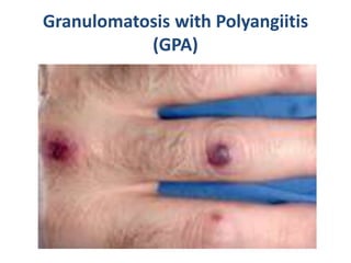
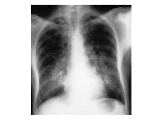
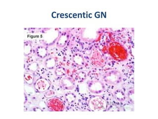
Vasculitis is characterized by inflammation of blood vessels. It can be primary or secondary to other diseases. Primary vasculitis syndromes include Granulomatosis with Polyangiitis (GPA, also known as Wegener's), which involves necrotizing granulomatous inflammation and vasculitis that commonly affects the lungs and kidneys. Diagnosis of GPA involves laboratory tests, biopsy of affected tissues showing vasculitis and granulomas, and testing positive for antineutrophil cytoplasmic antibodies (ANCA). Treatment is with corticosteroids and immunosuppressants to induce remission.
Vasculitis refers to a group of diseases characterized by inflammation of blood vessels. The document defines and classifies different types of vasculitis based on vessel size. It discusses the pathophysiology, clinical features, investigations and management of vasculitis. Giant cell arteritis is provided as an example of large vessel vasculitis that predominantly affects branches of the temporal and ophthalmic arteries in older individuals, with headaches, jaw pain and risk of vision loss as key clinical features.
The document outlines a 6 step approach to diagnosing and treating vasculitis:
1. Recognize features of vasculitis like purpura or organ ischemia.
2. Rule out secondary causes through tests for infections and malignancies.
3. Determine the size of blood vessels involved - large, medium, or small.
4. Consider characteristic presentations of primary vasculitides like temporal arteritis or Wegener's.
5. Make diagnoses through tests like ANCA, biopsy of affected tissues, and angiography.
6. Treat based on severity, using immunosuppression, addressing underlying causes, and monitoring for side effects.
Vasculitis refers to inflammation of blood vessels. This document discusses the approach to diagnosing and classifying different types of vasculitis. It describes:
1) The pathological changes in vasculitis which include thinning of vessel walls and narrowing/occlusion of affected vessels.
2) The different mechanisms that can cause vasculitis including immune complex formation, ANCA mediated, and T lymphocyte mediated responses.
3) How to classify vasculitis into primary disorders or those secondary to other medical conditions. Primary vasculitis are further broken down by vessel size affected.
4) The steps involved in evaluating a patient with suspected vasculitis which includes screening for mimics, assessing organ involvement
1. The document outlines a 6 step approach to diagnosing and treating vasculitis.
2. Step 1 is to learn to recognize vasculitis based on common features like purpura, pulmonary infiltrates, glomerulonephritis.
3. Step 2 is to rule out secondary causes of vasculitis like infections, malignancies, drugs.
4. Step 3 involves determining the pattern of vessel involvement - large, medium, or small vessels.
Vasculitis refers to inflammation of blood vessels. There are many types classified by the size of vessels involved and pathogenic mechanisms. Noninfectious vasculitis can be immune complex-mediated, associated with ANCAs, anti-endothelial cell antibodies, or autoreactive T cells. Infectious vasculitis involves direct invasion of vessels by pathogens. Common noninfectious vasculitides include giant cell arteritis, Takayasu arteritis, polyarteritis nodosa, Kawasaki disease, microscopic polyangiitis, Wegener's granulomatosis, Churg-Strauss syndrome, and Buerger's disease. Clinical manifestations depend on the organs involved. Treatment involves immunos
This document discusses gastrointestinal (GIT) vasculitis. It begins by noting that GIT vasculitis can affect blood vessels of all sizes and cause local or diffuse pathological changes in the GIT. Clinical features include ulcers, edema, hemorrhage, bowel ischemia, and perforation. The document then discusses various types of vasculitis that can involve the GIT, including large vessel vasculitis like Takayasu arteritis, medium vessel vasculitis like polyarteritis nodosa, and small vessel vasculitis like ANCA-associated vasculitis. Specific GIT manifestations of different types of vasculitis are described. The document emphasizes that diagnosis requires clinical suspicion along with radiological and histological confirmation via endoscopy
This document provides an overview of glomerular disease, including the pathogenesis, clinical evaluation, and treatment. It begins with an introduction to glomerular anatomy and physiology. Common causes of glomerular disease include genetic mutations, infections, autoimmunity, and atherosclerosis. Clinical presentations range from asymptomatic urine abnormalities to nephrotic syndrome. Evaluation involves history, physical exam, urine analysis, renal function tests, and sometimes renal biopsy. Main treatment approaches depend on the specific glomerular disease and include controlling hypertension and proteinuria, immunosuppression, and dialysis. Complications can include renal failure and chronic kidney disease if not properly treated.
Vasculitis refers to a group of disorders characterized by inflammation of blood vessels. The two main pathogenic mechanisms are immune-mediated inflammation and direct infection of blood vessels. Vasculitides are classified by the size of vessels affected, such as large-vessel vasculitis including giant cell arteritis, or small-vessel vasculitis including Wegener's granulomatosis. Diagnosis involves clinical features along with tests like ANCA. Treatment depends on the specific condition but often involves steroids and other immunosuppressants.
This document provides information on mixed connective tissue disease (MCTD). It discusses the definition, etiology, pathophysiology, diagnosis, treatment and prognosis of MCTD. MCTD is a rare autoimmune disease with overlapping features of at least two connective tissue diseases like SLE, SSc, PM and DM. It is characterized by the presence of anti-U1 RNP antibodies. Symptoms can affect multiple organ systems. Diagnosis involves assessing clinical features and antibody levels. Treatment aims to control symptoms and is tailored based on organ involvement. Prognosis varies, with some patients experiencing complete resolution while others face life-threatening complications like pulmonary hypertension.
Glomerular diseases are a heterogeneous group of kidney disorders that can be primary, secondary, or hereditary in nature. They involve pathological changes to the glomeruli, such as injury to the mesangial cells, endothelial cells, basement membrane, or podocytes. This can lead to clinical manifestations like proteinuria, hematuria, edema, hypertension, and renal failure. The mechanisms of glomerular injury are complex, often involving the immune system through immune complex deposition, complement activation, or cytotoxic antibodies. Glomerular diseases have diverse clinical presentations and outcomes depending on the specific cause and severity of involvement.
VEXAS syndromes , a diagnostic PuzzlepptxMarwa Besar
This document discusses VEXAS syndrome, a recently discovered autoinflammatory and hematologic condition caused by somatic mutations in the UBA1 gene. It provides details on the clinical manifestations (fevers, inflammatory symptoms affecting skin, joints, lungs, blood vessels), hematological abnormalities (anemia, cytopenias), and challenges in management due to the combined inflammatory and hematological involvement. Diagnosis is confirmed via genetic testing demonstrating UBA1 mutations. Treatment aims to suppress inflammation and eliminate mutant hematopoietic cells, but outcomes remain poor with high mortality.
- DRESS syndrome, also known as drug-induced hypersensitivity syndrome, is a severe cutaneous adverse reaction caused by certain medications. It is characterized by a rash, fever, lymphadenopathy, and involvement of internal organs like the liver, kidneys, lungs and heart.
- The onset of symptoms occurs 2-6 weeks after starting the culprit drug. Affected individuals typically develop a widespread, often purpuric rash along with systemic inflammatory signs. The liver and blood counts are often impacted.
- Diagnosis is based on clinical features and ruling out other conditions. Corticosteroids are the primary treatment but other immunosuppressants may be needed depending on severity. Identifying and stopping the
Vasculitis is inflammation of blood vessels that can involve different vessel sizes and patterns. It is characterized by leukocytic infiltration of vessel walls. The main causes are immune complex formation, ANCA mediated, and T lymphocyte mediated processes. Diagnosis involves evaluating clinical features, lab tests like ANCA and complement levels, and biopsy of affected tissues. Treatment focuses on immunosuppression with glucocorticoids and other agents depending on severity and organ involvement. Vasculitis often follows a chronic relapsing course.
The document provides information on different types of vasculitis:
[1] Any blood vessel can be affected by vasculitis, causing inflammation that leads to occlusion, aneurysm, ischemia and hemorrhage. The small vessel vasculitides include ANCA-associated disorders like Wegener's granulomatosis and types mediated by immune complexes.
[2] The approach to a patient with vasculitis involves considering signs and symptoms, performing tests and biopsies of affected organs to determine which organ systems are involved and the extent of involvement. Treatment depends on the specific type of vasculitis.
[3] Examples of specific vasculitides discussed include giant cell arteritis, poly
This document provides an overview of different types of vasculitis. It discusses the pathogenesis of vasculitis including immune complex formation and ANCA-mediated mechanisms. It then describes the clinical presentations and characteristics of various vasculitides that involve different vessel sizes such as giant cell arteritis, Takayasu's arteritis, polyarteritis nodosa, Kawasaki disease, ANCA-associated vasculitis (granulomatosis with polyangiitis, microscopic polyangiitis, Churg-Strauss syndrome), cutaneous small vessel vasculitis, and IgA vasculitis. It provides details on the patterns of organ involvement, diagnostic criteria and clinical course for each type of vasculitis.
This document provides information on acute glomerulonephritis (GN), including:
- GN is inflammation of the glomeruli that can cause hematuria, proteinuria, hypertension, and edema.
- It has an immunologically-mediated injury to glomeruli and renal interstitial damage.
- Common causes include post-streptococcal GN, lupus nephritis, ANCA vasculitis, and anti-glomerular basement membrane disease.
- Presentation, pathogenesis, treatment, and prognosis are described for different types of acute GN.
This document discusses several types of vasculitis:
1. Giant cell arteritis affects large arteries in the head and is characterized by granulomatous inflammation and headache along the temporal artery.
2. Polyarteritis nodosa involves medium and small arteries throughout the body and causes tissue ischemia. It is associated with hepatitis B/C.
3. Wegener's granulomatosis is defined by necrotizing granulomas of the respiratory tract, necrotizing vasculitis of small vessels, and renal disease. It is linked to C-ANCA antibodies.
4. Leukocytoclastic vasculitis mainly involves small skin blood vessels and is characterized by skin purpura
Non-infectious ocular inflammatory diseases by Dr. Iddi.pptxIddi Ndyabawe
This document provides an overview of various non-infectious ocular inflammatory diseases:
- Non-infectious scleritis can be caused by autoimmunity or trauma and is typically painful. Treatment involves corticosteroids and immunosuppressants.
- Acute anterior uveitis is the most common form of uveitis and often associated with HLA-B27 related diseases like ankylosing spondylitis. Corticosteroids are the main treatment.
- Post-operative inflammation can occur due to retained lens material or mechanical irritation from IOLs. Removal of causative material and corticosteroids are used.
Inflammatory disorders of the blood vessels, known as vasculitides, can affect arteries, veins, and capillaries through various immunological mechanisms. Three key types are summarized: (1) Large vessel vasculitis like giant cell arteritis typically involves the temporal artery and causes headaches and vision problems. (2) Medium vessel vasculitis such as polyarteritis nodosa involves arteries in organs like the kidneys and skin. (3) Small vessel vasculitis often presents as palpable purpura on the skin such as in Henoch-Schönlein purpura, a type of vasculitis more common in children.
Lupus nephritis is an inflammation of the kidneys caused by systemic lupus erythematosus (SLE), an autoimmune disease. Approximately 10-30% of SLE patients develop lupus nephritis, which can progress to end-stage renal disease requiring dialysis or transplantation if not properly treated. Lupus nephritis is classified into six classes based on pathological findings. Treatment involves medications to suppress the immune system such as corticosteroids, immunosuppressants, and biologic drugs depending on the class of lupus nephritis. Prognosis is generally good if treatment can control proteinuria, hypertension, and renal dysfunction.
Glomerulonephritis refers to a group of disorders involving inflammation of the glomeruli. It can be classified based on clinical presentation, histological appearance, etiology, and other factors. Common types include post-infectious glomerulonephritis, lupus nephritis, anti-GBM disease, IgA nephropathy, and rapidly progressive glomerulonephritis. The pathogenesis involves immunological injury to the glomerular capillary wall that can lead to scarring, loss of filtration, and decreased kidney function over time if not properly treated.
This document discusses sexually transmitted infections (STIs) caused by Treponema pallidum (syphilis), Neisseria gonorrhoeae (gonorrhea), and Chlamydia trachomatis (chlamydia). It describes the clinical manifestations of primary, secondary, and late syphilis. It also discusses gonococcal and chlamydial infections in males and females, including urethritis, cervicitis, pelvic inflammatory disease, and disseminated gonococcal infection. The document provides details on laboratory testing, treatment, and clinical presentation of these common STIs.
Systemic sclerosis (SSc) is an autoimmune disease characterized by fibrosis of the skin and internal organs. It results from autoimmune dysfunction leading to accumulation of T cells and cytokines in the skin that stimulate collagen deposition by fibroblasts. There are two main types: limited cutaneous SSc affects only the hands and forearms, while diffuse cutaneous SSc affects the skin over much of the body. SSc most commonly affects women ages 30-50 and can involve serious complications like scleroderma renal crisis resulting in high blood pressure, kidney damage, and other symptoms. Diagnosis involves clinical signs and presence of autoantibodies, and treatment depends on complications but may include ACE inhibitors for renal problems. Mixed connect
Vasculitis refers to inflammation of blood vessels. This document discusses the pathophysiology, classification, clinical presentation, diagnosis, and treatment of various types of vasculitis. The main types include large vessel vasculitis (e.g. giant cell arteritis, Takayasu arteritis), medium vessel vasculitis (e.g. polyarteritis nodosa, Kawasaki disease), small vessel vasculitis (ANCA-associated vasculitis like granulomatosis with polyangiitis, microscopic polyangiitis, eosinophilic granulomatosis with polyangiitis), and immune complex small vessel vasculitis (e.g. IgA vasculitis, antiglomer
Vasculitis is a condition characterized by inflammation and damage to blood vessels. There are several types of vasculitis classified by the size of vessels affected (large, medium, small). Imaging plays an important role in the diagnosis and monitoring of vasculitis by detecting vessel wall abnormalities and inflammation before lumen changes occur on angiography. Techniques like CT angiography, MRI, MRA, and PET are useful for revealing vessel wall alterations and inflammation. The choice of imaging depends on the suspected organ involvement.
1. Vasculitis is defined as inflammation of blood vessel walls and can be classified by the size of vessels involved - large, medium, or small.
2. Some examples of large vessel vasculitis include giant-cell arteritis, which commonly involves the temporal artery in older patients, and Takayasu arteritis, which usually occurs in younger patients.
3. Medium vessel vasculitis encompasses diseases like polyarteritis nodosa, characterized by necrotizing inflammation of renal arteries, and Kawasaki disease, an arteritis that can cause coronary aneurysms in children.
4. Small vessel vasculitis includes Wegener's granulomatosis, Churg-Stra
This document provides an overview of glomerular diseases. It begins by describing the anatomy of the nephron and glomerulus. It then discusses the cells that make up the glomerular filtration membrane and defines glomerular diseases as abnormalities in glomerular function caused by damage to glomerular components like the epithelium, basement membrane, or endothelium. Primary and secondary glomerular diseases are distinguished. Immunological and non-immunological mechanisms of glomerular injury are also summarized. Specific glomerular diseases like membranoproliferative glomerulonephritis, minimal change disease, focal segmental glomerulosclerosis, membranous glomerulonephritis, IgA nep
Basavarajeeyam is a Sreshta Sangraha grantha (Compiled book ), written by Neelkanta kotturu Basavaraja Virachita. It contains 25 Prakaranas, First 24 Chapters related to Rogas& 25th to Rasadravyas.
Here is the updated list of Top Best Ayurvedic medicine for Gas and Indigestion and those are Gas-O-Go Syp for Dyspepsia | Lavizyme Syrup for Acidity | Yumzyme Hepatoprotective Capsules etc
Histololgy of Female Reproductive System.pptxAyeshaZaid1
Dive into an in-depth exploration of the histological structure of female reproductive system with this comprehensive lecture. Presented by Dr. Ayesha Irfan, Assistant Professor of Anatomy, this presentation covers the Gross anatomy and functional histology of the female reproductive organs. Ideal for students, educators, and anyone interested in medical science, this lecture provides clear explanations, detailed diagrams, and valuable insights into female reproductive system. Enhance your knowledge and understanding of this essential aspect of human biology.
8 Surprising Reasons To Meditate 40 Minutes A Day That Can Change Your Life.pptxHolistified Wellness
We’re talking about Vedic Meditation, a form of meditation that has been around for at least 5,000 years. Back then, the people who lived in the Indus Valley, now known as India and Pakistan, practised meditation as a fundamental part of daily life. This knowledge that has given us yoga and Ayurveda, was known as Veda, hence the name Vedic. And though there are some written records, the practice has been passed down verbally from generation to generation.
Muktapishti is a traditional Ayurvedic preparation made from Shoditha Mukta (Purified Pearl), is believed to help regulate thyroid function and reduce symptoms of hyperthyroidism due to its cooling and balancing properties. Clinical evidence on its efficacy remains limited, necessitating further research to validate its therapeutic benefits.
Rasamanikya is a excellent preparation in the field of Rasashastra, it is used in various Kushtha Roga, Shwasa, Vicharchika, Bhagandara, Vatarakta, and Phiranga Roga. In this article Preparation& Comparative analytical profile for both Formulationon i.e Rasamanikya prepared by Kushmanda swarasa & Churnodhaka Shodita Haratala. The study aims to provide insights into the comparative efficacy and analytical aspects of these formulations for enhanced therapeutic outcomes.
Cell Therapy Expansion and Challenges in Autoimmune DiseaseHealth Advances
There is increasing confidence that cell therapies will soon play a role in the treatment of autoimmune disorders, but the extent of this impact remains to be seen. Early readouts on autologous CAR-Ts in lupus are encouraging, but manufacturing and cost limitations are likely to restrict access to highly refractory patients. Allogeneic CAR-Ts have the potential to broaden access to earlier lines of treatment due to their inherent cost benefits, however they will need to demonstrate comparable or improved efficacy to established modalities.
In addition to infrastructure and capacity constraints, CAR-Ts face a very different risk-benefit dynamic in autoimmune compared to oncology, highlighting the need for tolerable therapies with low adverse event risk. CAR-NK and Treg-based therapies are also being developed in certain autoimmune disorders and may demonstrate favorable safety profiles. Several novel non-cell therapies such as bispecific antibodies, nanobodies, and RNAi drugs, may also offer future alternative competitive solutions with variable value propositions.
Widespread adoption of cell therapies will not only require strong efficacy and safety data, but also adapted pricing and access strategies. At oncology-based price points, CAR-Ts are unlikely to achieve broad market access in autoimmune disorders, with eligible patient populations that are potentially orders of magnitude greater than the number of currently addressable cancer patients. Developers have made strides towards reducing cell therapy COGS while improving manufacturing efficiency, but payors will inevitably restrict access until more sustainable pricing is achieved.
Despite these headwinds, industry leaders and investors remain confident that cell therapies are poised to address significant unmet need in patients suffering from autoimmune disorders. However, the extent of this impact on the treatment landscape remains to be seen, as the industry rapidly approaches an inflection point.
Promoting Wellbeing - Applied Social Psychology - Psychology SuperNotesPsychoTech Services
A proprietary approach developed by bringing together the best of learning theories from Psychology, design principles from the world of visualization, and pedagogical methods from over a decade of training experience, that enables you to: Learn better, faster!
TEST BANK For Basic and Clinical Pharmacology, 14th Edition by Bertram G. Kat...rightmanforbloodline
TEST BANK For Basic and Clinical Pharmacology, 14th Edition by Bertram G. Katzung, Verified Chapters 1 - 66, Complete Newest Version.
2. Vasculitis
• DEFINITION AND PATHOGENESIS
• A clinicopathologic process characterized by inflammation
of and damage to blood vessels, compromise of vessel
lumen, and resulting ischemia.
• Clinical manifestations depend on size and location of
affected vessel.
• Most vasculitic syndromes appear to be mediated by
immunemechanisms.
• May be primary or sole manifestation of a disease or
secondary to another disease process.
• Unique vasculitic syndromes can differ greatly with regards
to clinical features, disease severity, histology, and
treatment.
3. PRIMARY VASCULITIS SYNDROMES
• Churg-Strauss Syndrome
• Granulomatous vasculitis of multiple organ
systems, particularly the lung; characterized by
asthma, peripheral eosinophilia, eosinophilic
tissue infiltration; glomerulonephritis can occur.
• Polyarteritis Nodosa (PAN)
• Medium-sized muscular arteries involved;
frequently associated with arteriographic
aneurysms; commonly affects renal arteries, liver,
GI tract, peripheral nerves, skin, heart; can be
associated with hepatitis B.
4. PRIMARY VASCULITIS SYNDROMES
• Microscopic Polyangiitis
• Small-vessel vasculitis that can affect the glomerulus
and lungs; mediumsized vessels also may be affected.
• Giant Cell Arteritis
• Inflammation of medium- and large-sized arteries;
primarily involves temporal artery but systemic and
large vessel involvement may occur; symptoms include
headache, jaw/tongue claudication, scalp tenderness,
fever, musculoskeletal symptoms (polymyalgia
rheumatica); sudden blindness from involvement of
optic vessels is a dreaded complication.
5. PRIMARY VASCULITIS SYNDROMES
• Takayasu’s Arteritis
• Vasculitis of the large arteries with strong
predilection for aortic arch and its branches; most
common in young women; presents with
inflammatory or ischemic symptoms in arms and
neck, systemic inflammatory symptoms, aortic
regurgitation.
• Henoch-Schönlein Purpura
• Characterized by involvement of skin, GI tract,
kidneys; more common in children; may recur
after initial remission.
6. PRIMARY VASCULITIS SYNDROMES
• Cryoglobulinemic Vasculitis
• Majority of cases are associated with hepatitis C where
an aberrant immune response leads to formation of
cryoglobulin; characterized by cutaneous vasculitis,
arthritis, peripheral neuropathy, and
glomerulonephritis.
• Idiopathic Cutaneous Vasculitis
• Cutaneous vasculitis is defined broadly as inflammation
of the blood vessels of the dermis; due to underlying
disease in >70% of cases (see “Secondary Vasculitis
Syndromes,” below) with 30% occurring idiopathically.
7. PRIMARY VASCULITIS SYNDROMES
• Miscellaneous Vasculitic Syndromes
• Kawasaki disease (mucocutaneous lymph
node syndrome)
• Isolated vasculitis of the central nervous
system
• Behçet’s syndrome
• Cogan’s syndrome
• Polyangiitis overlap syndrome
9. Vasculitis
• EVALUATION
• Thorough Hx and physical exam—special
reference to ischemic manifestations and
systemic inflammatory signs/symptoms.
• Laboratories—important in assessing organ
involvement: CBC with differential, ESR, renal
function tests, UA.
• Should also be obtained to rule out other
diseases: ANA, rheumatoid factor, anti-GBM,
hepatitis B/C serologies, HIV.
10. Vasculitis
• EVALUATION
• Antineutrophil cytoplasmic autoantibodies (ANCA)—associated
with granulomatosis with polyangiitis (Wegener’s), microscopic
polyangiitis, and some pts with Churg-Strauss syndrome; presence
of ANCA is adjunctive and should not be used in place of biopsy as a
means of diagnosis or to guide treatment decisions.
• Radiographs—CXR should be performed even in the absence of
symptoms.
• Diagnosis—can usually be made only by arteriogram or biopsy of
affected organ(s).
• DIFFERENTIAL DIAGNOSIS
• Guided by organ manifestations. In many instances includes
infections and neoplasms, which must be ruled out prior to
beginning immunosuppressive therapy. Consideration must also be
given for diseases that can mimic vasculitis
14. Vasculitis
• TREATMENT
• Therapy is based on the specific vasculitic
syndrome and the severity of its manifestations.
• Immunosuppressive therapy should be avoided in
disease that rarely results in irreversible organ
system dysfunction or that usually does not
respond to such agents (e.g., isolated cutaneous
vasculitis).
• Antiviral agents play an important role in treating
vasculitis occurring with hepatitis B or C.
15. Vasculitis
• TREATMENT
• Glucocorticoids alone may control giant cell
arteritis and Takayasu’s arteritis.
• Therapy that combines glucocorticoids with
another immunosuppressive agent is
particularly important in syndromes with life-
threatening organ system involvement,
especially active glomerulonephritis.
16. Vasculitis
• TREATMENT
• Frequently used agents:
• Prednisone
• Cyclophosphamide
• Rituximab
• Methotrexate
• Azathioprine
• Mycophenolate mofetil
• Plasmapheresis may have an adjunctive role in
rapidly progressive glomerulonephritis.
17. Granulomatosis with Polyangiitis (GPA)
(Wegener's Granulomatosis)
• Granulomatosis with polyangiitis is characterized by necrotizing
granulomatous inflammation, small- and medium-sized vessel
vasculitis, and focal necrotizing glomerulonephritis, often with
crescent formation.
• Typically, the upper and lower respiratory tract and the kidneys are
affected, but any organ may be.
• Symptoms vary depending on the organs and systems affected.
Patients may present with upper and lower respiratory tract
symptoms (eg, recurrent nasal discharge or epistaxis, cough),
followed by hypertension and edema, or with symptoms reflecting
multiorgan involvement.
• Diagnosis usually requires biopsy.
• Treatment is with corticosteroids plus an immunosuppressant.
Remission is usually possible, although relapses are common.
18. Granulomatosis with Polyangiitis (GPA)
(Wegener's Granulomatosis)
• Granulomatosis with polyangiitis (GPA) occurs
in about 1/25,000 people; it is most common
among whites but can occur in all ethnic
groups and at any age. Mean age at onset is
40.
• The cause of GPA is unknown, although
immunologic mechanisms play a role. Most
patients with active generalized disease have
antineutrophil cytoplasmic antibodies (ANCA).
19. Pathophysiology
• Characteristically, granulomas form with histiocytic
epithelioid cells and often with giant cells. Plasma cells,
lymphocytes, neutrophils, and eosinophils are present.
• Inflammation affects tissues as well as vessels; vasculitis
may be a small or large component of the disease.
• Micronecrosis, usually with neutrophils (microabscesses),
occurs early. Micronecrosis progresses to macronecrosis.
• A central area of necrosis (called geographic necrosis) is
rimmed by lymphocytes, plasma cells, macrophages, and
giant cells. A zone of fibroblastic proliferation with
palisading histiocytes may surround the area.
20. Pathophysiology
• Nonspecific chronic inflammation and tissue necrosis
occur in the nose.
• The lungs are most likely to display the full spectrum of
histopathologic abnormalities, but diagnostic features
are not typically identified on the small tissue samples
obtained by transbronchial biopsy.
• In the kidneys, the most common finding is a pauci-
immune crescentic focal glomerulonephritis with
necrosis and thrombosis of individual loops or larger
segments of the glomerulus. Vasculitic lesions and
disseminated granulomas occur only occasionally.
21. Histologic pattern
• May not correlate with the
clinical presentation
• Various histological types of
glomerulonephritis
23. Symptoms and Signs
• Onset of granulomatosis with polyangiitis may be insidious or
acute; the full spectrum of the disease may take years to evolve.
• Some patients present initially with upper and lower respiratory
tract symptoms; at some point later, the kidneys are affected.
• In other patients, onset of systemic manifestations is relatively
acute; several organs and systems, such as the upper respiratory
tract, peripheral nervous system (causing multiple
mononeuropathy [mononeuritis multiplex]), kidneys (causing
glomerulonephritis), and lower respiratory tract (causing
hemorrhage,
• lung nodules, cavities, or a combination), are simultaneously
affected.
24. Symptoms and Signs
• Upper respiratory tract:
• Sinus pain, serosanguineous or purulent discharge, and
epistaxis may occur.
• The mucosa appears granular (like cobblestones) and is
friable; ulcers, thick dark crusts, and septal perforation are
common.
• Nasal chondritis can occur with swelling, pain, and collapse of
the nasal bridge (saddle nose).
• Patients may report recurrent sinusitis that has responded
inadequately to multiple antibiotic regimens and has required
one or more sinus operations before diagnosis.
• Secondary infections (eg, due to Staphylococcus aureus) may
develop. Subglottic stenosis may develop, causing symptoms
such as pain in the larynx, hoarseness, dyspnea, wheezing,
and stridor.
25.
26. Symptoms and Signs
• Ears:
• Otitis, sensorineural hearing loss, vertigo, and chondritis
may occur. The middle ear, inner ear, and mastoids are
often affected.
• Eyes:
• Eyes may appear red and swollen. Nasolacrimal duct
inflammation and obstruction, conjunctivitis, scleritis,
uveitis, or retinal vasculitis may also occur. Inflammatory
infiltrates in the retro-orbital space (orbital pseudotumor)
can cause proptosis, compression of the optic nerve, and
blindness. Extension into the extraocular muscles leads to
diplopia. If serious eye symptoms develop, evaluation and
treatment are required immediately to prevent permanent
vision loss.
27.
28. Symptoms and Signs
• Lower respiratory tract:
• Respiratory manifestations are common. Inflammation of
the major bronchi and branches can cause localized
wheezing, postobstructive pneumonia, and atelectasis.
Single or multiple pulmonary nodules, with or without
cavitation, and parenchymal infiltrates, sometimes cause
symptoms, such as chest pain, shortness of breath, and
productive cough. Dyspnea with bilateral infiltrates, with
or without hemoptysis, may indicate alveolar hemorrhage
and must be evaluated immediately.
• Heart: Coronary artery disease may occur but rarely.
• Musculoskeletal system: Patients frequently present with
myalgias, arthralgias, or nonerosive inflammatory arthritis.
29.
30.
31.
32. Symptoms and Signs
• Skin: Palpable purpura, tender subcutaneous nodules, papules,
livedo reticularis, or ulcers may develop.
• Nervous system: Vasculitis may cause ischemic peripheral
neuropathy, brain lesions, or extension of lesions from contiguous
sites. Lesions that originate in the sinuses or middle ear may extend
directly to the retropharyngeal area and base of the skull, leading to
cranial neuropathy, proptosis, diabetes insipidus, or meningitis.
• Kidneys: Symptoms and signs of glomerulonephritis develop.
Urinary sediment is frequently abnormal, and serum creatinine may
increase rapidly. Edema and hypertension may result. Rapidly
progressive glomerulonephritis, which is life threatening, can
develop.
• Venous system: Deep venous thrombosis can affect the lower
extremities mostly when granulomatosis with polyangiitis is active.
• Other organs: Occasionally, an inflammatory mass occurs in the
breasts, kidneys, prostate, or other organs.
35. Diagnosis
• Routine laboratory tests, including urinalysis
• Tests for antineutrophil cytoplasmic antibodies
• Biopsy for definitive diagnosis
• Granulomatosis with polyangiitis should be
suspected in patients with chronic, unexplained
respiratory symptoms and signs (including otitis
media in adults), particularly if manifestations in
other organ systems, especially the kidneys, also
suggest the disorder. Routine laboratory tests are
done, but ANCA testing and biopsy yield the
most specific findings.
36. Diagnosis
• Routine laboratory tests include:
• ESR,
• C-reactive protein,
• CBC with differential,
• Serum albumin and total protein,
• Serum creatinine,
• Urinalysis,
• 24-hour urine protein,
• Chest x-ray.
• Chest CT without contrast is nearly always necessary because the chest x-
ray may miss nodules, masses, and/or cavitary lesions caused by
granulomatosis with polyangiitis.
• In most patients with active disease, ESR and C-reactive protein are
elevated, and serum albumin and total protein are decreased; anemia,
thrombocytosis, and mild-to-moderate eosinophilia are detected.
• Dysmorphic RBCs and RBC casts, detected during urinalysis, indicate
glomerular involvement. Proteinuria may be detected. Serum creatinine
may be increased.
37. Diagnosis
• Serologic testing to detect antineutrophil
cytoplasmic antibodies (ANCA) is followed by
enzyme-linked immunosorbent assay (ELISA)
to check for specific antibodies.
• Most patients with active disease have
cytoplasmic ANCA (c-ANCA), with antibodies
against proteinase-3 (PR3); these findings plus
characteristic clinical findings suggest GPA.
38. Diagnosis
• Some patients with other disorders (eg, bacterial
endocarditis, cocaine abuse, systemic lupus erythematosus,
amebiasis, tuberculosis) test positive for ANCA.
• Because tests for rare diseases are likely to be falsely
positive when ordered for the general population and the
positive predictive value of a positive ANCA test is around
50%,
• ANCA testing should be reserved for patients in whom the
pretest probability for GPA or another ANCA-associated
vasculitis is at least moderately high (eg, patients with
alveolar hemorrhage, glomerulonephritis, or multiple
mononeuropathy plus other features of microscopic
polyangiitis or GPA).
39. Diagnosis
• A positive ANCA test does not rule out mycobacterial and
fungal infections; thus, patients with positive ANCA results
and cavitary lung lesions still require bronchoscopy and
adequate cultures and other tests for tuberculosis and
fungal infections.
• ANCA testing (titre) should not be used to guide
subsequent treatment.
• During apparent remission, ANCA may increase or ANCA
test results may change from negative to positive.
• In some of these patients, symptoms do not recur; in
others, symptoms recur or worsen soon after the test is
done or during the next few weeks, months, or sometimes
years.
40. Diagnosis
• Biopsy should be done if possible to confirm the diagnosis of GPA.
• Clinically abnormal sites may be biopsied first.
• Biopsy of affected lung tissue is most likely to reveal characteristic
findings; open thoracotomy provides the best access.
• Biopsies of lung or sinus tissue are cultured to exclude infection.
• Renal biopsy that shows pauci-immune necrotizing focal crescentic
or noncrescentic glomerulonephritis strongly supports the
diagnosis.
• Biopsy results of various tissues may also provide histologic
information that can help guide treatment (eg, renal fibrosis).
• Differential diagnosis includes other vasculitic disorders that affect
small- and medium-sized vessels.
• Infections, especially those due to slow-growing fungi or acid-fast
organisms, should be ruled out by staining and culture of the
sampled tissues.
•
41. Treatment
• To induce remission in life- or organ-threatening GPA, high-
dose corticosteroids plus
either cyclophosphamide or rituximab
• To induce remission in less severe GPA, corticosteroids and
either methotrexate or rituximab
• To maintain remission, rituximab alone or another drug
such as methotrexate, azathioprine,
or mycophenolate mofetil (rituximab plus another of these
drugs, sometimes with a low dose of a corticosteroid, if
patients have multiple relapses or GPA is difficult to control)
• Kidney transplantation if necessary.
• Treatment of granulomatosis with polyangiitis depends on
the severity of disease. A multidisciplinary approach is
required for multiorgan disease, often including a
rheumatologist, otorhinolaryngologist, pulmonologist, and
nephrologist.
42. Treatment
• Patients who have severe life-threatening or organ-
threatening manifestations (eg, alveolar hemorrhage,
rapidly progressive glomerulonephritis, multiple
mononeuropathy with motor involvement) require
immediate hospital admission for treatment to induce
remission. These patients require high-dose corticosteroids
and cyclophosphamide or rituximab.
• Efficacies of rituximab and cyclophosphamide appear to be
similar for inducing and maintaining remission.
• Although the evidence supporting use of plasma exchange
is weaker than that for the other interventions, plasma
exchange can be added to the standard treatment regimen
in patients with severe acute renal insufficiency
(particularly if the anti–glomerular basement membrane
antibody test is not known to be negative, so that rapidly
progressive glomerulonephritis has not been excluded) or
alveolar hemorrhage.
43. Treatment
• Rituximab seems to be particularly helpful in patients with
recurrent disease. In one study that included patients with
GPA and other ANCA-associated vasculitides, major
relapses occurred in only 5% of patients treated
with rituximab but occurred in 29% of patients treated
with azathioprine.
• Whether rituximab should be given alone or in combination
with another drug and the dose and frequency
of rituximab are not entirely clear. However, in one
retrospective study, relapse rates were lower
when rituximab was combined
with methotrexate, azathioprine,
or mycophenolate mofetil than when rituximab was used
alone.
• The optimal dosage of rituximab for maintenance therapy
has not been established. A corticosteroid, given at a low
dose, is often used to help maintain remission.
44. Treatment
• For less severe disease, corticosteroids
and methotrexate are used to induce remission.
• Rituximab may be used instead of methotrexate.
• For upper respiratory tract
manifestations, rituximab appears to maintain
remission better
than cyclophosphamide, methotrexate,
or azathioprine.
• Corticosteroids are tapered to as low a dose as
possible or discontinued.
• Irrigation of sinuses with saline, with or
without mupirocin 2% nasal ointment, helps minimize
crusting and secondary staphylococcal infections.
45. Treatment
• Treatment of subglottic stenosis is difficult. Systemic
immunosuppressants may not be effective.
Intralesional injection of long-acting corticosteroids,
with gentle progressive dilation, markedly improves
outcome and limits the need for tracheostomy.
• Patients should be taught about the disorder so that
relapses can be detected early. Patients should learn
how to test their urine for blood and protein and be
instructed to notify their physician of any sign of
hematuria.
• Kidney transplantation has been successful; the risk of
relapse after transplantation is reduced compared with
maintenance dialysis treatment (possibly due in part to
use of immunosuppressants to prevent rejection).
46. Goodpasture's Syndrome;
Anti-GBM Antibody Disease
• Goodpasture syndrome, a subtype of pulmonary-renal syndrome,
is an autoimmune syndrome of alveolar
hemorrhage and glomerulonephritis caused by circulating anti-
glomerular basement membrane (anti-GBM) antibodies.
• Goodpasture syndrome most often develops in genetically
susceptible people who smoke cigarettes, but hydrocarbon
exposure and viral respiratory infections are additional possible
triggers.
• Symptoms are dyspnea, cough, fatigue, hemoptysis, and hematuria.
• Goodpasture syndrome is suspected in patients with hemoptysis or
hematuria and is confirmed by the presence of anti-GBM antibodies
in the blood or in a renal biopsy specimen.
• Prognosis is good when treatment is begun before onset of
respiratory or renal failure. Treatment includes plasma exchange,
corticosteroids, and immunosuppressants, such
as cyclophosphamide.
47. Pathophysiology
• Goodpasture syndrome is the combination of
glomerulonephritis with alveolar hemorrhage
and anti-GBM antibodies. Goodpasture syndrome
most often manifests as diffuse alveolar
hemorrhage and glomerulonephritis together
but can occasionally cause glomerulonephritis
(10 to 20%) or pulmonary disease (10%) alone.
• Men are affected more often than women.
• Anti-GBM antibodies are directed against the
noncollagenous (NC-1) domain of the alpha3
chain of type IV collagen, which occurs in highest
concentration in the basement membranes of
renal and pulmonary capillaries.
48.
49.
50.
51. Pathophysiology
• Environmental exposures—cigarette smoking,
viral URI, and hydrocarbon solvent inhalation
most commonly and pneumonia less
commonly—expose alveolar capillary antigens
to circulating antibody in genetically
susceptible people, most notably those with
HLA-DRw15, -DR4, and -DRB1 alleles.
• Circulating anti-GBM antibodies bind to
basement membranes, fix complement, and
trigger a cell-mediated inflammatory
response, causing glomerulonephritis,
pulmonary capillaritis, or both.
52. Symptoms and Signs
• Hemoptysis is the most prominent symptom; however,
hemoptysis may not occur in patients with alveolar
hemorrhage, and patients may present with only chest
x-ray infiltrates or with infiltrates and respiratory
distress, respiratory failure, or both.
• Other common symptoms include:
• Dyspnea
• Cough
• Fatigue
• Fever
• Weight loss
• Hematuria
53. Symptoms and Signs
• Up to 40% of patients have gross hematuria,
although pulmonary hemorrhage may
precede renal manifestations by weeks to
years.
• Signs vary over time and range from clear
lungs on auscultation to crackles and rhonchi.
• Some patients have peripheral edema due to
renal failure and pallor due to anemia.
54. Diagnosis
• Serum anti-GBM antibody tests
• Sometimes renal biopsy
• Patients are tested for serum anti-GBM
antibodies by indirect immunofluorescence
testing or, when available, direct enzyme-linked
immunosorbent assay (ELISA) with recombinant
or human NC-1 alpha3.
• Presence of these antibodies confirms the
diagnosis.
• Antineutrophil cytoplasmic antibodies (ANCA)
testing is positive (in a peripheral pattern) in only
25% of patients with Goodpasture syndrome.
55. Diagnosis
• If anti-GBM antibodies are absent and patients have
evidence of glomerulonephritis (hematuria, proteinuria,
red cell casts detected with urinalysis, renal insufficiency, or
a combination of these findings), renal biopsy is indicated
to confirm the diagnosis.
• A rapidly progressive focal segmental necrotizing
glomerulonephritis with crescent formation is found in
biopsy specimens in patients with Goodpasture syndrome
and all other causes of pulmonary-renal syndrome.
• Immunofluorescence staining of renal or lung tissue
classically shows linear IgG deposition along the glomerular
or alveolar capillaries. IgG deposition also occurs in the
kidneys of patients with diabetes or with fibrillary
glomerulonephritis (a rare disorder causing the pulmonary-
renal syndrome), but GBM binding of antibodies in these
disorders is nonspecific and does not occur in linear
patterns.
56. Treatment
• Plasma exchange
• Corticosteroids and cyclophosphamide
• Immediate survival in patients with pulmonary
hemorrhage and respiratory failure is linked to
airway control; endotracheal
intubation and mechanical ventilation are
recommended for patients with borderline
ABGs and impending respiratory failure.
• Patients with significant renal impairment may
require dialysis or kidney transplantation.
57. Treatment
• Treatment is daily or every-other-day plasma
exchange for 2 to 3 wk using 4-L exchanges to remove
anti-GBM antibodies, combined with a corticosteroid
(usually methylprednisolone 1 g IV over 20 min
once/day or every other day for 3 doses followed
by prednisone (1 mg/kg po once/day for 3 wk, then
titrated down to 20 mg po once/day for 6 to 12 mo)
and cyclophosphamide (2 mg/kg po or IV once/day for
6 to 12 mo) to prevent formation of new antibodies.
• Therapy can be tapered when pulmonary and renal
function stop improving.
• Rituximab could be used in some patients who have
severe adverse effects due to cyclophosphamide or
refuse cyclophosphamide as treatment, but it has not
been studied in patients with Goodpasture syndrome.
58. Idiopathic pulmonary hemosiderosis
• Idiopathic pulmonary hemosiderosis is a rare
disease that causes recurrent diffuse alveolar
hemorrhage with no detectable underlying
disorder; it occurs mainly in children < 10 years.
In patients with idiopathic pulmonary
hemosiderosis, repeated alveolar bleeding can
eventually result in pulmonary hemosiderosis and
fibrosis.
• The disease is thought to be due to a defect in
the alveolar capillary endothelium, possibly due
to autoimmune injury. Many affected patients
have celiac disease.
59. Idiopathic pulmonary hemosiderosis
• Symptoms and Signs
• Symptoms and signs of idiopathic pulmonary
hemosiderosis in children include recurrent
episodes of dyspnea and cough, particularly
nonproductive cough initially. Hemoptysis occurs
later.
• Children with idiopathic pulmonary
hemosiderosis may present with only failure to
thrive and iron deficiency anemia.
• The most common symptoms in adults are
exertional dyspnea and fatigue due to pulmonary
hemorrhage and iron deficiency anemia.
60. Idiopathic pulmonary hemosiderosis
• Diagnosis
• Bronchoalveolar lavage
• Diagnosis of idiopathic pulmonary hemosiderosis
involves demonstration of a combination of
characteristic clinical findings, iron deficiency
anemia, and hemosiderin-laden macrophages in
bronchoalveolar lavage (BAL) fluid or lung biopsy
specimens plus no evidence of small-vessel
vasculitis (pulmonary capillaritis) or another
explanatory diagnosis; it is confirmed by lung
biopsy if other findings are inconclusive.
61. Idiopathic pulmonary hemosiderosis
• Treatment
• Corticosteroids
• Corticosteroids may reduce the morbidity and
mortality of acute episodes of alveolar
bleeding and may control the disease
progression of pulmonary fibrosis.
• Some patients may require additional
immunosuppressive drugs.
• Patients with celiac disease should be on a
gluten-free diet.