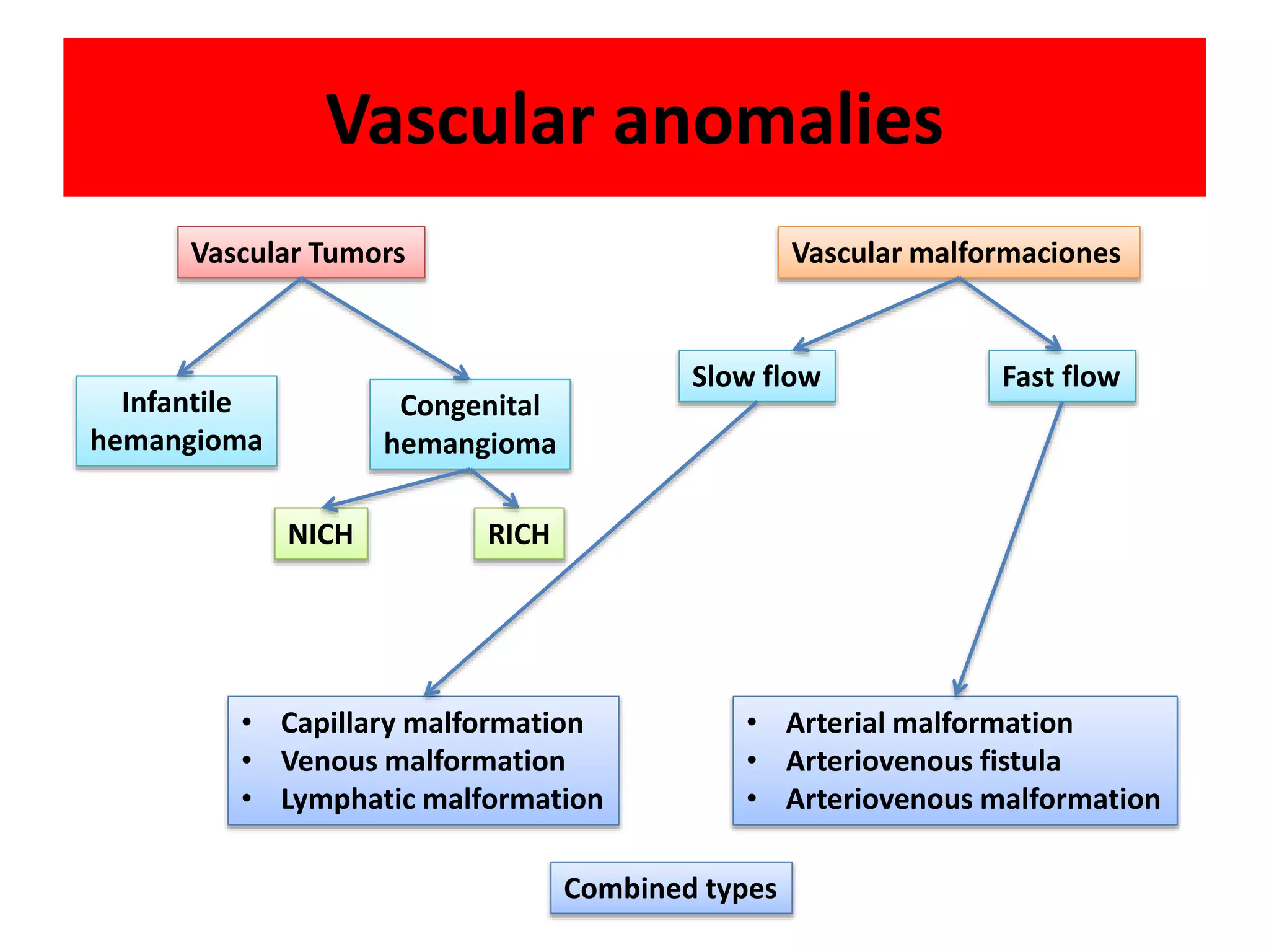
Vascular tumors and malformations are common skin lesions in infants and children. This document discusses several types of vascular tumors: infantile hemangiomas, congenital hemangiomas, kaposiform hemangioendothelioma, and pyogenic granuloma. Infantile hemangiomas are the most common tumor of infancy, appearing as red lesions that grow rapidly during the first year of life and slowly involute over subsequent years. Congenital hemangiomas are fully developed at birth and do not undergo postnatal growth. Kaposiform hemangioendothelioma is a rare but locally aggressive vascular tumor. Pyogenic granuloma is a small red papule that grows rapidly to form a
Hemangiomas and Vascular Malformations /certified fixed orthodontic courses b...Indian dental academy
The Indian Dental Academy is the Leader in continuing dental education , training dentists in all aspects of dentistry and
offering a wide range of dental certified courses in different formats.for more details please visit
www.indiandentalacademy.com
hemangioma , detailed ,with images,slides of hemangioma ,tumor, Infantile hemangiomas are benign vascular neoplasms that have a characteristic clinical course marked by early proliferation and followed by spontaneous involution. Hemangiomas are the most common tumors of infancy and usually are medically insignificant
This presentation is intended for diagnosing various vascular lesions on the basis of history and clinical examination. It covers a broad range of anomalies plus you can test yourself in the end.
What are Vascular Anomalies?
Hemangioma
Rendu Osler Weber Disease
Sturge Weber syndrome
Lymphangioma
Cystic Hygroma
Hemangiomas: lesions demonstrating endothelial hyperplasia.
Vascular Malformations : lesions with normal endothelial turnover.
A hemangioma is a benign and usually self-involuting tumor of the endothelial cells that line blood vessels, and is characterised by increased number of normal or abnormal vessels filled with blood.
May be present at Birth or arise during early childhood.
El sarcoma de Kaposi es una enfermedad por la que se forman tumores malignos (cancerosos) en la piel, las membranas mucosas, los ganglios linfáticos y otros órganos.
Para detectar (encontrar) y diagnosticar el sarcoma de Kaposi se utilizan pruebas que examinan la piel, los pulmones y el tubo digestivo.
Después de que se diagnostica un sarcoma de Kaposi, se realizan prueban para determinar si las células cancerosas se diseminaron a otras partes del cuerpo.
Ciertos factores afectan el pronóstico (probabilidad de recuperación) y las opciones de tratamiento.
PAPEL DE LOS ESTUDIOS NO INVASIVOS EN EL DIAGNOSTICO Y TRATAMIENTO DE LA ENFE...CLINICA VASCULAR DE CALI
Descripcion basada en la evidencia, acerca de los metodos diagnosticos en la patologia carotidea (duplex, tomografia, resonancia magnetica, arteriografia)
Sarcomas clinical and radiographic features /prosthodontic coursesIndian dental academy
The Indian Dental Academy is the Leader in continuing dental education , training dentists in all aspects of dentistry and
offering a wide range of dental certified courses in different formats.
,patologia benigna de mama ,mastalgia ,matopatía no ciclíca ,mastitis puerperal ,mastitis no puerperal ,mastitis necrosante ,fibroadenoma ,tumor filodes ,papilomatosis ,fistulizacion periareolar ,enfermedad de mondor
Tumores Benignos y Malignos comunes en PielJuan Meléndez
En esta presentación se podrán aprecias varios de los tumores comunes en piel, entre ellos:
-Acrocordones
-Queratosis
-Fibromas
No olvidar leer las notas
-
Nephroblastoma also known as Wilms tumor, is the most common renal malignancy affecting one in 10,000 children <15 years old
Children with bilateral disease are diagnosed at an earlier age (median age, girls at 31 months and boys at 24 months): Patients with associated congenital anomalies are also diagnosed at an earlier age
Accounts for 6-7% of cases of childhood cancer in the developed world and 12% in South Africa
In Tanzania the prevalence is 6.7% ( Mgaya E et al., 2000), “third from leukemia and lymphoma” (Shakilu J, 2017)
The overall survival rate of nephroblastoma approaches 90% in the developed world but in developing countries the survival rates are much less and in some sub-Saharan countries it is only 40% at 8 months after diagnosis
These may occur in 1% of infantile kidneys but typically regress during childhood
Nephrogenic rests normally occur in 1% of newborn kidneys and regress early in childhood: in contrast, they are present in 35% of kidneys with unilateral Wilms tumor and almost 100% of kidneys with bilateral disease
Has been associated with loss of function mutations of a number of tumor suppressor and transcription genes
These include mutations of the WT1, p53, FWT1, and FWT2 genes and at the 11p15.5 locus
Histologically, the classic favorable histology Wilms tumor is comprised of three cell types:
Blastemal cells – Undifferentiated cells
Stromal cells – Immature spindle cells and heterologous skeletal muscle, cartilage, osteoid or fat
Epithelial cells – Glomeruli and tubules
Grossly, Wilms tumors are usually well-circumscribed and have a pseudo-capsule
Histologically, Wilms is divided into "Favorable" and "Unfavorable" histologies
"Favorable" Histology: 90% of Wilms tumors will demonstrate "favorable" histology which generally has a better prognosis
Most children with Wilms tumor present with an abdominal mass or swelling without other signs or symptoms
The definitive diagnosis of Wilms tumor is made by histologic confirmation at the time of either surgical excision or biopsy
Stage I indicates the tumor was completely contained within the kidney without any breaks or spillage outside the renal capsule and no vascular invasion
Stage II would be a tumor that has grown outside the kidney
Stage IIIunresectable tumor
Stage IV-Metastasis
Stage V-bilateral kidney
Surgery is the main treatment
Infantile and congenital hemangiomas managment.pptxMohammad Daboos
This product discuss the definitions, theories, pathogenisis, pathology and methods of diagnosis of Infantile and Congenital hemangiomas, and explain when and how to treat both infantile and Congenital Hemangiomas. In addition to describe hemangioma in espicial sites as [parotid, GIT and Hepatic Hemangioma] So, i advice all pediatric, plastic, and dermatologist to revise this lecture and update his knowledge.
Neuroblastoma diagnosis, treatment, complications, and further management. The main contents of this review have been accessed from MedScape. Please do not reprint or copy this material without permission from the copyright owner.
Presentación realizada en el "XLIII Congreso Internacional en Salud - IOCIM" realizado en Acapulco - Mexico, el 30 de Agosto de 2019.
Tema presentado: "El reto del salvamento de extremidades"
Presentación que busca integrar el concepto de enfermedad arterial periférica en la gestión del riesgo cardiovascular.
Revision de la literatura y presentacion de caso manejado con tratamiento hibrido con endoprotesis fenestrada en aneurisma micotico y drenaje de coleccion por via abierta
Efecto de la flebectomía ambulatoria estética sin safenectomía en el diámetro...CLINICA VASCULAR DE CALI
ABSTRACT
It has been traditionally accepted that reflux in the Greater Saphenous Vein (GSV) starts at the Saphenofemoral junction and extends downward, resulting in the transmission of reflux tributary veins.
Another hypothesis describes a multifocal origin and ascending reflux that begins in tributary veins and transmitted to the GSV.
Paul Pittaluga et al. published in 2005, describing a procedure called ASVAL (Ablation selective des varices sous anestesie locale), where they perform selective resection of varicose veins and incompetent GSV is preserved.
Objective: To describe the effect of selective phlebectomy to diameter and reflux in the GSV. As a secondary objective, describe the safety, effectiveness and acceptability of this procedure by patients.
Methods: Patients with reflux in the GSV and tributaries varices, reflux confirmed by duplex ultrasound (DU) before the procedure, operated by selective phlebectomy, preserving the incompetent VSM. Postoperatively the presence of reflux and the diameter of the VSM with DU will appreciate.
Results: The study was conducted in 202 patients and followed up to 5 years. In 75% of limbs reflux disappeared in the GSV and the diameter decreased in 70% of these veins. They no major complications were reported and there was great acceptance by patients.
Conclusions: Selective phlebectomy without saphenectomy is an effective method to abolish reflux in the GSV; decreases symptoms, safe and accepted by patients.
DESCRIPCION BASADA EN LA EVIDENCIA DE UNA NUEVA TERAPEUTICA ENDOVASCULAR EN EL TRATEMIENTO DE LA HIPERTENSION ARTERIAL REFRACTARIA AL TRATAMIENTO CONVENCIONAL
Revision basada en la evidencia de los metodos endovasculares para ablacion de la vena safena, en pacientes con insuficiencia venosa cronica.
Evidence-based review of endovascular methods for ablation of the saphenous vein in patients with chronic venous insufficiency.
Title: Sense of Taste
Presenter: Dr. Faiza, Assistant Professor of Physiology
Qualifications:
MBBS (Best Graduate, AIMC Lahore)
FCPS Physiology
ICMT, CHPE, DHPE (STMU)
MPH (GC University, Faisalabad)
MBA (Virtual University of Pakistan)
Learning Objectives:
Describe the structure and function of taste buds.
Describe the relationship between the taste threshold and taste index of common substances.
Explain the chemical basis and signal transduction of taste perception for each type of primary taste sensation.
Recognize different abnormalities of taste perception and their causes.
Key Topics:
Significance of Taste Sensation:
Differentiation between pleasant and harmful food
Influence on behavior
Selection of food based on metabolic needs
Receptors of Taste:
Taste buds on the tongue
Influence of sense of smell, texture of food, and pain stimulation (e.g., by pepper)
Primary and Secondary Taste Sensations:
Primary taste sensations: Sweet, Sour, Salty, Bitter, Umami
Chemical basis and signal transduction mechanisms for each taste
Taste Threshold and Index:
Taste threshold values for Sweet (sucrose), Salty (NaCl), Sour (HCl), and Bitter (Quinine)
Taste index relationship: Inversely proportional to taste threshold
Taste Blindness:
Inability to taste certain substances, particularly thiourea compounds
Example: Phenylthiocarbamide
Structure and Function of Taste Buds:
Composition: Epithelial cells, Sustentacular/Supporting cells, Taste cells, Basal cells
Features: Taste pores, Taste hairs/microvilli, and Taste nerve fibers
Location of Taste Buds:
Found in papillae of the tongue (Fungiform, Circumvallate, Foliate)
Also present on the palate, tonsillar pillars, epiglottis, and proximal esophagus
Mechanism of Taste Stimulation:
Interaction of taste substances with receptors on microvilli
Signal transduction pathways for Umami, Sweet, Bitter, Sour, and Salty tastes
Taste Sensitivity and Adaptation:
Decrease in sensitivity with age
Rapid adaptation of taste sensation
Role of Saliva in Taste:
Dissolution of tastants to reach receptors
Washing away the stimulus
Taste Preferences and Aversions:
Mechanisms behind taste preference and aversion
Influence of receptors and neural pathways
Impact of Sensory Nerve Damage:
Degeneration of taste buds if the sensory nerve fiber is cut
Abnormalities of Taste Detection:
Conditions: Ageusia, Hypogeusia, Dysgeusia (parageusia)
Causes: Nerve damage, neurological disorders, infections, poor oral hygiene, adverse drug effects, deficiencies, aging, tobacco use, altered neurotransmitter levels
Neurotransmitters and Taste Threshold:
Effects of serotonin (5-HT) and norepinephrine (NE) on taste sensitivity
Supertasters:
25% of the population with heightened sensitivity to taste, especially bitterness
Increased number of fungiform papillae
These simplified slides by Dr. Sidra Arshad present an overview of the non-respiratory functions of the respiratory tract.
Learning objectives:
1. Enlist the non-respiratory functions of the respiratory tract
2. Briefly explain how these functions are carried out
3. Discuss the significance of dead space
4. Differentiate between minute ventilation and alveolar ventilation
5. Describe the cough and sneeze reflexes
Study Resources:
1. Chapter 39, Guyton and Hall Textbook of Medical Physiology, 14th edition
2. Chapter 34, Ganong’s Review of Medical Physiology, 26th edition
3. Chapter 17, Human Physiology by Lauralee Sherwood, 9th edition
4. Non-respiratory functions of the lungs https://academic.oup.com/bjaed/article/13/3/98/278874
Ethanol (CH3CH2OH), or beverage alcohol, is a two-carbon alcohol
that is rapidly distributed in the body and brain. Ethanol alters many
neurochemical systems and has rewarding and addictive properties. It
is the oldest recreational drug and likely contributes to more morbidity,
mortality, and public health costs than all illicit drugs combined. The
5th edition of the Diagnostic and Statistical Manual of Mental Disorders
(DSM-5) integrates alcohol abuse and alcohol dependence into a single
disorder called alcohol use disorder (AUD), with mild, moderate,
and severe subclassifications (American Psychiatric Association, 2013).
In the DSM-5, all types of substance abuse and dependence have been
combined into a single substance use disorder (SUD) on a continuum
from mild to severe. A diagnosis of AUD requires that at least two of
the 11 DSM-5 behaviors be present within a 12-month period (mild
AUD: 2–3 criteria; moderate AUD: 4–5 criteria; severe AUD: 6–11 criteria).
The four main behavioral effects of AUD are impaired control over
drinking, negative social consequences, risky use, and altered physiological
effects (tolerance, withdrawal). This chapter presents an overview
of the prevalence and harmful consequences of AUD in the U.S.,
the systemic nature of the disease, neurocircuitry and stages of AUD,
comorbidities, fetal alcohol spectrum disorders, genetic risk factors, and
pharmacotherapies for AUD.
Knee anatomy and clinical tests 2024.pdfvimalpl1234
This includes all relevant anatomy and clinical tests compiled from standard textbooks, Campbell,netter etc..It is comprehensive and best suited for orthopaedicians and orthopaedic residents.
Ozempic: Preoperative Management of Patients on GLP-1 Receptor Agonists Saeid Safari
Preoperative Management of Patients on GLP-1 Receptor Agonists like Ozempic and Semiglutide
ASA GUIDELINE
NYSORA Guideline
2 Case Reports of Gastric Ultrasound
NVBDCP.pptx Nation vector borne disease control programSapna Thakur
NVBDCP was launched in 2003-2004 . Vector-Borne Disease: Disease that results from an infection transmitted to humans and other animals by blood-feeding arthropods, such as mosquitoes, ticks, and fleas. Examples of vector-borne diseases include Dengue fever, West Nile Virus, Lyme disease, and malaria.
Tom Selleck Health: A Comprehensive Look at the Iconic Actor’s Wellness Journeygreendigital
Tom Selleck, an enduring figure in Hollywood. has captivated audiences for decades with his rugged charm, iconic moustache. and memorable roles in television and film. From his breakout role as Thomas Magnum in Magnum P.I. to his current portrayal of Frank Reagan in Blue Bloods. Selleck's career has spanned over 50 years. But beyond his professional achievements. fans have often been curious about Tom Selleck Health. especially as he has aged in the public eye.
Follow us on: Pinterest
Introduction
Many have been interested in Tom Selleck health. not only because of his enduring presence on screen but also because of the challenges. and lifestyle choices he has faced and made over the years. This article delves into the various aspects of Tom Selleck health. exploring his fitness regimen, diet, mental health. and the challenges he has encountered as he ages. We'll look at how he maintains his well-being. the health issues he has faced, and his approach to ageing .
Early Life and Career
Childhood and Athletic Beginnings
Tom Selleck was born on January 29, 1945, in Detroit, Michigan, and grew up in Sherman Oaks, California. From an early age, he was involved in sports, particularly basketball. which played a significant role in his physical development. His athletic pursuits continued into college. where he attended the University of Southern California (USC) on a basketball scholarship. This early involvement in sports laid a strong foundation for his physical health and disciplined lifestyle.
Transition to Acting
Selleck's transition from an athlete to an actor came with its physical demands. His first significant role in "Magnum P.I." required him to perform various stunts and maintain a fit appearance. This role, which he played from 1980 to 1988. necessitated a rigorous fitness routine to meet the show's demands. setting the stage for his long-term commitment to health and wellness.
Fitness Regimen
Workout Routine
Tom Selleck health and fitness regimen has evolved. adapting to his changing roles and age. During his "Magnum, P.I." days. Selleck's workouts were intense and focused on building and maintaining muscle mass. His routine included weightlifting, cardiovascular exercises. and specific training for the stunts he performed on the show.
Selleck adjusted his fitness routine as he aged to suit his body's needs. Today, his workouts focus on maintaining flexibility, strength, and cardiovascular health. He incorporates low-impact exercises such as swimming, walking, and light weightlifting. This balanced approach helps him stay fit without putting undue strain on his joints and muscles.
Importance of Flexibility and Mobility
In recent years, Selleck has emphasized the importance of flexibility and mobility in his fitness regimen. Understanding the natural decline in muscle mass and joint flexibility with age. he includes stretching and yoga in his routine. These practices help prevent injuries, improve posture, and maintain mobilit
- Video recording of this lecture in English language: https://youtu.be/lK81BzxMqdo
- Video recording of this lecture in Arabic language: https://youtu.be/Ve4P0COk9OI
- Link to download the book free: https://nephrotube.blogspot.com/p/nephrotube-nephrology-books.html
- Link to NephroTube website: www.NephroTube.com
- Link to NephroTube social media accounts: https://nephrotube.blogspot.com/p/join-nephrotube-on-social-media.html
Prix Galien International 2024 Forum ProgramLevi Shapiro
June 20, 2024, Prix Galien International and Jerusalem Ethics Forum in ROME. Detailed agenda including panels:
- ADVANCES IN CARDIOLOGY: A NEW PARADIGM IS COMING
- WOMEN’S HEALTH: FERTILITY PRESERVATION
- WHAT’S NEW IN THE TREATMENT OF INFECTIOUS,
ONCOLOGICAL AND INFLAMMATORY SKIN DISEASES?
- ARTIFICIAL INTELLIGENCE AND ETHICS
- GENE THERAPY
- BEYOND BORDERS: GLOBAL INITIATIVES FOR DEMOCRATIZING LIFE SCIENCE TECHNOLOGIES AND PROMOTING ACCESS TO HEALTHCARE
- ETHICAL CHALLENGES IN LIFE SCIENCES
- Prix Galien International Awards Ceremony
2. DR. LEONARDO BALLESTAS MALDONADO
GENERAL SURGERY RESIDENT
UNIVERSIDAD DEL VALLE
DR. RENE TIMARAN
VASCULAR SURGERY TEACHER
HOSPITAL SAN VICENTE DE PAUL
UNIVERSIDAD DE ANTIOQUIA
3. Clinic case
• Female
• 2 weeks after birth
• Premature antecedent
• GLUT-1 positive
DIAGNOSIS
4. “Not every skin lesion looking like a
strawberry is a hemangioma; not all
hemangiomas look like strawberries”
J.B. Mulliken, MD
5. Evidence
• A search of the evidence in the database:
Medline, Embase, Cohcrane, Tripdatabase,
Scielo
• Key words: Hemangioma, vascular anomalies,
kaposiform hemangioendothelioma, infantile
hemangioma, pyogenic granuloma, congenital
hemangioma
6.
7. Introduction
• Vascular anomalies are the most common skin and soft
tissue lesions observed in infants and children
• Older nomenclature continues to cause confusion,
misunderstood diagnoses, and potencial mismagement
• In 1982, Milliken and Glowacki proposed a
classification system for vascular anomalies based on
their clinical behavior and endothelial cell
characteristic
Kilcline C, Frieden IJ. Infantile hemangiomas: how common are they? A systematic review of the medical literature. Pediatr
Dermatol 2008;25(2):168–73.
8. ISSVA Classification
Society for the Study of Vascular Anomalies
Vascular tumors Vascular malformation Vascullar anomalliies
Hemangioma Vascular malformation
10. Differentiating Features
TUMOR
• True tumors, with
proliferation of the vascular
endothelium
• >3:1 female : male
• Small or absent at birth
• Rapid growth during infancy
• Self-limited
• Diagnosis: Clinical +
appearence
MALFORMATION
• No tumor, comprised of
dysplastic vessels
• 1:1 female : male
• Present at birth
• Growth proportional to
child
• Never disappear
• Diagnosis: MRI, Doppler
ultrasonography,
angiography
12. Infantile Hemangioma (IH)
• Bening endothelial cell tumor
• Most common tumor of infancy/childhood
• Incidence of 4.5% overall
• It is more frequent in caucasian, premature infants and
females (3:1 to 5:1)
• Perinatal characteristics associated with a higher risk of IH
include preeclampsia, multiple gestation, and low birth
weight
Hemangioma Investigator Group, Haggstrom AN, Drolet BA, Baselga E, et al. Prospective study of infan- tile hemangiomas:
demographic, prenatal, and peri- natal characteristics. J Pediatr 2007;150(3):291–4.
13. Pathogenesis
• Despite the frequency of this tumor,
understanding of the pathogenetic mechanisms
• Generally believed to be a complex interaction of
both genetic and environmental factors
• Histologically, have markedly increased cellularity
with clusters of plump cells that are positive for
markers of immature endothelial cells
Drolet BA, Swanson EA, Frieden IJ, et al. Infantile hemangiomas: an emerging health issue linked to an increased rate of low birth
weight infants. J Pediatr 2008;153(5):712 No-715.
14. Pathogenesis
• Endothelial-like cells of the hemangioma
expressed GLUT-1, the erythrocyte-type
glucose transporter protein that has been
shown to be upregulated in zones of hypoxia
• GLUT-1 seems to be an exclusive marker for IH
and is an invaluable tool used to distinguish
hemangiomas from other vascular lesions.
NorthPE,WanerM,MizerackiA,etal.GLUT1:anewly discovered immunohistochemical marker for juvenile hemangiomas. Hum Pathol
2000;31(1):11–22
15. Diagnosis
• History and physical
examination
• Radiographic imaging
• Histopathology – GLUT-1
16. History
• The diagnosis is typically made clinically based on
its appearance and characteristic behavior
• Between 30% and 50% of lesions are visible at
birth as a small, pale spot, telangiectatic stain or
ecchymotic area
• Appears weeks/months afther birth (the median
age appearance is 2 weeks)
17. Localitation
• Most hemangiomas are
single (80%)
Head an neck 60%
Trunk 25%
Extremities 15%
23. Classification based on morphology
• Localized or
segmental or
indeterminate
• Segmental
hemangiomas are at
higher risk of
complications and
associated anomalies
24. Ultrasound
• Differentiate deep hemangiomas to venous
malformations
• To evaluate the response to drug treatment
• Findings
Dense parenchyma rapid flow
Minor arterial resistance
Increased venous velocity
Effect of soft tissue mass
•Connors III JP, Mulliken JB. Vascular Tumors and Malformations in Childhood. Vascular Surgery 6th ed 2005
25. Magnetic resonance
• Test "reference”
• Findings
Intermediate intensity parenchymal tissue (T1), and
moderate hyperintensity (T2)
Empty Flow (arteriovenous shunts)
Glasses high flow or low flow
Reducing the size and number of vessels irrigating /
draining
lobularity
Body fat avascular
•Connors III JP, Mulliken JB. Vascular Tumors and Malformations in Childhood. Vascular Surgery 6th ed 2005
26. Complications
• Although most IHs are uncomplicated and do
not require treatment
• 24% of those referred to tertiary institutions
had complications
• Size, location, and subtype (localized vs
segmental) are major factors to consider in
evaluating an infant’s risk
Haggstrom AN, Drolet BA, Baselga E, et al. Prospective study of infantile hemangiomas: clinical characteristics predicting
complications and treat- ment. Pediatrics 2006;118(3):882–7.
27.
28. Ulceration
• Is the most common complication and can result
in pain, infection, bleeding, and permanent
scarring
• Larger in size or of the segmental subtype are
more likely to develop ulceration
• The cause of ulceration is not well understood,
but maceration and friction are likely contributing
factors given the higher frequency in locations
prone to this
Chamlin SL, Haggstrom AN, Drolet BA, et al. Multi- center prospective study of ulcerated hemangi- omas. J Pediatr
2007;151(6):684–9, 689-e1.
30. Visual compromise
• Threat to vision is a common reason for
treatment in the IH population
• Infants are at particular risk because stimulus
deprivation for as little as 1 week can interrupt
visual development and result in permanent
visual impairment
• Periocular IH may cause ptosis, strabismus, and
anisometropia, each of which may result in
astigmatism, amblyopia, or blindness
Al Dhaybi R, Superstein R, Milet A, et al. Treatment of periocular infantile hemangiomas with propranolol: case series of 18
children. Ophthalmology 2011; 118(6):1184–8.
31. Visceral involvement and
complications
• Hemangiomatosis – 30%
• The lesions are usually less
than 5 mm in diameter and
domelike
• Are recognized to have a
higher risk of visceral
hemangiomas, with liver and
gastrointestinal involvement
being most common
• Should be screened with
ultrasound to rule out lesions
in internal organs
32. Anomalies associated with anatomic
localization of HI
• The presence of IH in
particular locations can
be a marker for
underlying or
associated anomalies
• The “beard”
distribution has been
associated with the
presence of airway
hemangiomas
Orlow SJ, Isakoff MS, Blei F. Increased risk of symp- tomatic hemangiomas of the airway in association with cutaneous
hemangiomas in a “beard” distribution. J Pediatr 1997;131(4):643–6.
33. Anomalies associated with anatomic
localization of HI
In the lumbosacral area
have also been reported in
association with occult
spinal dysraphism
Girard C, Bigorre M, Guillot B, et al. PELVIS syndrome. Arch Dermatol 2006;142(7):884–8.
34. PHACE syndrome
The neurocutaneous
disorder characterized by
posterior fossa
abnormalities [P], large
facial hemangiomas [H],
arterial anomalies [A],
cardiac defects [C], and
eye anomalies [E]
Frieden IJ, Reese V, Cohen D. PHACE syndrome. The association of posterior fossa brain malforma- tions, hemangiomas, arterial
anomalies, coarctation of the aorta and cardiac defects, and eye abnormal- ities. Arch Dermatol 1996;132(3):307–11.
35. Treatment Objetives
• Prevent or reverse the life-threatening
complications or function
• Prevent or minimize disfigurement
• Minimize psychosocial stress for the patient and
family
• Properly treat ulceration to minimize scarring,
bleeding, infection and pain
36. Conservative treatment
• Observation is the mainstay of management
because 90% of IHs are small and localized
and do not involve aesthetically or
functionally important areas
• Up to 38% of hemangiomas referred to
tertiary care specialists require systemic
treatment because of complications
37. B-Blockers
• The use of propranolol for IH quickly gained favor,
because it has been perceived to have a lower
side-effect profile than other systemic therapies
• Its mechanism of action in the treatment of IH is
unknown
• Possible mechanisms of action purported include
vasocon- striction, inhibition of angiogenesis, and
induction of apoptosis
Leaute-Labreze C, Dumas de la Roque E, Hubiche T, et al. Propranolol for severe hemangiomas of infancy. N Engl J Med
2008;358(24):2649–51
38. Systemic Corticosteroids
• Response to treatment is variable, regression in one
third of cases, stabilization of growth in another third
of cases, and minimal to no response in the final third
of cases
• Adverse effects are common and include irritability,
gastrointestinal upset, sleep disturbance, cushingoid
facies, adrenal suppression, immunosuppression,
hypertension, bone demineralization, cardiomyopathy,
and growth retardation
• The duration of treatment is variable
Barrio VR, Drolet BA. Treatment of hemangiomas of infancy. Dermatol Ther 2005;18(2):151–9.
39. Intralesional and topical
corticosteroids
• Reported to decrease the size or slow growth
• Are most effective for small and localized
cutaneous hemangiomas
• The efficacy is limited by the depth of its
penetration compared with the depth of
hemangioma involvement
Barrio VR, Drolet BA. Treatment of hemangiomas of infancy. Dermatol Ther 2005;18(2):151–9.
40. Vincristine
• Effective in the treatment of IH and has historically
been reserved for those IHs resistant to corticosteroids
or in patients intolerant of corticosteroids
• Constipation is the most common side effect, but
neuromyopathy, most commonly presenting as foot
drop, is a potentially serious side effect
• Administration of vincristine requires placement of a
central line; therefore, risks associated with this must
be considered as well
Barrio VR, Drolet BA. Treatment of hemangiomas of infancy. Dermatol Ther 2005;18(2):151–9.
41. Interferon
• Recombinant interferon-a an inhibitor of
angiogenesis, administered as a subcutaneous
injection
• Side effects include flulike symptoms of fever,
irritability, and malaise
• Its use is not recommended in earlier age
42. Laser therapy
• The mechanism for this is not well understood
• Controversy exists surrounding its use for the treatment of
proliferating IH as adverse outcomes including ulceration
and scarring have been described
• Tuse of laser for intact IH is limited by the depth of the
laser’s penetration (1 mm)
• A few studies describing its benefit in the treatment of
ulcerated hemangiomas in terms of both increasing
reepithelialization and decreasing pain
Witman PM, Wagner AM, Scherer K, et al. Complica- tions following pulsed dye laser treatment of super- ficial hemangiomas.
Lasers SurgMed 2006;38(2): 116–23.
43. Surgery
• Surgical excision may be an option for
function- threatening or life-threatening
hemangiomas when medical therapy fails or is
not tolerated
• More commonly its role is for removal of
residual fibrofatty tissue or correction of
scarring after involution
44.
45. Congenital Hemangioma (CH)
• Unlike IH, CH is fully
developed at birth and does
not undergo postnatal
growth
• Rare (compared to infantile)
• Blue/gray hue, pale halo
(skin)
• Is more common in the
extremities and has an
equal sex distribution
Vascullarr anomalliies
Hemangioma
47. Growth patterns of hemangiomas
GROWTH NICH
RICH
IH
BIRTH 1 YR 2 YRS
AGE
RICH:rapidly involuting congenital hemangioma; NICH:noninvoluting congenital hemangioma. IH: Infantile Hemangioma
48. Treatment
• Most CHs are managed by observation
• RICHs begin to involute shortly after birth, and
most have resolved by 12 months of age
• NICH is a stable lesion that does not involute
or respond to pharmac logic treatment. Thus,
NICH causing disfigurement or functional
problems is treated by excision
49. Kaposiform Hemangioendothelioma
• Rare vascular neoplasm that is
locally aggressive but does not
metastasize
• Fifty percent of lesions are
present at birth and are
diagnosed during infancy
(58%), early childhood (1 to 10
years; 32%), or late child hood
(10 to 20 years; 10%)
• Has an equal sex distribution,
is solitary, and affects the head
or neck (40%), trunk (30%), or
extremity (30%)
51. Treatment
• A localized tumor can be resected, depending on
its location
• Patients with KMP require systemic treatment to
prevent lifethreatening complications
• Responds best to vincristine (90%), followed by
IFN (50%) and corticosteroid (10%)
52. Pyogenic granuloma
• Is a solitary a small red papule that grows rapidly, forming a stalk
• The malefemale ratio is 2 : 1
• It is commonly complicated by bleeding (64.2%) and epidermal ulceration
(36.3%)
• The presentation is inversely correlated with age
• They are distributed on the head or neck (62%), trunk (19%), upper
extremity (13%), or lower extremity (5%)
• Twentyfive percent of patients have a history of trauma or an underlying
cutaneous condition (including capillary malformation, dermatologic
disorder, viral infection, or insect bite).
53. Treatment
• Numerous treatment
methods have been
described: curettage, shave
excision, laser therapy, and
excision
• These modalities have a
recurrence rate of 43.5%
• Definitive treatment
requires fullthickness skin
excision, which has an
approximately 100% cure
rate
54. Clinic case
• Female
• 2 weeks after birth
• Premature antecedent
INFANTILE HEMANGIOMA
Ecografía
Diferenciar los hemangiomas profundos de las malformaciones venosas
Evaluar la respuesta al tratamiento farmacológico
Hallazgos:
Parénquima denso y un flujo rápido
Menor resistencia arterial
Aumento de la velocidad venosa
Efecto de masa de tejido blando
Resonancia magnética
Prueba “de referencia”
Hallazgos:
Tejido parenquimatoso de intensidad intermedia (T1), y de hiperintensidad moderada (T2)
Vacios de flujo (shunts arteriovenosos)
Vasos de alto flujo o bajo flujo
Reducción del tamaño y número de vasos irrigantes/drenantes
Lobularidad
Masa adiposa avascular
The pulsed dye laser (PDL) has been successfully used for vascular birthmarks, namely, capillary mal- formations or “port-wine stains” for years, and its efficacy in this setting is well-established