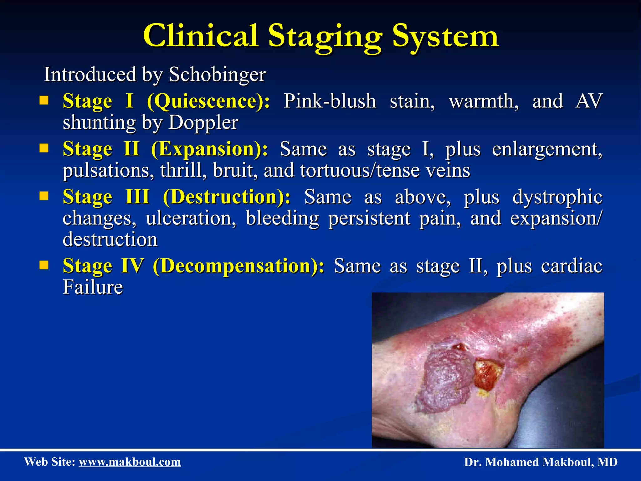
Vascular Anomalies; Haemangiomas and  Vascular Malformations Dr. Mohamed Makboul, MD Plastic Surgery Department Assiut University
The term  hemangioma   refers to the common tumor of infancy that has a proliferative endothelium, and exhibits rapid postnatal growth and slow regression during childhood; this tumor  never  appears in an adolescent or adult.  Whereas vascular malformations are comprised of abnormally formed channels that are lined by stable endothelium, present at birth, never regress and often expand.
Vascular anomalies can be classified into two unique groups:- Hemangiomas Vascular malformations which subcategories into : Slow-flow Capillary Lymphatic Venous Fast-flow Arterial arterial (aneurysm, stenosis, ectasia) Arteriovenous fistulae (AVF) Arteriovenous malformation (AVM)
Haemangioma
Haemangioma Hemangioma is the most common tumor of infancy, appears in neonatalhood, usually within the first 2 weeks and has a defined natural history.
Clinical Features  The natural history of Hemangioma is characterized by three phases: Proliferating Phase Involuting Phase Involuted Phase
Proliferating Phase Hemangioma grows rapidly during the first 6 to 8 months of infancy, the skin becomes raised, bosselated, and a vivid crimson color.  Palpation at this stage reveals a tense and noncompressible mass.
Day of Birth 1 month 3 month 2 month
Involuting Phase Hemangioma reaches its peak before the first year; and for a time thereafter, growth is proportionate to that of the child.  The first signs of the involuting phase appear as the crimson color fades to a dull purplish color, the skin gradually pales, and the tumor feels less tense.  The involuting phase continues until the child is 5 to 10 years of age.
Involuted Phase Regression is complete in 50% of children by age 5 years and in 70% by age 7 years, with continued improvement until age 10 to 12 years.  The hemangioma may persist in the form of residual tumor, loose skin, telangiectasias, or scarring.
Hemangiomas can be superficial, deep, or visceral in location Superficial lesions:  often exhibit the classic crimson color of the so-called strawberry hemangioma.  Deep lesions:  Are those within the deep dermis or subcutaneous tissues often present as pale blue or purple masses that may be confused with venous malformations. Visceral lesions:   Are not apparent on physical examination, and in 50% of cases, there are no accompanying cutaneous hemangiomas. In this setting, their presence may only be suggested by physiologic findings such as hepatomegaly, congestive heart failure, or stridor.
DIAGNOSTIC IMAGING Hemangiomas are readily distinguished from other tumors by: Ultrasonography (US) Computerized tomography (CT) Magnetic resonance imaging (MRI) Arteriography
DIFFERENTIAL DIAGNOSIS Hemangiomas must be differentiated from: Macular stains,  Vascular malformations, and  Other vascular tumors of infancy.
Macular Stains The most common type of vascular birthmark is the macular stain.  These are flat lesions, ranging in color from pink to red, seen in as many as 40 percent of newborns.  Typically seen in the neck, glabella, eyelid, and forehead, these probably are physiologic phenomena that will resolve with time.
Vascular Malformations The natural history of vascular malformations is distinct from hemangiomas.  Boys and girls are affected equally.  All malformations are present at birth.  The physical appearance of vascular malformations is dependent on the type of vessels involved.  Venous malformations may appear as bluish masses and be confused with subcutaneous hemangiomas, but they are distinguished by the ability to be emptied of blood with compression.
Malformations grow commensurately with the child and do not undergo the rapid proliferative growth phase exhibited by hemangiomas.  The greatest distinction between hemangiomas and malformations is that the former spontaneously involute and the latter do not. Hemangiomas, unlike vascular malformations, rarely cause bony distortion or hypertrophy. Vascular Malformations (cont.)
Other Vascular Tumors of Infancy Pyogenic granuloma Is an acquired vascular lesion that closely resembles hemangioma upon clinical and microscopic examination.  They tend to occur on the skin and mucosa of older children and young adults, with a mean age of 6.7 years. Pyogenic granulomas arise suddenly and usually without a history of trauma.  Frequently located on the cheeks, eyelids, extremities. The natural history is one of superficial ulceration and repetitive episodes of bleeding.
COMPLICATIONS Ulceration Infection Visual Impairment Airway Obstruction Auditory Canal Obstruction Congestive Heart Failure : usually occurs in one of two settings: diffuse neonatal hemangiomatosis or large visceral hemangiomas
MANAGEMENT In most instances, parental education and reassurance will suffice. Because most hemangiomas undergo spontaneous involution, treatment may be reserved for those lesions of functional or psychological concern.  Hemangiomas obstructing the visual axis, airway, and auditory canals, or those associated with congestive heart failure, ulceration, or bleeding, should be considered for treatment.
A number of options are available for the treatment of hemangiomas.  Effective treatments may be divided into:  Those that are nonspecific (e.g., excision, cryotherapy);  Antiangiogenic (e.g., corticosteroids, interferon); and  Antiproliferative (e.g., chemotherapy, radiation).
Surgical Excision Surgical excision may be indicated for:  Infancy for obstructing effect,  Ulcerating, or  Large hemangiomas unresponsive to pharmacologic therapy.
Capillary Malformation (CM)
CLINICAL PRESENTATION Port wine stains are intradermal capillary or venular malformations that are present at birth.
CM is a macular, red, vascular stain that presents at birth and persists throughout life.  CM can be localized or extensive, on the face, trunk, or limbs. The incidence rate in newborns is 0.3%.
Two-thirds of these lesions darken in color over time.  Most CMs are harmless cutaneous birthmarks, but some are red flags that signal underlying abnormalities.  CM in a limb often is associated with axial and transverse hypertrophy.
Treatment CM is treated by flashlamp pulsed-dye laser; the results are better if initiated in infancy and childhood.  Soft-tissue and skeletal hypertrophy also can require surgical therapy.  Laser therapy cannot correct soft-tissue overgrowth.
Laser treatment Pre Post
Hypertrophy of the upper lip, treated by partial debulking
Venous Malformation (VM)
CLINICAL PRESENTATION Venous malformations are localized or extensive, minor or distorting, single or multiple, and located anywhere on the head, limbs, or trunk.  Most VMs are sporadic, blue color, soft, compressible on palpation, a slow refill, and increased size with dependency are pathognomonic for venous malformation.
DIAGNOSIS Clinical picture. MRI
TREATMENT Sclerotherapy is the primary treatment for VMs, although subsequent surgical resection is often needed. VM Upper lip, Post Sclerotherapy and excision
VM Rt. Cheek, Post Sclerotherapy
Extensive venous malformation of the buttock and right thigh. Staged surgical reduction of the right buttock and thigh
Lymphatic Malformation (LM)
CLINICAL PRESENTATION LMs can be described as microcystic, macrocystic, and combined lymphaticovenous (LVM) forms.  Lymphatic anomalies present in a wide spectrum, from lymphedema to large cystic malformations.  LMs are generally detected at birth or shortly, thereafter LMs grow with the child.
Lymphatic malformations can affect all areas of the body, but most are located in the cervicofacial region and axilla.  LMs are the most common cause of congenital tongue enlargement (macroglossia), lip enlargement (macrocheilia), and ear enlargement (macrotia).  Skeletal hypertrophy and distortion in 80% of cervicofacial LMs occur by 10 years of age.   LM never regresses.
TREATMENT Sclerotherapy has assumed a major role in the management of LM.  Surgical resection is the only way to potentially cure LM.
Large lymphatic malformation of the right lateral forehead and temple region. After excision.
Arteriovenous Malformation (AVM)
AVM is a high-flow vascular malformation comprised of micro- and macro-aneriovenous fistulas (AVFs). The epicenter of an AVM is called the  nidus   and consists of arterial feeders, micro- and macroarteriovenous fistulas (AVFs), and ectatic veins.
Clinical Staging System Introduced by Schobinger Stage I (Quiescence):  Pink-blush stain, warmth, and AV shunting by Doppler  Stage II (Expansion):  Same as stage I, plus enlargement, pulsations, thrill, bruit, and tortuous/tense veins Stage III (Destruction):  Same as above, plus dystrophic changes, ulceration, bleeding persistent pain, and expansion/ destruction Stage IV (Decompensation):  Same as stage II, plus cardiac Failure
Diagnosis Confirmation of clinical diagnosis is helpful with Color Doppler examination, Magnetic Resonance Imaging, and Angiography.
Treatment Usually AVM is treated whenever endangering signs and symptoms arise, such as ischemic pain, ulceration, bleeding, or increased cardiac output (Stages III and IV ).  Arterial embolization is done to  temporarily occlude  the nidus in preparation for resection 24 to 72 hours later.
Questions???
 

Haemangiomas And Vascular Malformations

  • 1.
  • 2.
    Vascular Anomalies; Haemangiomasand Vascular Malformations Dr. Mohamed Makboul, MD Plastic Surgery Department Assiut University
  • 3.
    The term hemangioma refers to the common tumor of infancy that has a proliferative endothelium, and exhibits rapid postnatal growth and slow regression during childhood; this tumor never appears in an adolescent or adult. Whereas vascular malformations are comprised of abnormally formed channels that are lined by stable endothelium, present at birth, never regress and often expand.
  • 4.
    Vascular anomalies canbe classified into two unique groups:- Hemangiomas Vascular malformations which subcategories into : Slow-flow Capillary Lymphatic Venous Fast-flow Arterial arterial (aneurysm, stenosis, ectasia) Arteriovenous fistulae (AVF) Arteriovenous malformation (AVM)
  • 5.
  • 6.
    Haemangioma Hemangioma isthe most common tumor of infancy, appears in neonatalhood, usually within the first 2 weeks and has a defined natural history.
  • 7.
    Clinical Features The natural history of Hemangioma is characterized by three phases: Proliferating Phase Involuting Phase Involuted Phase
  • 8.
    Proliferating Phase Hemangiomagrows rapidly during the first 6 to 8 months of infancy, the skin becomes raised, bosselated, and a vivid crimson color. Palpation at this stage reveals a tense and noncompressible mass.
  • 9.
    Day of Birth1 month 3 month 2 month
  • 10.
    Involuting Phase Hemangiomareaches its peak before the first year; and for a time thereafter, growth is proportionate to that of the child. The first signs of the involuting phase appear as the crimson color fades to a dull purplish color, the skin gradually pales, and the tumor feels less tense. The involuting phase continues until the child is 5 to 10 years of age.
  • 11.
    Involuted Phase Regressionis complete in 50% of children by age 5 years and in 70% by age 7 years, with continued improvement until age 10 to 12 years. The hemangioma may persist in the form of residual tumor, loose skin, telangiectasias, or scarring.
  • 12.
    Hemangiomas can besuperficial, deep, or visceral in location Superficial lesions: often exhibit the classic crimson color of the so-called strawberry hemangioma. Deep lesions: Are those within the deep dermis or subcutaneous tissues often present as pale blue or purple masses that may be confused with venous malformations. Visceral lesions: Are not apparent on physical examination, and in 50% of cases, there are no accompanying cutaneous hemangiomas. In this setting, their presence may only be suggested by physiologic findings such as hepatomegaly, congestive heart failure, or stridor.
  • 13.
    DIAGNOSTIC IMAGING Hemangiomasare readily distinguished from other tumors by: Ultrasonography (US) Computerized tomography (CT) Magnetic resonance imaging (MRI) Arteriography
  • 14.
    DIFFERENTIAL DIAGNOSIS Hemangiomasmust be differentiated from: Macular stains, Vascular malformations, and Other vascular tumors of infancy.
  • 15.
    Macular Stains Themost common type of vascular birthmark is the macular stain. These are flat lesions, ranging in color from pink to red, seen in as many as 40 percent of newborns. Typically seen in the neck, glabella, eyelid, and forehead, these probably are physiologic phenomena that will resolve with time.
  • 16.
    Vascular Malformations Thenatural history of vascular malformations is distinct from hemangiomas. Boys and girls are affected equally. All malformations are present at birth. The physical appearance of vascular malformations is dependent on the type of vessels involved. Venous malformations may appear as bluish masses and be confused with subcutaneous hemangiomas, but they are distinguished by the ability to be emptied of blood with compression.
  • 17.
    Malformations grow commensuratelywith the child and do not undergo the rapid proliferative growth phase exhibited by hemangiomas. The greatest distinction between hemangiomas and malformations is that the former spontaneously involute and the latter do not. Hemangiomas, unlike vascular malformations, rarely cause bony distortion or hypertrophy. Vascular Malformations (cont.)
  • 18.
    Other Vascular Tumorsof Infancy Pyogenic granuloma Is an acquired vascular lesion that closely resembles hemangioma upon clinical and microscopic examination. They tend to occur on the skin and mucosa of older children and young adults, with a mean age of 6.7 years. Pyogenic granulomas arise suddenly and usually without a history of trauma. Frequently located on the cheeks, eyelids, extremities. The natural history is one of superficial ulceration and repetitive episodes of bleeding.
  • 19.
    COMPLICATIONS Ulceration InfectionVisual Impairment Airway Obstruction Auditory Canal Obstruction Congestive Heart Failure : usually occurs in one of two settings: diffuse neonatal hemangiomatosis or large visceral hemangiomas
  • 20.
    MANAGEMENT In mostinstances, parental education and reassurance will suffice. Because most hemangiomas undergo spontaneous involution, treatment may be reserved for those lesions of functional or psychological concern. Hemangiomas obstructing the visual axis, airway, and auditory canals, or those associated with congestive heart failure, ulceration, or bleeding, should be considered for treatment.
  • 21.
    A number ofoptions are available for the treatment of hemangiomas. Effective treatments may be divided into: Those that are nonspecific (e.g., excision, cryotherapy); Antiangiogenic (e.g., corticosteroids, interferon); and Antiproliferative (e.g., chemotherapy, radiation).
  • 22.
    Surgical Excision Surgicalexcision may be indicated for: Infancy for obstructing effect, Ulcerating, or Large hemangiomas unresponsive to pharmacologic therapy.
  • 23.
  • 24.
    CLINICAL PRESENTATION Portwine stains are intradermal capillary or venular malformations that are present at birth.
  • 25.
    CM is amacular, red, vascular stain that presents at birth and persists throughout life. CM can be localized or extensive, on the face, trunk, or limbs. The incidence rate in newborns is 0.3%.
  • 26.
    Two-thirds of theselesions darken in color over time. Most CMs are harmless cutaneous birthmarks, but some are red flags that signal underlying abnormalities. CM in a limb often is associated with axial and transverse hypertrophy.
  • 27.
    Treatment CM istreated by flashlamp pulsed-dye laser; the results are better if initiated in infancy and childhood. Soft-tissue and skeletal hypertrophy also can require surgical therapy. Laser therapy cannot correct soft-tissue overgrowth.
  • 28.
  • 29.
    Hypertrophy of theupper lip, treated by partial debulking
  • 30.
  • 31.
    CLINICAL PRESENTATION Venousmalformations are localized or extensive, minor or distorting, single or multiple, and located anywhere on the head, limbs, or trunk. Most VMs are sporadic, blue color, soft, compressible on palpation, a slow refill, and increased size with dependency are pathognomonic for venous malformation.
  • 32.
  • 33.
    TREATMENT Sclerotherapy isthe primary treatment for VMs, although subsequent surgical resection is often needed. VM Upper lip, Post Sclerotherapy and excision
  • 34.
    VM Rt. Cheek,Post Sclerotherapy
  • 35.
    Extensive venous malformationof the buttock and right thigh. Staged surgical reduction of the right buttock and thigh
  • 36.
  • 37.
    CLINICAL PRESENTATION LMscan be described as microcystic, macrocystic, and combined lymphaticovenous (LVM) forms. Lymphatic anomalies present in a wide spectrum, from lymphedema to large cystic malformations. LMs are generally detected at birth or shortly, thereafter LMs grow with the child.
  • 38.
    Lymphatic malformations canaffect all areas of the body, but most are located in the cervicofacial region and axilla. LMs are the most common cause of congenital tongue enlargement (macroglossia), lip enlargement (macrocheilia), and ear enlargement (macrotia). Skeletal hypertrophy and distortion in 80% of cervicofacial LMs occur by 10 years of age. LM never regresses.
  • 39.
    TREATMENT Sclerotherapy hasassumed a major role in the management of LM. Surgical resection is the only way to potentially cure LM.
  • 40.
    Large lymphatic malformationof the right lateral forehead and temple region. After excision.
  • 41.
  • 42.
    AVM is ahigh-flow vascular malformation comprised of micro- and macro-aneriovenous fistulas (AVFs). The epicenter of an AVM is called the nidus and consists of arterial feeders, micro- and macroarteriovenous fistulas (AVFs), and ectatic veins.
  • 43.
    Clinical Staging SystemIntroduced by Schobinger Stage I (Quiescence): Pink-blush stain, warmth, and AV shunting by Doppler Stage II (Expansion): Same as stage I, plus enlargement, pulsations, thrill, bruit, and tortuous/tense veins Stage III (Destruction): Same as above, plus dystrophic changes, ulceration, bleeding persistent pain, and expansion/ destruction Stage IV (Decompensation): Same as stage II, plus cardiac Failure
  • 44.
    Diagnosis Confirmation ofclinical diagnosis is helpful with Color Doppler examination, Magnetic Resonance Imaging, and Angiography.
  • 45.
    Treatment Usually AVMis treated whenever endangering signs and symptoms arise, such as ischemic pain, ulceration, bleeding, or increased cardiac output (Stages III and IV ). Arterial embolization is done to temporarily occlude the nidus in preparation for resection 24 to 72 hours later.
  • 46.
  • 47.