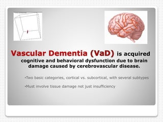
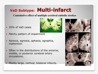
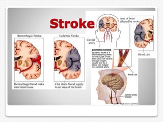
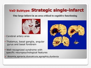
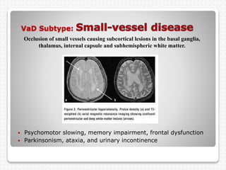
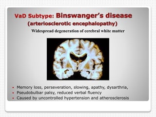
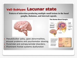
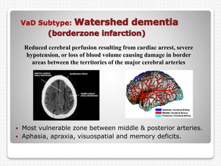
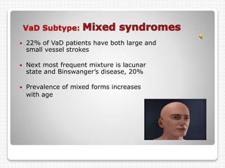
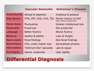
Vascular Dementia
in its many forms
Dr. Drew Chenelly
Vascular Dementia is acquired
cognitive and behavioral dysfunction due to brain
damage caused by cerebrovascular disease.
•Two basic categories, cortical vs. subcortical, with several subtypes
•Must involve tissue damage not just insufficiency
Who does VaD strike ?
 Dementia affects 10% of people over 65
and 20% of people over 80
 Second most common form of dementia
 10% to 40% of dementia patients
 Mixed with DAT in 10% to 25% of patients
 Onset most often in the 7th decade
 Men more than woman
 Blacks and Asians more than whites
What happens after VaD strikes?
 Survival lower than other dementias, 3 to 6
years
 Death usually from cardiovascular disease or
stroke
 Comorbid depression in 25% to 50% of patients
 Delusional thinking in 50% of patients
 Anxiety in 70% of patients
Signs and Symptoms
 a “patchy” pattern of intellectual impairment and “stuttering
course”.
 memory impairment, poor judgment, dilapidation of cognitive
functioning perseveration
 psychomotor slowing, lack of initiative, spontaneity and
perseverance, fatigue, loss of vigor, apathy.
 irritability, paranoia, disinhibition
 gait problems and falls, weakness, ataxia, rigidity, dysarthria,
parkinsonism, urinary incontinence.
 nocturnal confusion, fluctuating mental status, vulnerability to
delirium and “delirium-like” episodes
VaD Risk factors
 Uncontrolled hypertension 80%
 Diabetes Mellitus 20%
 Cardiac disease and heart surgery
 Hx of stroke or TIA
 Hyperlipidemia
 Smoking
 Substance abuse
 Obstructive sleep apnea
 Family history
 Pulmonary disease
See attached comprehensive list of VaD causes.
VaD Subtype:
 20% of VaD cases
 Patchy pattern of impairment
 Apraxia, agnosia, aphasia, agraphia,
inattention
 Often in the distributions of the anterior,
middle, or posterior cerebral artery
circulations.
 Mostly large, cortical, bilateral infarcts.
Cumulative effect of multiple cerebral embolic strokes
Stroke animations
VaD Subtype:
 Anomia,apraxia,dyscalcuia,agraphia,dyslexia
One large infarct in an area critical to cognitive functioning
• Cerebral artery area
• Thalamus, basal ganglia, angular
gyrus and basal forebrain
• Well-recognized syndrome with
specific neuropsychological features
VaD Subtype:
 Psychomotor slowing, memory impairment, frontal dysfunction
 Parkinsonism, ataxia, and urinary incontinence
Occlusion of small vessels causing subcortical lesions in the basal ganglia,
thalamus, internal capsule and subhemispheric white matter.
Thrombus and occlusion animations
VaD Subtype:
 Memory loss, perseveration, slowing, apathy, dysarthria,
 Pseudobulbar palsy, reduced verbal fluency
 Caused by uncontrolled hypertension and atherosclerosis
Widespread degeneration of cerebral white matter
VaD Subtype:
 Pseudobulbar palsy, gaze abnormalities,
arousal problems and psychomotor slowing.
 Pyramidal and extrapyramidal disorders.
 Prominent frontal systems dysfunction
Pattern of infarction producing multiple small lesions in the basal
ganglia, thalamus, and internal capsule.
VaD Subtype:
 Most vulnerable zone between middle & posterior arteries.
 Aphasia, apraxia, visuospatial and memory deficits.
Reduced cerebral perfusion resulting from cardiac arrest, severe
hypotension, or loss of blood volume causing damage in border
areas between the territories of the major cerebral arteries
VaD Subtype:
 22% of VaD patients have both large and
small vessel strokes
 Next most frequent mixture is lacunar
state and Binswanger’s disease, 20%
 Prevalence of mixed forms increases
with age
Differential Diagnosis
Vascular Dementia Alzheimer's Disease
Onset &Course Abrupt & stepwise Insidious & gradual
Risk factors HTN, DM, CVD, CVA, TIA Family history of DAT
Early memory impairment
Mental Status Fluctuating Greater intellectual loss
Personality Preserved Not preserved
Language Better fluency Worse fluency
Behavior Apathy & lability Loss of insight
Neuro exam Focal findings Non-focal findings
Neuroimaging CVA, white matter loss Generalized atrophy
Hallucinations “Delirium-like” episodes Late & vague
Neurological Ataxia, weakness Late rigidity
Evaluation
 History
 ADLs
 Memory
 Language
 Perception
 Executive functions
 Emotions
 Attention & arousal
 Motor functions
gait..praxis..reflexes
 Neuroimaging:
CT...MRI...PET
 Labs:
CBC...electrolytes
renal...liver...thyroid
glucose...Folate...B12
Treatment & prevention
Where possible Rx the underlying cause
 Antihypertensives
 Vasodialators
 Antiplatelet agents (aspirin)
 Anticoagulants (Coumadin)
 Lipid lowering agents
 Acetylcholinesterase inhibitors no
 NMDA antagonist (Namenda)
 Smoking cessation and blood sugar control
 Estrogen replacement in woman
 Carotid endarterectomy & other surgeries
Management of
 Antidopaminergic drug… Risperdal
 Engage around intact intellectual functions
 Low stimulation
 Schedule activities at peak arousal
 Consistent daily routine
 Compensate for memory deficits
 Explain disinhibition syndrome
 Limit activity expectations
 Attend to physical comfort

Vascular Dementia

  • 1.
    Vascular Dementia in itsmany forms Dr. Drew Chenelly
  • 2.
    Vascular Dementia isacquired cognitive and behavioral dysfunction due to brain damage caused by cerebrovascular disease. •Two basic categories, cortical vs. subcortical, with several subtypes •Must involve tissue damage not just insufficiency
  • 3.
    Who does VaDstrike ?  Dementia affects 10% of people over 65 and 20% of people over 80  Second most common form of dementia  10% to 40% of dementia patients  Mixed with DAT in 10% to 25% of patients  Onset most often in the 7th decade  Men more than woman  Blacks and Asians more than whites
  • 4.
    What happens afterVaD strikes?  Survival lower than other dementias, 3 to 6 years  Death usually from cardiovascular disease or stroke  Comorbid depression in 25% to 50% of patients  Delusional thinking in 50% of patients  Anxiety in 70% of patients
  • 5.
    Signs and Symptoms a “patchy” pattern of intellectual impairment and “stuttering course”.  memory impairment, poor judgment, dilapidation of cognitive functioning perseveration  psychomotor slowing, lack of initiative, spontaneity and perseverance, fatigue, loss of vigor, apathy.  irritability, paranoia, disinhibition  gait problems and falls, weakness, ataxia, rigidity, dysarthria, parkinsonism, urinary incontinence.  nocturnal confusion, fluctuating mental status, vulnerability to delirium and “delirium-like” episodes
  • 6.
    VaD Risk factors Uncontrolled hypertension 80%  Diabetes Mellitus 20%  Cardiac disease and heart surgery  Hx of stroke or TIA  Hyperlipidemia  Smoking  Substance abuse  Obstructive sleep apnea  Family history  Pulmonary disease See attached comprehensive list of VaD causes.
  • 7.
    VaD Subtype:  20%of VaD cases  Patchy pattern of impairment  Apraxia, agnosia, aphasia, agraphia, inattention  Often in the distributions of the anterior, middle, or posterior cerebral artery circulations.  Mostly large, cortical, bilateral infarcts. Cumulative effect of multiple cerebral embolic strokes
  • 9.
  • 10.
    VaD Subtype:  Anomia,apraxia,dyscalcuia,agraphia,dyslexia Onelarge infarct in an area critical to cognitive functioning • Cerebral artery area • Thalamus, basal ganglia, angular gyrus and basal forebrain • Well-recognized syndrome with specific neuropsychological features
  • 11.
    VaD Subtype:  Psychomotorslowing, memory impairment, frontal dysfunction  Parkinsonism, ataxia, and urinary incontinence Occlusion of small vessels causing subcortical lesions in the basal ganglia, thalamus, internal capsule and subhemispheric white matter.
  • 12.
  • 13.
    VaD Subtype:  Memoryloss, perseveration, slowing, apathy, dysarthria,  Pseudobulbar palsy, reduced verbal fluency  Caused by uncontrolled hypertension and atherosclerosis Widespread degeneration of cerebral white matter
  • 14.
    VaD Subtype:  Pseudobulbarpalsy, gaze abnormalities, arousal problems and psychomotor slowing.  Pyramidal and extrapyramidal disorders.  Prominent frontal systems dysfunction Pattern of infarction producing multiple small lesions in the basal ganglia, thalamus, and internal capsule.
  • 15.
    VaD Subtype:  Mostvulnerable zone between middle & posterior arteries.  Aphasia, apraxia, visuospatial and memory deficits. Reduced cerebral perfusion resulting from cardiac arrest, severe hypotension, or loss of blood volume causing damage in border areas between the territories of the major cerebral arteries
  • 16.
    VaD Subtype:  22%of VaD patients have both large and small vessel strokes  Next most frequent mixture is lacunar state and Binswanger’s disease, 20%  Prevalence of mixed forms increases with age
  • 17.
    Differential Diagnosis Vascular DementiaAlzheimer's Disease Onset &Course Abrupt & stepwise Insidious & gradual Risk factors HTN, DM, CVD, CVA, TIA Family history of DAT Early memory impairment Mental Status Fluctuating Greater intellectual loss Personality Preserved Not preserved Language Better fluency Worse fluency Behavior Apathy & lability Loss of insight Neuro exam Focal findings Non-focal findings Neuroimaging CVA, white matter loss Generalized atrophy Hallucinations “Delirium-like” episodes Late & vague Neurological Ataxia, weakness Late rigidity
  • 18.
    Evaluation  History  ADLs Memory  Language  Perception  Executive functions  Emotions  Attention & arousal  Motor functions gait..praxis..reflexes  Neuroimaging: CT...MRI...PET  Labs: CBC...electrolytes renal...liver...thyroid glucose...Folate...B12
  • 19.
    Treatment & prevention Wherepossible Rx the underlying cause  Antihypertensives  Vasodialators  Antiplatelet agents (aspirin)  Anticoagulants (Coumadin)  Lipid lowering agents  Acetylcholinesterase inhibitors no  NMDA antagonist (Namenda)  Smoking cessation and blood sugar control  Estrogen replacement in woman  Carotid endarterectomy & other surgeries
  • 20.
    Management of  Antidopaminergicdrug… Risperdal  Engage around intact intellectual functions  Low stimulation  Schedule activities at peak arousal  Consistent daily routine  Compensate for memory deficits  Explain disinhibition syndrome  Limit activity expectations  Attend to physical comfort