Renal Physiology andSkin
Excretion is the process by which the unwanted substances and metabolic wastes are eliminated from the body.
A large amount of waste materials and carbon dioxide are produced in the tissues during metabolic process.
In addition, residue of undigested food, heavy metals, drugs, toxic substances and pathogenic organisms like bacteria
are also present in the body.
All these substances must be removed to keep the body in healthy condition. Various systems/organs in the body are
involved in performing the excretory function, viz.
1. Digestive system excretes food residues in the form of feces. Some bacteria and toxic substances also are excreted
through feces
2. Lungs remove carbon dioxide and water vapor
3. Skin excretes water, salts and some wastes. It also removes heat from the body
4. Liver excretes many substances like bile pigments, heavy metals, drugs, toxins, bacteria, etc. through bile.
Although various organs are involved in removal of wastes from the body, their excretory capacity is limited.
But renal system or urinary system has maximum excretory capacity and so it plays a major role in homeostasis.
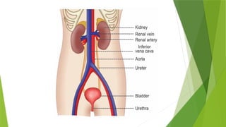
Renal system includes:
1. A pair of kidneys 2. Ureters 3. Urinary bladder 4. Urethra.
Kidneys produce the urine. Ureters transport the urine to urinary bladder. Urinary bladder stores the urine until it is
voided (emptied). Urine is voided from bladder through urethra
4.
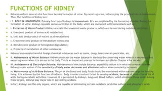
FUNCTIONS OF KIDNEY
Kidneys perform several vital functions besides formation of urine. By excreting urine, kidneys play the principal role in homeostasis.
Thus, the functions of kidney are:
„ 1. ROLE IN HOMEOSTASIS: Primary function of kidneys is homeostasis. It is accomplished by the formation of urine. During the
formation of urine, kidneys regulate various activities in the body, which are concerned with homeostasis such as:
i. Excretion of Waste Products Kidneys excrete the unwanted waste products, which are formed during metabolic activities:
a. Urea (end product of amino acid metabolism)
b. Uric acid (end product of nucleic acid metabolism)
c. Creatinine (end product of metabolism in muscles)
d. Bilirubin (end product of hemoglobin degradation)
e. Products of metabolism of other substances.
Kidneys also excrete harmful foreign chemical substances such as toxins, drugs, heavy metals pesticides, etc.
ii. Maintenance of Water Balance: Kidneys maintain the water balance in the body by conserving water when it is decreased and
excreting water when it is excess in the body. This is an important process for homeostasis (Refer Chapter 4 for details).
iii. Maintenance of Electrolyte Balance: Maintenance of electrolyte balance, especially sodium is in relation to water balance.
Kidneys retain sodium if the osmolarity of body water decreases and eliminate sodium when osmolarity increases.
iv. Maintenance of Acid–Base Balance: The pH of the blood and body fluids should be maintained within narrow range for healthy
living. It is achieved by the function of kidneys . Body is under constant threat to develop acidosis, because of production of lot of
acids during metabolic activities. However, it is prevented by kidneys, lungs and blood buffers, which eliminate these acids. Among
these organs, kidneys play major role in preventing acidosis.
In fact, kidneys are the only organs, which are capable of eliminating certain metabolic acids like sulfuric and phosphoric acids.
5.
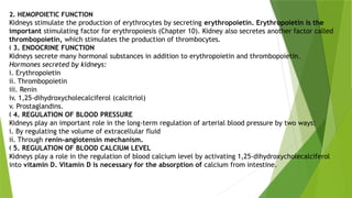
2. HEMOPOIETIC FUNCTION
Kidneysstimulate the production of erythrocytes by secreting erythropoietin. Erythropoietin is the
important stimulating factor for erythropoiesis (Chapter 10). Kidney also secretes another factor called
thrombopoietin, which stimulates the production of thrombocytes.
„ 3. ENDOCRINE FUNCTION
Kidneys secrete many hormonal substances in addition to erythropoietin and thrombopoietin.
Hormones secreted by kidneys:
i. Erythropoietin
ii. Thrombopoietin
iii. Renin
iv. 1,25-dihydroxycholecalciferol (calcitriol)
v. Prostaglandins.
„ 4. REGULATION OF BLOOD PRESSURE
Kidneys play an important role in the long-term regulation of arterial blood pressure by two ways:
i. By regulating the volume of extracellular fluid
ii. Through renin-angiotensin mechanism.
„ 5. REGULATION OF BLOOD CALCIUM LEVEL
Kidneys play a role in the regulation of blood calcium level by activating 1,25-dihydroxycholecalciferol
into vitamin D. Vitamin D is necessary for the absorption of calcium from intestine.
6.
FUNCTIONAL ANATOMY OFKIDNEY
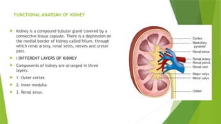
Kidney is a compound tubular gland covered by a
connective tissue capsule. There is a depression on
the medial border of kidney called hilum, through
which renal artery, renal veins, nerves and ureter
pass.
„ DIFFERENT LAYERS OF KIDNEY
Components of kidney are arranged in three
layers:
1. Outer cortex
2. Inner medulla
3. Renal sinus.
7.
1. Outer Cortex:Cortex is dark and granular in appearance. It contains renal corpuscles and
convoluted tubules. At intervals, cortical tissue penetrates medulla in the form of columns,
which are called renal columns or columns of Bertini.
2. Inner Medulla: Medulla contains tubular and vascular structures arranged in parallel radial
lines. Medullary mass is divided into 8 to 18 medullary or Malpighian pyramids.
Broad base of each pyramid is in contact with cortex and the apex projects into minor calyx.
3. Renal Sinus: Renal sinus consists of the following structures:
i. Upper expanded part of ureter called renal pelvis
ii. Subdivisions of pelvis: 2 or 3 major calyces and about 8 minor calyces
iii. Branches of nerves, arteries and tributaries of veins
iv. Loose connective tissues and fat.
TUBULAR STRUCTURES OF KIDNEY: Kidney is made up of closely arranged tubular structures
called uriniferous tubules. Blood vessels and interstitial connective tissues are interposed
between these tubules. Uriniferous tubules include:
1. Terminal or secretary tubules called nephrons, which are concerned with formation of
urine
2. Collecting ducts or tubules, which are concerned with transport of urine from nephrons
to pelvis of ureter.
Collecting ducts unite to form ducts of Bellini, which open into minor calyces through
papilla.
8.
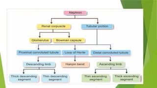
Nephron
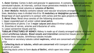
Nephron isdefined as the
structural and functional unit of
kidney. Each kidney consists of 1
to 1.3 millions of nephrons. The
number of nephrons starts
decreasing after about 45 to 50
years of age at the rate of 0.8% to
1% every year.
Each nephron is formed by two
parts :
1. A blind end called renal
corpuscle or Malpighian corpuscle
2. A tubular portion called renal
tubule.
9.
RENAL CORPUSCLE
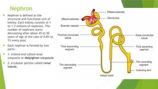
Renalcorpuscle or Malpighian corpuscle is a spheroidal and slightly flattened
structure with a diameter of about 200 μ.
Function of the renal corpuscle is the filtration of blood which forms the first phase
of urine formation.
„ SITUATION OF RENAL CORPUSCLE AND TYPES OF NEPHRON
Renal corpuscle is situated in the cortex of the kidney either near the periphery or
near the medulla.
Classification of Nephrons
Based on the situation of renal corpuscle, the nephrons are classified into two types:
1. Cortical nephrons or superficial nephrons: Nephrons having the corpuscles in
outer cortex of the kidney near the periphery . In human kidneys, 85% nephrons are
cortical nephrons.
2. Juxtamedullary nephrons: Nephrons having the corpuscles in inner
cortex near medulla or corticomedullary junction.
10.
STRUCTURE OF RENALCORPUSCLE
Renal corpuscle is formed by two portions:
1. Glomerulus
2. Bowman capsule.
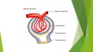
Glomerulus: Glomerulus is a tuft of capillaries enclosed by Bowman capsule. It consists of
glomerular capillaries interposed between afferent arteriole on one end and efferent arteriole on
the other end. Thus, the vascular system in the glomerulus is purely arterial . Glomerular
capillaries arise from the afferent arteriole. After entering the Bowman capsule, the afferent
arteriole divides into 4 or 5 large capillaries. Each large capillary subdivides into many small
capillaries. These small capillaries are arranged in irregular loops and form anastomosis. All the
smaller capillaries finally reunite to form the efferent arteriole, which leaves the Bowman
capsule. Diameter of the efferent arteriole is less than that of afferent arteriole. This difference
in diameter has got functional significance.
Functional histology
Glomerular capillaries are made up of single layer of endothelial cells, which are attached to a
basement membrane. Endothelium has many pores called fenestrae or filtration pores.
Diameter of each pore is 0.1 μ. Presence of the fenestra is the evidence of the filtration function
of the glomerulus.
12.
Bowman Capsule
Bowmancapsule is a capsular structure, which encloses the glomerulus. It is formed by two layers:
i. Inner visceral layer
ii. Outer parietal layer.
Visceral layer covers the glomerular capillaries. It is continued as the parietal layer at the visceral pole.
Parietal layer is continued with the wall of the tubular portion of nephron. The cleftlike space between
the visceral and parietal layers is continued as the lumen of the tubular portion. Functional anatomy of
Bowman capsule resembles a funnel with filter paper. Diameter of Bowman capsule is 200 μ.
Functional histology
Both the layers of Bowman capsule are composed of a single layer of flattened epithelial cells resting on a
basement membrane. Basement membrane of the visceral layer fuses with the basement membrane of
glomerular capillaries on which the capillary endothelial cells are arranged. Thus, the basement
membranes, which are fused together, form the separation between the glomerular capillary endothelium
and the epithelium of visceral layer of Bowman capsule. Epithelial cells of the visceral layer fuse with the
basement membrane but the fusion is not complete. Each cell is connected with basement membrane by
cytoplasmic extensions of epithelial cells called pedicles or feet. These pedicles are arranged in an
interdigitating manner leaving small cleft like spaces in between. The Cleft like space is called slit pore.
Epithelial cells with pedicles are called podocytes
13.
TUBULAR PORTION OFNEPHRON
Tubular portion of nephron is the continuation of Bowman capsule.
It is made up of three parts:
1. Proximal convoluted tubule 2. Loop of Henle 3. Distal convoluted tubule
PROXIMAL CONVOLUTED TUBULE: Proximal convoluted tubule is the coiled portion arising from
Bowman capsule. It is situated in the cortex. It is continued as descending limb of loop of Henle. Length
of proximal convoluted tubule is 14 mm and the diameter is 55 μ. Proximal convoluted tubule is
continued as loop of Henle.
Functional histology: Proximal convoluted tubule is formed by single layer of cuboidal epithelial cells.
Characteristic feature of these cells is the presence of hairlike projections directed towards the lumen
of the tubule. Because of the presence of these projections, the epithelial cells are called brush-
bordered cells.
„ LOOP OF HENLE: Loop of Henle consists of:
i. Descending limb
ii. Hairpin bend
iii. Ascending limb.
14.
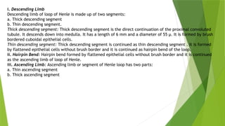
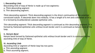
i. Descending Limb
Descendinglimb of loop of Henle is made up of two segments:
a. Thick descending segment
b. Thin descending segment.
Thick descending segment: Thick descending segment is the direct continuation of the proximal convoluted
tubule. It descends down into medulla. It has a length of 6 mm and a diameter of 55 μ. It is formed by brush
bordered cuboidal epithelial cells.
Thin descending segment: Thick descending segment is continued as thin descending segment . It is formed
by flattened epithelial cells without brush border and it is continued as hairpin bend of the loop.
ii. Hairpin Bend: Hairpin bend formed by flattened epithelial cells without brush border and it is continued
as the ascending limb of loop of Henle.
iii. Ascending Limb: Ascending limb or segment of Henle loop has two parts:
a. Thin ascending segment
b. Thick ascending segment
15.
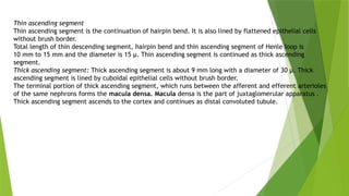
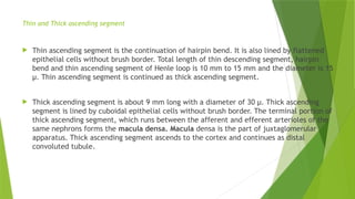
Thin ascending segment
Thinascending segment is the continuation of hairpin bend. It is also lined by flattened epithelial cells
without brush border.
Total length of thin descending segment, hairpin bend and thin ascending segment of Henle loop is
10 mm to 15 mm and the diameter is 15 μ. Thin ascending segment is continued as thick ascending
segment.
Thick ascending segment: Thick ascending segment is about 9 mm long with a diameter of 30 μ. Thick
ascending segment is lined by cuboidal epithelial cells without brush border.
The terminal portion of thick ascending segment, which runs between the afferent and efferent arterioles
of the same nephrons forms the macula densa. Macula densa is the part of juxtaglomerular apparatus .
Thick ascending segment ascends to the cortex and continues as distal convoluted tubule.
16.
i. Descending Limb
Descendinglimb of loop of Henle is made up of two segments:
a. Thick descending segment
b. Thin descending segment.
Thick descending segment: Thick descending segment is the direct continuation of the proximal
convoluted tubule. It descends down into medulla. It has a length of 6 mm and a diameter of 55 μ.
It is formed by brushbordered cuboidal epithelial cells.
Thin descending segment: Thick descending segment is continued as thin descending segment. It is
formed by flattened epithelial cells without brush border and it is continued as hairpin bend of the
loop.
ii. Hairpin Bend
Hairpin bend formed by flattened epithelial cells without brush border and it is continued as the
ascending limb of loop of Henle.
iii. Ascending Limb
Ascending limb or segment of Henle loop has two parts:
a. Thin ascending segment
b. Thick ascending segment.
17.
Thin and Thickascending segment
Thin ascending segment is the continuation of hairpin bend. It is also lined by flattened
epithelial cells without brush border. Total length of thin descending segment, hairpin
bend and thin ascending segment of Henle loop is 10 mm to 15 mm and the diameter is 15
μ. Thin ascending segment is continued as thick ascending segment.
Thick ascending segment is about 9 mm long with a diameter of 30 μ. Thick ascending
segment is lined by cuboidal epithelial cells without brush border. The terminal portion of
thick ascending segment, which runs between the afferent and efferent arterioles of the
same nephrons forms the macula densa. Macula densa is the part of juxtaglomerular
apparatus. Thick ascending segment ascends to the cortex and continues as distal
convoluted tubule.
19.
Length and Extentof Loop of Henle
Length and the extent of the loop of Henle vary in different nephrons:
i. In cortical nephrons, it is short and the hairpin bend penetrates only
up to outer medulla
ii. In juxtamedullary nephrons, this is long and the hairpin bend extends
deep into the inner medulla. In some nephrons it even runs up to the
papilla.
20.
DISTAL CONVOLUTED TUBULEAND COLLECTING DUCT
Distal convoluted tubule is the continuation of thick ascending segment and occupies the cortex of kidney.
It is continued as collecting duct. The length of the distal convoluted tubule is 14.5 to 15 mm. It has a
diameter of 22 to 50 μ.
Functional histology
Distal convoluted tubule is lined by single layer of cuboidal epithelial cells without brush border. Epithelial
cells in distal convoluted tubule are called intercalated cells (I cells).
COLLECTING DUCT
Distal convoluted tubule continues as the initial or arched collecting duct, which is in cortex. The lower
part of the collecting duct lies in medulla. Seven to ten initial
collecting ducts unite to form the straight collecting duct, which passes through medulla.
Length of the collecting duct is 20 to 22 mm and its diameter varies between 40 and 200 μ. Collecting duct
is formed by cuboidal or columnar epithelial cells.
Functional histology
Collecting duct is formed by two types of epithelial cells:
1. Principal or P cells
2. Intercalated or I cells.
21.
PASSAGE OF URINE
At the inner zone of medulla, the straight collecting ducts from each medullary
pyramid unite to form papillary ducts or ducts of Bellini, which open into a
‘V’ shaped area called papilla. Urine from each medullary pyramid is
collected in the papilla. From here it is drained into a minor calyx. Three or
four minor calyces unite to form one major calyx. Each kidney has got about
8 minor calyces and 2 to 3 major calyces.
From minor calyces urine passes through major calyces, which open into the
pelvis of the ureter. Pelvis is the expanded portion of ureter present in the
renal sinus. From renal pelvis, urine passes through remaining portion of ureter
and reaches urinary bladder.
22.
Juxtaglomerular Apparatus
DEFINITION
Juxtaglomerular apparatus is a specialized organ situated near the glomerulus of each nephron (juxta =
near).
„ STRUCTURE OF JUXTAGLOMERULAR APPARATUS Juxtaglomerular apparatus is formed by three different
structures:
1. Macula densa
2. Extraglomerular mesangial cells
3. Juxtaglomerular cells.
„ MACULA DENSA: Macula densa is the end portion of thick ascending segment before it opens into distal
convoluted tubule. It is situated between afferent and efferent arterioles of the same nephron. It is very
close to afferent arteriole. Macula densa is formed by tightly packed cuboidal epithelial cells.
„ EXTRAGLOMERULAR MESANGIAL CELLS: Extraglomerular mesangial cells are situated in the triangular
region bound by afferent arteriole, efferent arteriole and macula densa. These cells are also called
agranular cells, lacis cells or Goormaghtigh cells.
23.
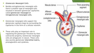
Glomerular MesangialCells
Besides extraglomerular mesangial cells
there is another type of mesangial cells
situated in between glomerular capillaries
called glomerular mesangial or
intraglomerular mesangial cells.
Glomerular mesangial cells support the
glomerular capillary loops by surrounding the
capillaries in the form of a cellular network.
These cells play an important role in
regulating the glomerular filtration by their
contractile property. Glomerular mesangial
cells are phagocytic in nature. These cells
also secrete glomerular interstitial matrix,
prostaglandins and cytokines.
24.
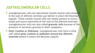
JUXTAGLOMERULAR CELLS
Juxtaglomerularcells are specialized smooth muscle cells situated
in the wall of afferent arteriole just before it enters the Bowman
capsule. These smooth muscle cells are mostly present in tunica
media and tunica adventitia of the wall of the afferent arteriole.
Juxtaglomerular cells are also called granular cells because of the
presence of secretary granules in their cytoplasm.
Polar Cushion or Polkissen: Juxtaglomerular cells form a thick
cuff called polar cushion or polkissen around the afferent
arteriole before it enters the Bowman capsule.
25.
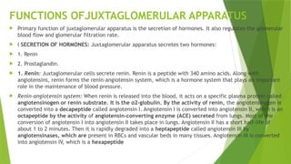
FUNCTIONS OFJUXTAGLOMERULAR APPARATUS
Primary function of juxtaglomerular apparatus is the secretion of hormones. It also regulates the glomerular
blood flow and glomerular filtration rate.
„ SECRETION OF HORMONES: Juxtaglomerular apparatus secretes two hormones:
1. Renin
2. Prostaglandin.
1. Renin: Juxtaglomerular cells secrete renin. Renin is a peptide with 340 amino acids. Along with
angiotensins, renin forms the renin-angiotensin system, which is a hormone system that plays an important
role in the maintenance of blood pressure.
Renin-angiotensin system: When renin is released into the blood, it acts on a specific plasma protein called
angiotensinogen or renin substrate. It is the α2-globulin. By the activity of renin, the angiotensinogen is
converted into a decapeptide called angiotensin I. Angiotensin I is converted into angiotensin II, which is an
octapeptide by the activity of angiotensin-converting enzyme (ACE) secreted from lungs. Most of the
conversion of angiotensin I into angiotensin II takes place in lungs. Angiotensin II has a short half-life of
about 1 to 2 minutes. Then it is rapidly degraded into a heptapeptide called angiotensin III by
angiotensinases, which are present in RBCs and vascular beds in many tissues. Angiotensin III is converted
into angiotensin IV, which is a hexapeptide
26.
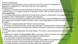
Actions of Angiotensins
AngiotensinI is physiologically inactive and serves only as the precursor of angiotensin II.
Angiotensin II: Angiotensin II is the most active form. Its actions are:
On blood vessels:
i. Angiotensin II increases arterial blood pressure by directly acting on the blood vessels and causing
vasoconstriction. It is a potent constrictor of arterioles. Earlier, when its other actions were not found it was
called hypertensin.
ii. It increases blood pressure indirectly by increasing the release of noradrenaline from postganglionic
sympathetic fibers. Noradrenaline is a general vasoconstrictor.
On adrenal cortex:It stimulates zona glomerulosa of adrenal cortex to secrete aldosterone. Aldosterone acts on
renal tubules and increases retention of sodium, which is also responsible for elevation of blood pressure.
On kidney:
i. Angiotensin II regulates glomerular filtration rate by two ways:
a. It constricts the efferent arteriole, which causes decrease in filtration after an initial increase
b. It contracts the glomerular mesangial cells leading to decrease in surface area of glomerular capillaries and
filtration.
ii. It increases sodium reabsorption from renal tubules. This action is more predominant on proximal tubules.
On brain:
i. Angiotensin II inhibits the baroreceptor reflex and thereby indirectly increases the blood pressure.
Baroreceptor reflex is responsible for decreasing the blood pressure.
ii. It increases water intake by stimulating the thirst center
iii. It increases the secretion of orticotropin-releasing hormone (CRH) from hypothalamus. CRH in
turn increases secretion of adrenocorticotropic hormone (ACTH) from pituitary
iv. It increases secretion of antidiuretic hormone (ADH) from hypothalamus.
27.
Renal Circulation
Bloodvessels of kidneys are highly specialized to facilitate the functions of nephrons in the
formation of urine. In the adults, during resting conditions both the
kidneys receive 1,300 mL of blood per minute or about 26% of the cardiac output.
Maximum blood supply to kidneys has got the functional significance. Renal arteries supply blood
to the kidneys.
28.
RENAL BLOOD VESSELS
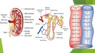
Renal Artery
Renal artery arises directly from abdominal aorta and enters the kidney through the hilus. While passing through renal
sinus, the renal artery divides into many segmental arteries.
Segmental Artery
Segmental artery subdivides into interlobar arteries.
Interlobar Artery
Interlobar artery passes in between the medullary pyramids. At the base of the pyramid, it turns and runs parallel to the
base of pyramid forming arcuate artery.
Arcuate Artery
Each arcuate artery gives rise to interlobular arteries.
Interlobular Artery
Interlobular arteries run through the renal cortex perpendicular to arcuate artery. From each interlobular artery,
numerous afferent arterioles arise.
Afferent Arteriole
Afferent arteriole enters the Bowman capsule and forms glomerular capillary tuft. After entering the Bowman capsule,
the afferent arteriole divides into 4 or 5 large capillaries.
29.
Glomerular Capillaries
Each largecapillary divides into small glomerular capillaries, which form the loops. And, the
capillary
loops unite to form the efferent arteriole, which leaves the Bowman capsule.
Efferent Arteriole
Efferent arterioles form a second capillary network called peritubular capillaries, which surround
the tubular portions of the nephrons. Thus, the renal circulation forms a portal system by the
presence of two sets of capillaries namely glomerular capillaries and peritubular capillaries.
Peritubular Capillaries and Vasa Recta
Peritubular capillaries are found around the tubular portion of cortical nephrons only. The tubular
portion of juxtamedullary nephrons is supplied by some specialized capillaries called vasa recta.
These capillaries are straight blood vessels hence the name vasa recta. Vasa recta arise directly
from the efferent arteriole of the juxtamedullary nephrons and run parallel to the renal tubule into
the medulla and ascend up towards the cortex .
Venous System Peritubular capillaries and vasa recta drain into the venous system. Venous system
starts with peritubular venules and continues as interlobular veins, arcuate veins, interlobar veins,
segmental veins and finally the renal vein.
Renal vein leaves the kidney through the hilus and joins inferior vena cava.
31.
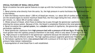
SPECIAL FEATURES OFRENAL CIRCULATION
Renal circulation has some special features to cope up with the functions of the kidneys. Such special features
are:
1. Renal arteries arise directly from the aorta. So, the high pressure in aorta facilitates the high blood flow
to the kidneys.
2. Both the kidneys receive about 1,300 mL of blood per minute, i.e. about 26% of cardiac output. Kidneys
are the second organs to receive maximum blood flow, the first organ being the liver, which receives 1,500 mL
per minute, i.e. about 30% of cardiac output.
3. Whole amount of blood, which flows to kidney has to pass through the glomerular capillaries before
entering the venous system. Because of this, the blood is completely filtered at the renal glomeruli.
4. Renal circulation has a portal system, i.e. a double network of capillaries, the glomerular capillaries and
peritubular capillaries.
5. Renal glomerular capillaries form high pressure bed with a pressure of 60 mm Hg to 70 mm Hg. It is
much greater than the capillary pressure elsewhere in the body, which is only about 25 mm Hg to 30 mm
Hg. High pressure is maintained in the glomerular capillaries because the diameter of afferent arteriole
is more than that of efferent arteriole. The high capillary pressure augments glomerular filtration.
6. Peritubular capillaries form a low pressure bed with a pressure of 8 mm Hg to 10 mm Hg. This low
pressure helps tubular reabsorption.
7. Autoregulation of renal blood flow is well established.
32.
Urine Formation
Urineformation is a blood cleansing function. Normally, about 1,300 mL of blood (26% of cardiac output)
enters the kidneys. Kidneys excrete the unwanted substances along with water from the blood as urine.
Normal urinary output is 1 L/day to 1.5 L/day.
Processes of Urine Formation
When blood passes through glomerular capillaries, the plasma is filtered into the Bowman capsule. This
process is called glomerular filtration. Filtrate from Bowman capsule passes through the tubular portion of
the nephron. While passing through the tubule, the filtrate undergoes various changes both in quality and
in quantity. Many wanted substances like glucose, amino acids, water and electrolytes are reabsorbed
from the tubules. This process is called tubular reabsorption. And, some unwanted substances are
secreted into the tubule from peritubular blood vessels. This process is called tubular secretion or
excretion. Thus, the urine formation includes three processes:
A. Glomerular filtration
B. Tubular reabsorption
C. Tubular secretion.
Among these three processes filtration is the function of the glomerulus. Reabsorption and secretion are
the functions of tubular portion of the nephron.
33.
GLOMERULAR FILTRATION
Glomerularfiltration is the process by which the blood is filtered while passing through the glomerular
capillaries by filtration membrane. It is the first process of urine formation. The structure of filtration
membrane is well suited for filtration.
Filtration Membrane: Filtration membrane is formed by three layers:
1. Glomerular capillary membrane
2. Basement membrane
3. Visceral layer of Bowman capsule.
1. Glomerular capillary membrane Glomerular capillary membrane is formed by single layer of endothelial
cells, which are attached to the basement membrane. The capillary membrane has many pores called
fenestrae or filtration pores with a diameter of 0.1 μ.
2. Basement membrane: Basement membrane of glomerular capillaries and the basement membrane of
visceral layer of Bowman capsule fuse together. The fused basement membrane separates the endothelium of
glomerular capillary and the epithelium of visceral layer of Bowman capsule.
3. Visceral layer of Bowman capsule: This layer is formed by a single layer of flattened epithelial cells resting
on a basement membrane. Each cell is connected with the basement membrane by cytoplasmic extensions
called pedicles or feet. Epithelial cells with pedicles are called podocytes. Pedicles interdigitate leaving
small cleftlike spaces in between. The cleftlike space is called slit pore or filtration slit. Filtration takes
place through these slit pores.
34.
Process of GlomerularFiltration
and ultrafiltration
When blood passes through glomerular capillaries, the plasma is filtered into the Bowman capsule.
All the substances of plasma are filtered except the plasma proteins. The filtered fluid is called
glomerular filtrate.
Ultrafiltration
Glomerular filtration is called ultrafiltration because even the minute particles are filtered. But,
the plasma proteins are not filtered due to their large molecular size. The protein molecules are
larger than the slit pores present in the endothelium of capillaries. Thus, the glomerular filtrate
contains all the substances present in plasma except the plasma proteins.
GLOMERULAR FILTRATION RATE: Glomerular filtration rate (GFR) is defined as the total quantity of
filtrate formed in all the nephrons of both the kidneys in the given unit of time. Normal GFR is 125
mL/minute or about 180 L/day.
„ FILTRATION FRACTION:Filtration fraction is the fraction (portion) of the renal plasma, which
becomes the filtrate. It is the ratio between renal plasma flow and glomerular filtration rate. It is
expressed in percentage. Normal filtration fraction varies from 15% to 20%.
35.
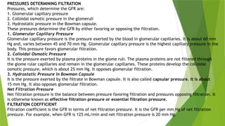
PRESSURES DETERMINING FILTRATION
Pressures,which determine the GFR are:
1. Glomerular capillary pressure
2. Colloidal osmotic pressure in the glomeruli
3. Hydrostatic pressure in the Bowman capsule.
These pressures determine the GFR by either favoring or opposing the filtration.
1. Glomerular Capillary Pressure
Glomerular capillary pressure is the pressure exerted by the blood in glomerular capillaries. It is about 60 mm
Hg and, varies between 45 and 70 mm Hg. Glomerular capillary pressure is the highest capillary pressure in the
body. This pressure favors glomerular filtration.
2. Colloidal Osmotic Pressure
It is the pressure exerted by plasma proteins in the glome ruli. The plasma proteins are not filtered through
the glome rular capillaries and remain in the glomerular capillaries. These proteins develop the colloidal
osmotic pressure, which is about 25 mm Hg. It opposes glomerular filtration.
3. Hydrostatic Pressure in Bowman Capsule
It is the pressure exerted by the filtrate in Bowman capsule. It is also called capsular pressure. It is about
15 mm Hg. It also opposes glomerular filtration.
Net Filtration Pressure
Net filtration pressure is the balance between pressure favoring filtration and pressures opposing filtration. It
is otherwise known as effective filtration pressure or essential filtration pressure.
FILTRATION COEFFICIENT
Filtration coefficient is the GFR in terms of net filtration pressure. It is the GFR per mm Hg of net filtration
pressure. For example, when GFR is 125 mL/min and net filtration pressure is 20 mm Hg.
36.
TUBULAR REABSORPTION
Tubularreabsorption is the process by which water and other substances are transported
from renal tubules back to the blood. When the glomerular filtrate flows through the
tubular portion of nephron, both quantitative and qualitative changes occur. Large quantity
of water (more than 99%), electrolytes and other substances are reabsorbed by the tubular
epithelial cells. The reabsorbed substances move into the interstitial fluid of renal
medulla. And, from here, the substances move into the blood in peritubular capillaries.
Since the substances are taken back into the blood from the glomerular filtrate, the entire
process is called tubular reabsorption.
37.
SELECTIVE REABSORPTION
Tubularreabsorption is known as selective reabsorption because the tubular cells reabsorb only the
substances necessary for the body. Essential substances such as glucose, amino acids and vitamins are
completely reabsorbed from renal tubule. Whereas the unwanted substances like metabolic waste products
are not reabsorbed and excreted through urine.
MECHANISM OF REABSORPTION: Basic transport mechanisms involved in tubular reabsorption are of two
types:
1. Active reabsorption
2. Passive reabsorption.
1. Active Reabsorption
Active reabsorption is the movement of molecules against the electrochemical (uphill) gradient. It needs
liberation of energy, which is derived from ATP. Substances reabsorbed actively Substances reabsorbed
actively from the renal tubule are sodium, calcium, potassium, phosphates, sulfates,bicarbonates, glucose,
amino acids, ascorbic acid, uricacid and ketone bodies.
2. Passive Reabsorption
Passive reabsorption is the movement of moleculesalong the electrochemical (downhill) gradient. This
process does not need energy. Substances reabsorbed passively Substances reabsorbed passively are chloride,
ureaand water.
38.
ROUTES OF REABSORPTION
Reabsorption of substances from tubular lumen into the peritubular capillary occurs by two routes:
1. Trancelluar route
2. Paracellular route.
1. Transcellular Route
In this route the substances move through the cell. It includes transport of substances from:
a. Tubular lumen into tubular cell through apical (luminal) surface of the cell membrane
b. Tubular cell into interstitial fluid
c. Interstitial fluid into capillary.
2. Paracelluar Route
In this route, the substances move through the intercellular space. It includes transport of substances
from:
i. Tubular lumen into interstitial fluid present in lateral intercellular space through the tight junction
between the cells
ii. Interstitial fluid into capillary
39.
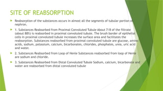
SITE OF REABSORPTION
Reabsorption of the substances occurs in almost all the segments of tubular portion of
nephron.
1. Substances Reabsorbed from Proximal Convoluted Tubule About 7/8 of the filtrate
(about 88%) is reabsorbed in proximal convoluted tubule. The brush border of epithelial
cells in proximal convoluted tubule increases the surface area and facilitates the
reabsorption. Substances reabsorbed from proximal convoluted tubule are glucose, amino
acids, sodium, potassium, calcium, bicarbonates, chlorides, phosphates, urea, uric acid
and water.
2. Substances Reabsorbed from Loop of Henle Substances reabsorbed from loop of Henle
are sodium and chloride.
3. Substances Reabsorbed from Distal Convoluted Tubule Sodium, calcium, bicarbonate and
water are reabsorbed from distal convoluted tubule
40.
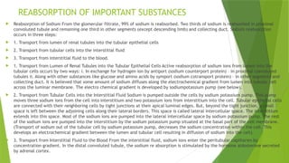
REABSORPTION OF IMPORTANTSUBSTANCES
Reabsorption of Sodium From the glomerular filtrate, 99% of sodium is reabsorbed. Two thirds of sodium is reabsorbed in proximal
convoluted tubule and remaining one third in other segments (except descending limb) and collecting duct. Sodium reabsorption
occurs in three steps:
1. Transport from lumen of renal tubules into the tubular epithelial cells
2. Transport from tubular cells into the interstitial fluid
3. Transport from interstitial fluid to the blood.
1. Transport from Lumen of Renal Tubules into the Tubular Epithelial Cells Active reabsorption of sodium ions from lumen into the
tubular cells occurs by two ways: i. In exchange for hydrogen ion by antiport (sodium counterport protein) – in proximal convoluted
tubules ii. Along with other substances like glucose and amino acids by symport (sodium cotransport protein) – in other segments and
collecting duct. It is believed that some amount of sodium diffuses along the electrochemical gradient from lumen into tubular cell
across the luminar membrane. The electro chemical gradient is developed by sodiumpotassium pump (see below).
2. Transport from Tubular Cells into the Interstitial Fluid Sodium is pumped outside the cells by sodium potassium pump. This pump
moves three sodium ions from the cell into interstitium and two potassium ions from interstitium into the cell. Tubular epithelial cells
are connected with their neighboring cells by tight junctions at their apical luminal edges. But, beyond the tight junction, a small
space is left between the adjoining cells along their lateral borders. This space is called lateral intercellular space. The interstitium
extends into this space. Most of the sodium ions are pumped into the lateral intercellular space by sodium potassium pump. The rest
of the sodium ions are pumped into the interstitium by the sodium potassium pump situated at the basal part of the cell membrane.
(Transport of sodium out of the tubular cell by sodium potassium pump, decreases the sodium concentration within the cell. This
develops an electrochemical gradient between the lumen and tubular cell resulting in diffusion of sodium into the cell).
3. Transport from Interstitial Fluid to the Blood From the interstitial fluid, sodium ions enter the peritubular capillaries by
concentration gradient. In the distal convoluted tubule, the sodium re absorption is stimulated by the hormone aldosterone secreted
by adrenal cortex.
41.
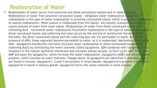
Reabsorption of Water
Reabsorption of water occurs from proximal and distal convoluted tubules and in collecting duct.
Reabsorption of water from proximal convoluted tubule – obligatory water reabsorption Obligatory
reabsorption is the type of water reabsorption in proximal convoluted tubule, which is secondary (obligatory)
to sodium reabsorption. When sodium is reabsorbed from the tubule, the osmotic pressure decreases. It
causes osmosis of water from renal tubule. Reabsorption of water from distal convoluted tubule and
collecting duct – facultative water reabsorption Facultative reabsorption is the type of water reabsorption in
distal convoluted tubule and collecting duct that occurs by the activity of antidiuretic hormone (ADH).
Normally, the distal convoluted tubule and the collecting duct are not permeable to water. But in the
presence of ADH, these segments become permeable to water, so it is reabsorbed. Mechanism of action of
ADH – Aquaporins Antidiuretic hormone increases water reabsorption in distal convoluted tubules and
collecting ducts by stimulating the water channels called aquaporins. ADH combines with vasopressin (V2)
receptors in the tubular epithelial membrane and activates adenyl cyclase, to form cyclic AMP. This cyclic
AMP activates the aquaporins, which increase the water reabsorption. Aquaporins (AQP) are the membrane
proteins, which function as water channels. Though about 10 aquaporins are identified in mammals only 5
are found in humans. Aquaporin1, 2 and 3 are present in renal tubules. Aquaporin4 is present in brain and
aquaporin5 is found in salivary glands. Aquaporin2 forms the water channels in renal tubules.
42.
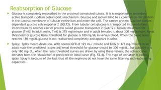
Reabsorption of Glucose
Glucose is completely reabsorbed in the proximal convoluted tubule. It is transported by secondary
active transport (sodium cotransport) mechanism. Glucose and sodium bind to a common carrier protein
in the luminal membrane of tubular epithelium and enter the cell. The carrier protein is called sodium-
dependant glucose cotransporter 2 (SGLT2). From tubular cell glucose is transported into medullary
interstitium by another carrier protein called glucose transporter 2 (GLUT2). Tubular maximum for
glucose (TmG) In adult male, TmG is 375 mg/minute and in adult females it about 300 mg/minute. Renal
threshold for glucose Renal threshold for glucose is 180 mg/dL in venous blood. When the blood level
reaches 180 mg/dL glucose is not reabsorbed completely and appears in urine.
Splay: Splay means deviation. With normal GFR of 125 mL/ minute and TmG of 375 mg/minute in an
adult male the predicted (expected) renal threshold for glucose should be 300 mg/dL. But actually it is
only 180 mg/dL. When the renal threshold curves are drawn by using these values, the actual curve
deviates from the ‘should be’ or predicted or ideal curve (Fig. 52.4). This type of deviation is called
splay. Splay is because of the fact that all the nephrons do not have the same filtering and reabsorbing
capacities.
43.
Reabsorption of AminoAcids
Amino acids are also reabsorbed completely in proximal convoluted tubule.
Amino acids are reabsorbed actively by the secondary active transport
mechanism along with sodium.
44.
Reabsorption of Bicarbonates
Bicarbonate is reabsorbed actively, mostly in proximal tubule . It is
reabsorbed in the form of carbon dioxide. Bicarbonate is mostly present as
sodium bicarbonate in the filtrate. Sodium bicarbonate dissociates into
sodium and bicarbonate ions in the tubular lumen. Sodium diffuses into
tubular cell in exchange of hydrogen. Bicarbonate combines with hydrogen to
form carbonic acid.
Carbonic acid dissociates into carbon dioxide and water in the presence of
carbonic anhydrase. Carbon dioxide and water enter the tubular cell. In the
tubular cells, carbon dioxide combines with water to form carbonic acid. It
immediately dissociates into hydrogen and bicarbonate. Bicarbonate from the
tubular cell enters the interstitium. There it combines with sodium to form
sodium bicarbonate
45.
TUBULAR SECRETION
Tubularsecretion is the process by which the substances are transported from blood into renal tubules.
It is also called tubular excretion. In addition to reabsorption from renal tubules, some substances are
also secreted into the lumen from the peritubular capillaries through the tubular epithelial cells. Dye
phenol red was the first substance found to be secreted in renal tubules in experimental conditions.
Later many other substances were found to be secreted.
Such substances are:
1. Paraaminohippuric acid (PAH)
2.Diodrast
3. 5hydroxyindoleacetic acid (5HIAA)
4. Amino derivatives
5. Penicillin.
46.
SUBSTANCES SECRETED INDIFFERENT SEGMENTS
OF RENAL TUBULES
1. Potassium is secreted actively by sodium-potassium pump in proximal and distal convoluted
tubules and collecting ducts
2. Ammonia is secreted in the proximal convoluted tubule
3.Hydrogen ions are secreted in the proximal and distal convoluted tubules. Maximum
hydrogen ion secretion occurs in proximal tubule
4.Urea is secreted in loop of Henle.
47.
Concentration of Urine
Every day 180 L of glomerular filtrate is formed with large quantity of water. If this much of water is excreted in urine, body will
face serious threats. So the concentration of urine is very essential. 53 tion of concentrated urine is not as simple as that of
dilute urine. It involves two processes: 1. Development and maintenance of medullary gradient by countercurrent system 2.
Secretion of ADH. Osmolarity of glomerular filtrate is same as that of plasma and it is 300 mOsm/L. But, normally urine is
concentrated and its osmolarity is four times more than that of plasma, i.e. 1,200 mOsm/L. Osmolarity of urine depends upon
two factors: 1. Water content in the body 2. Antidiuretic hormone (ADH). Mechanism of urine formation is the same for dilute
urine and concentrated urine till the fluid reaches the distal convoluted tubule. However, dilution or concentration of urine
depends upon water content of the body.
FORMATION OF DILUTE URINE When, water content in the body increases, kidney excretes dilute urine. This is achieved by
inhibition of ADH secretion from posterior pituitary (Chapter 66). So water reabsorption from renal tubules does not take place
leading to excretion of large amount of water. This makes the urine dilute.
FORMATION OF CONCENTRATED URINE When the water content in body decreases, kidney retains water and excretes
concentrated urine. Formation of concentrated urine is not as simple as that of dilute urine. It involves two processes: 1.
Development and maintenance of medullary gradient by countercurrent system 2. Secretion of ADH.
MEDULLARY GRADIENT
MEDULLARY HYPEROSMOLARITY Cortical interstitial fluid is isotonic to plasma with the osmolarity of 300 mOsm/L. Osmolarity of
medullary interstitial fluid near the cortex is also 300 mOsm/L. However, while proceeding from outer part towards the inner
part of medulla, the osmolarity increases gradually and reaches the maximum at the inner most part of medulla near renal sinus.
Here, the interstitial f luid is hypertonic with osmolarity of 1,200 mOsm/L. This type of gradual increase in the osmolarity of the
medullary interstitial fluid is called the medullary gradient. It plays an important role in the concentration of urine.
DEVELOPMENT AND MAINTENANCE OF MEDULLARY GRADIENT
Kidney has some unique mechanism called counter current mechanism, which is responsible for the develop ment and
maintenance of medullary gradient and hyper osmolarity of interstitial fluid in the inner medulla.
48.
COUNTERCURRENT MECHANISM
COUNTERCURRENTFLOW: A countercurrent system is a system of ‘U’shaped tubules (tubes) in which, the flow of
fluid is in opposite direction in two limbs of the ‘U’shaped tubules. Divisions of Countercurrent System
Countercurrent system has two divisions: 1. Countercurrent multiplier formed by loop of Henle 2. Countercurrent
exchanger formed by vasa recta. COUNTERCURRENT MULTIPLIER Loop of Henle Loop of Henle functions as
countercurrent multiplier. It is responsible for development of hyperosmolarity of medullary interstitial fluid and
medullary gradient.
Role of Loop of Henle in Development of Medullary:
Gradient Loop of Henle of juxtamedullary nephrons plays a major role as countercurrent multiplier because loop
of these nephrons is long and extends upto the deeper parts of medulla. Main reason for the hyperosmolarity of
medullary interstitial fluid is the active reabsorption of sodium chloride and other solutes from ascending limb of
Henle loop into the medullary interstitium. These solutes accumulate in the medullary interstitium and increase
the osmolarity. Now, due to the concentration gradient, the sodium and chlorine ions diffuse from medullary
interstitium into the descending limb of Henle loop and reach the ascending limb again via hairpin bend. Thus,
the sodium and chlorine ions are repeatedly re circulated between the descending limb and ascending limb of
Henle loop through medullary interstitial fluid leaving a small portion to be excreted in the urine. Apart from this
there is regular addition of more and more new sodium and chlorine ions into descending limb by constant
filtration. Thus, the reabsorption of sodium chloride from ascending limb and addition of new sodium chlorine
ions into the filtrate increase or multiply the osmolarity of medullary interstitial fluid and medullary gradient.
Hence, it is called countercurrent multiplier.
49.
Micturition
Micturition isa process by which urine is voided from the urinary bladder. It is a reflex process. However, in grown up children and
adults, it can be controlled voluntarily to some extent. The functional anatomy and nerve supply of urinary bladder are essential
for the process of micturition.
FUNCTIONAL ANATOMY OF URINARY BLADDER AND URETHRA
URINARY BLADDER Urinary bladder is a triangular hollow organ located in lower abdomen. It consists of a body and neck. Wall of
the bladder is formed by smooth muscle. It consists of three ill-defined layers of muscle fibers called detrusor muscle, viz. the
inner longitudinal layer, middle circular layer and outer longitudinal layer. Inner surface of urinary bladder is lined by mucus
membrane. In empty bladder, the mucosa falls into many folds called rugae. At the posterior surface of the bladder wall, there is
a triangular area called trigone. At the upper angles of this trigone, two ureters enter the bladder. Lower part of the bladder is
narrow and forms the neck. It opens into urethra via internal urethral sphincter.
URETHRA Male urethra has both urinary function and reproductive function. It carries urine and semen. Female urethra has only
urinary function and it carries only urine. So, male urethra is structurally different from female urethra. Male Urethra Male
urethra is about 20 cm long. After origin from bladder it traverses the prostate gland, which lies below the bladder and then runs
through the penis Throughout its length, the urethra has mucus glands called glands of Littre. Male urethra is divided into three
parts: 1. Prostatic urethra 2. Membranous urethra 3. Spongy urethra. 1. Prostatic urethra Prostatic urethra is 3 cm long and it runs
through prostate gland. The prostatic fluid is emptied into this part of urethra through prostatic sinuses. Sperms from vas
deferens and the fluid from seminal vesicles are also emptied into prostatic urethra via ejaculatory ducts (Chapter 74). Part of
the urethra after taking origin from neck of bladder before entering the prostate gland is known as preprostatic urethra. Its
length is about 0.5 to 1.5 cm. This part of urethra is considered as part of prostatic urethra. 2. Membranous urethra Membranous
urethra is about 1 to 2 cm long. It runs from base of the prostate gland through urogenital diaphragm up to the bulb of urethra. 3.
Spongy urethra Spongy urethra is also known as cavernous urethra and its length is about 15 cm. Spongy urethra is surrounded by
corpus spongiosum of penis. It is divided into a proximal bulbar urethra and a distal penil urethra. Penile urethra is narrow with a
length of about 6 cm. It ends with external urethral meatus or orifice, which is located at the end of penis. The bilateral
bulbourethral glands open into spongy urethra. Bulbourethral glands are also called Cowper glands.
50.
Female UrethraFemale urethra is narrower and shorter than male urethra. It is about 3.5 to
4 cm long. After origin from bladder it traverses through urogenital diaphragm and runs
along anterior wall of vagina. Then it terminates at external orifice of urethra, which is
located between clitoris and vaginal opening
URETHRAL SPHINCTERS There are two urethral sphincters in urinary tract:
1. Internal urethral sphincter
2. External urethral sphincter.
1. Internal Urethral sphincter This sphincter is situated between neck of the bladder and
upper end of urethra. It is made up of smooth muscle fibers and formed by thickening of
detrusor muscle. It is innervated by autonomic nerve fibers. This sphincter closes the urethra
when bladder is emptied.
2. External Urethral sphincter, the sympathetic nerve is called nerve External sphincter is
located in the urogenital diaphragm. This sphincter is made up of circular skeletal muscle
fibers, which are innervated by somatic nerve fibers.
51.
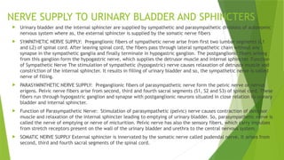
NERVE SUPPLY TOURINARY BLADDER AND SPHINCTERS
Urinary bladder and the internal sphincter are supplied by sympathetic and parasympathetic divisions of autonomic
nervous system where as, the external sphincter is supplied by the somatic nerve fibers
SYMPATHETIC NERVE SUPPLY: Preganglionic fibers of sympathetic nerve arise from first two lumbar segments (L1
and L2) of spinal cord. After leaving spinal cord, the fibers pass through lateral sympathetic chain without any
synapse in the sympathetic ganglia and finally terminate in hypogastric ganglion. The postganglionic fibers arising
from this ganglion form the hypogastric nerve, which supplies the detrusor muscle and internal sphincter. Function
of Sympathetic Nerve The stimulation of sympathetic (hypogastric) nerve causes relaxation of detrusor muscle and
constriction of the internal sphincter. It results in filling of urinary bladder and so, the sympathetic nerve is called
nerve of filling.
PARASYMPATHETIC NERVE SUPPLY: Preganglionic fibers of parasympathetic nerve form the pelvic nerve or nervus
erigens. Pelvic nerve fibers arise from second, third and fourth sacral segments (S1, S2 and S3) of spinal cord. These
fibers run through hypogastric ganglion and synapse with postganglionic neurons situated in close relation to urinary
bladder and internal sphincter.
Function of Parasympathetic Nerve: Stimulation of parasympathetic (pelvic) nerve causes contraction of detrusor
muscle and relaxation of the internal sphincter leading to emptying of urinary bladder. So, parasympathetic nerve is
called the nerve of emptying or nerve of micturition. Pelvic nerve has also the sensory fibers, which carry impulses
from stretch receptors present on the wall of the urinary bladder and urethra to the central nervous system.
SOMATIC NERVE SUPPLY External sphincter is innervated by the somatic nerve called pudendal nerve. It arises from
second, third and fourth sacral segments of the spinal cord.
52.
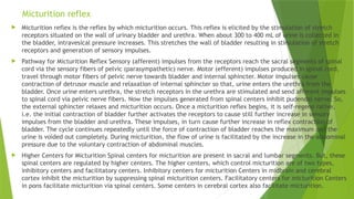
Micturition reflex
Micturitionreflex is the reflex by which micturition occurs. This reflex is elicited by the stimulation of stretch
receptors situated on the wall of urinary bladder and urethra. When about 300 to 400 mL of urine is collected in
the bladder, intravesical pressure increases. This stretches the wall of bladder resulting in stimulation of stretch
receptors and generation of sensory impulses.
Pathway for Micturition Reflex Sensory (afferent) impulses from the receptors reach the sacral segments of spinal
cord via the sensory fibers of pelvic (parasympathetic) nerve. Motor (efferent) impulses produced in spinal cord,
travel through motor fibers of pelvic nerve towards bladder and internal sphincter. Motor impulses cause
contraction of detrusor muscle and relaxation of internal sphincter so that, urine enters the urethra from the
bladder. Once urine enters urethra, the stretch receptors in the urethra are stimulated and send afferent impulses
to spinal cord via pelvic nerve fibers. Now the impulses generated from spinal centers inhibit pudendal nerve. So,
the external sphincter relaxes and micturition occurs. Once a micturition reflex begins, it is self-regene rative,
i.e. the initial contraction of bladder further activates the receptors to cause still further increase in sensory
impulses from the bladder and urethra. These impulses, in turn cause further increase in reflex contraction of
bladder. The cycle continues repeatedly until the force of contraction of bladder reaches the maximum and the
urine is voided out completely. During micturition, the flow of urine is facilitated by the increase in the abdominal
pressure due to the voluntary contraction of abdominal muscles.
Higher Centers for Micturition Spinal centers for micturition are present in sacral and lumbar segments. But, these
spinal centers are regulated by higher centers. The higher centers, which control micturition are of two types,
inhibitory centers and facilitatory centers. Inhibitory centers for micturition Centers in midbrain and cerebral
cortex inhibit the micturition by suppressing spinal micturition centers. Facilitatory centers for micturition Centers
in pons facilitate micturition via spinal centers. Some centers in cerebral cortex also facilitate micturition.
53.
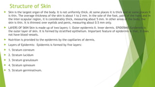
Structure of Skin
Skin is the largest organ of the body. It is not uniformly thick. At some places it is thick and at some places it
is thin. The average thickness of the skin is about 1 to 2 mm. In the sole of the foot, palm of the hand and in
the inter-scapular region, it is considerably thick, measuring about 5 mm. In other areas of the body, the
skin is thin. It is thinnest over eyelids and penis, measuring about 0.5 mm only.
LAYERS OF SKIN Skin is made up of two layers: I. Outer epidermis II. Inner dermis. EPIDERMIS Epidermis is
the outer layer of skin. It is formed by stratified epithelium. Important feature of epidermis is that, it does
not have blood vessels.
Nutrition is provided to the epidermis by the capillaries of dermis.
Layers of Epidermis: Epidermis is formed by five layers:
1. Stratum corneum
2. Stratum lucidum
3. Stratum granulosum
4. Stratum spinosum
5. Stratum germinativum.
54.
1. STRATUM CORNEUM
Stratum corneum is also known as horny layer. It is the outermost layer and
consists of dead cells, which are called corneocytes. These cells lose their
nucleus due to pressure and become dead cells. The cytoplasm is flat tened
with fibrous protein known as keratin. Apart from this, these cells also
contain phospholipids and glycogen.
55.
2. STRATUM LUCIDUM
Stratum lucidum is made up of flattened epithelial cells. Many cells have
degenerated nucleus and in some cells, the nucleus is absent. As these cells
exhibit shiny character, the layer looks like a homogeneous translucent zone.
So, this layer is called stratum lucidum (lucid = clear).
56.
3. STRATUM GRANULOSUM
Stratum granulosum is a thin layer with two to five rows of flattened
rhomboid cells. Cytoplasm contains granules of a protein called keratohyalin.
Keratohyalin is the precursor of keratin.
57.
4. STRATUM SPINOSUM
Stratum spinosum is also known as prickle cell layer because, the cells of this
layer possess some spinelike protoplasmic projections. By these projections,
the cells are connected to one another.
58.
5. STRATUM GERMINATIVUM
Stratum germinativum is a thick layer made up of polygonal cells,
superficially and columnar or cuboidal epithelial cells in the deeper parts.
Here, new cells are constantly formed by mitotic division. The newly formed
cells move continuously towards the stratum corneum. The stem cells, which
give rise to new cells, are known as keratinocytes. Another type of cells
called melanocytes are scattered between the keratinocytes. Melanocytes
produce the pigment called melanin. The color of the skin depends upon
melanin. From this layer, some projections called rete ridges extend down up
to dermis. These projections provide anchoring and nutritional function.
59.
DERMIS
Dermis isthe inner layer of the skin. It is a connective tissue layer, made up
of dense and stout collagen fibers, fibroblasts and histiocytes. Collagen fibers
exhibit elastic property and are capable of storing or holding water. Collagen
fibers contain the enzyme collagenase, which is responsible for wound
healing.
Layers of Dermis Dermis is made up of two layers: 1. Superficial papillary
layer 2. Deeper reticular layer.
60.
SUPERFICIAL PAPILLARY LAYER
Superficial papillary layer projects into the epidermis. It contains blood
vessels, lymphatics and nerve fibers. This layer also has some
pigmentcontaining cells known as chromatophores. Dermal papillae are finger
like projections, arising from the superficial papillary dermis. Each papilla
contains a plexus of capillaries and lymphatics, which are oriented
perpendicular to the skin surface. The papillae are surrounded by rete ridges,
extending from the epidermis.
61.
RETICULAR LAYER
Reticularlayer is made up of reticular and elastic fibers. These fibers are found
around the hair bulbs, sweat glands and sebaceous glands. The reticular layer also
contains mast cells, nerve endings, lymphatics, epidermal appendages and
fibroblasts. Immediately below the dermis, subcutaneous tissue is present. It is a
loose connective tissue, which connects the skin with the internal structures of the
body. It serves as an insulator to protect the body from excessive heat and cold of the
environment. Lot of smooth muscles called arrector pili are also found in skin around
the hair follicles.
62.
APPENDAGES OF SKIN
Hair follicles with hair, nails, sweat glands, sebaceous glands and mammary
glands are considered as append ages of the skin.
63.
COLOR OF SKIN
Color of skin depends upon two important factors: 1. Pigmentation of skin 2.
Hemoglobin in the blood.
64.
PIGMENTATION OF SKIN
Cells of the skin contain a brown pigment called melanin, which is responsible
for the color of the skin. It is synthesized by melanocytes, which are present
mainly in the stratum germinativum and stratum spinosum of epidermis. After
synthesis, this pigment spreads to the cells of the other layers. Melanin
Melanin is the skin pigment and it forms the major color determinant of
human skin. Skin becomes dark when melanin content increases. It is protein
in nature and it is synthesized from the amino acid tyrosine via
dihydroxyphenylalanine (DOPA). Deficiency of melanin leads to albinism
(hypopig mentary congenital disorder).
65.
HEMOGLOBIN IN THEBLOOD
Amount and nature of hemoglobin that circulates in the cutaneous blood
vessels play an important role in the coloration of the skin. Skin becomes: i.
ii. iii. Pale, when hemoglobin content decreases Pink, when blood rushes to
skin due to cutaneous vasodilatation (blushing) Bluish during cyanosis, which
is caused by excess amount of reduced hemoglobin.
66.
FUNCTIONS OF SKIN
Primary function of skin is protection of organs. However, it has many other important functions also.
1. PROTECTIVE FUNCTION Skin forms the covering of all the organs of the body and protects these organs from
the following factors: i. Bacteria and toxic substances ii. Mechanical blow iii. Ultraviolet rays. i. Protection
from Bacteria and Toxic Substances Skin covers the organs of the body and protects the organs from having
direct contact with external environment. Thus, it prevents the bacterial infection. Lysozyme secreted in skin
destroys the bacteria. Keratinized stratum corneum of epidermis is responsible for the protective function of
skin. This layer also offers resistance against toxic chemicals like acids and alkalis. If the skin is injured,
infection occurs due to invasion of bacteria from external environment. During injury or skin infection, the
keratinocytes secrete: a. Cytokines like interleukins, α-tumor necrosis factor and γ-interferon, which play
important role in inflammation, immunological reactions, tissue repair and wound healing b. Antimicrobial
peptides like β-defensins, which prevent invasion of microbes.
ii. Protection from Mechanical Blow Skin is not tightly placed over the underlying organs or tissues. It is
somewhat loose and moves over the underlying subcutaneous tissues. So, the mechanical impact of any blow
to the skin is not transmitted to the underlying tissues.
iii. Protection from Ultraviolet Rays Skin protects the body from ultraviolet rays of sunlight. Exposure to
sunlight or to any other source of ultraviolet rays increases the production of melanin pigment in skin. Melanin
absorbs ultraviolet rays. At the same time, the thickness of stratum corneum increases. This layer of epidermis
also absorbs the ultraviolet rays.
67.
2. SENSORY FUNCTION
Skin is considered as the largest sense organ in the body. It has many nerve
endings, which form the specialized cutaneous receptors .These receptors are
stimulated by sensations of touch, pain, pressure or temperature sensation
and convey these sensations to the brain via afferent nerves. At the brain
level, perception of different sensations occurs
68.
3. STORAGE FUNCTIONSkin stores fat, water, chloride and sugar. It can also store blood by the dilatation
of the cutaneous blood vessels.
4. SYNTHETIC FUNCTION Vitamin D3 is synthesized in skin by the action of ultraviolet rays from sunlight
on cholesterol.
5. REGULATION OF BODY TEMPERATURE Skin plays an important role in the regulation of body
temperature. Excess heat is lost from the body through skin by radiation, conduction, convection and
evaporation. Sweat glands of the skin play an active part in heat loss, by secreting sweat. The lipid
content of sebum prevents loss of heat from the body in cold environment.
6. REGULATION OF WATER AND ELECTROLYTE BALANCE Skin regulates water balance and electrolyte
balance by excreting water and salts through sweat.
7. EXCRETORY FUNCTION Skin excretes small quantities of waste materials like urea, salts and fatty
substance.
8. ABSORPTIVE FUNCTION Skin absorbs fat-soluble substances and some ointments.
9. SECRETORY FUNCTION Skin secretes sweat through sweat glands and sebum through sebaceous glands.
By secreting sweat, skin regulates body temperature and water balance. Sebum keeps the skin smooth
and moist.