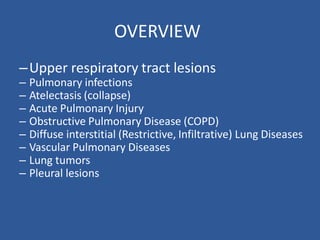
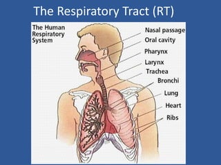
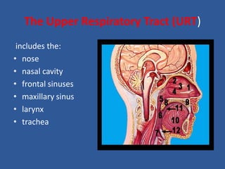
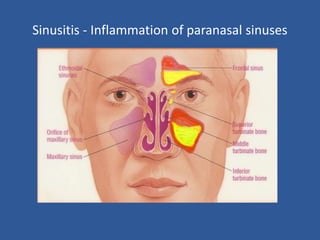
Upper Respiratory Tract
Pathology
Respiratory Pathology
OVERVIEW
–Upper respiratory tract lesions
– Pulmonary infections
– Atelectasis (collapse)
– Acute Pulmonary Injury
– Obstructive Pulmonary Disease (COPD)
– Diffuse interstitial (Restrictive, Infiltrative) Lung Diseases
– Vascular Pulmonary Diseases
– Lung tumors
– Pleural lesions
The Respiratory Tract (RT)
The Upper Respiratory Tract (URT)
includes the:
• nose
• nasal cavity
• frontal sinuses
• maxillary sinus
• larynx
• trachea
Overview
• Acute infections
• Nasal polyps
• Nasopharyngeal carcinoma
• Laryngeal lesions
Acute infections
• Common cold
• Pharyngitis
• Epiglottitis
• Laryngitis
• Laryngotracheobronchitis (croup)
• Sinusitis
Common cold
• Acute infections of the URT mostly viral and self-
limiting but can predispose to secondary
bacterial infection
• Nasal congestion, watery nasal discharge
(rhinorrhea), sneezing, dry sore throat and a
slight increase in temperature especially in young
children, malaise, myalgia and headache,
• More than 200 viruses with seasonal variation
Transmission -
• Direct contact with infected secretions
• Hand - to - hand
• Hand - to environmental surface - to hand
• Spread by aerosols
Pathogenesis
• Incubation period 1 - 4 days
• Begins in posterior pharynx
• Viral shedding days 3 - 4
• May be complicated by otitis media or sinusitis
• Use of antibiotics – no benefit, do not reduce bacterial
complications, emergence of resistant organisms
Localized infections of the URT
• Acute pharyngitis
• Mild accompanying common cold
• Severe cases need to be seriously taken –
• beta-haemolytic streptococcal infection is associated
with tonsillitis;
• Coxsackievirus A causes pharyngeal vesicles and ulcers;
• Infectious mononucleosis is caused by Epstein-Barr
virus and is another cause of pharyngitis
Diffuse tonsillar and
pharyngeal erythema
seen here as a non-
specific finding that
can be produced by a
variety of pathogens
Exudative tonsillitis
seen with either
Group A
Beta-hemolytic
streptococcal
or Epstein-Barr virus
infection.
Acute bacterial epiglottitis –
• usually in young children
• caused by H. influenzae
• can be fatal due to airway obstruction
Epiglottitis
This 2-yr old was in
severe distress
and was too
exhausted to hold
its head up.
The epiglottis
appears intensely
red and swollen.
• Acute laryngitis
Common cold, allergic reaction or due to inhalation of
irritants
• Tuberculous laryngitis
• Diphtheria caused by Corynebacterium diphtheriae,
implants in the mucosa and produces an exotoxin that
results in necrosis and a dense fibrinopurulent
exudate forming the classic pseudomembrane of
diphteria with the hazard of airway obstruction.
Absorption of the exotoxin causes myocarditis and
peripheral neuropathy.
• Laryngotracheobronchitis (croup)
• In children mostly caused by parainfluenza
virus
• Self-limiting but can cause severe inspiratory
stridor and harsh persistent cough
•
Paranasal Sinuses
Lateral view of paranasal sinuses
Sinusitis
• Bacterial or viral
• Fever (50%), purulent nasal discharge, swelling, facial
pain worse on percussion, headache, nasal
obstruction, loss of smell
• Children: facial pain, swelling, malodorous breath
(50%), cough (80%), nasal discharge (76%), fever (63%),
sore throat (23%)
• Maxillary toothache, colored nasal discharge, poor
response to nasal decongestants, abnormal
transillumination, purulent secretions, cough > 7 days
• Frontal sinusitis with tenderness and headache -
thin barrier to CNS, treat for 10-14 days
• Ethmoid sinusitis: edema of eyelids, tearing,
retroorbital pain, proptosis
• Sphenoid sinusitis: intractable headache,
hypo/hyperesthesia of ophthalmic or maxillary
branches of trigeminal n. (30%)
Sinusitis - Inflammation of paranasal sinuses
Diagnosis of sinusitis
Dr.Emad Ibrahim Osman
• Nasal swabs not helpful
• Transillumination of maxillary and frontal sinuses
• Sinus x-rays: air-fluid level, complete opacity,
mucosal thickening
• CT scan not indicated - unless chronic infection,
immunocompromised, suspected intracranial or
orbital complication
• Direct sinus aspiration
10/2/98
Factors that predispose to sinusitis
• Impaired mucociliary function
• Obstruction of sinus ostia
• Immune defects
• Increased risk of microbial invasion
10/2/98
Chronic sinusitis
• Symptoms for > 3 months
• Allergies, inadequately treated
• Aerobes and anaerobes
• ENT evaluation for endoscopy or CT scan
• Antibiotics for 3-4 weeks
Intracranial Complications
of Sinusitis
Complication Clinical Signs
• Meningitis Headache, fever,
stiff neck lethargy,
rapid death
• Osteomyelitis Pott’s puffy tumor
• Epidural abscess Headache, fever
• Subdural empyema Headache, seizures
hemiplegia, rapid death
• Cerebral abscess Convulsions, headache,
personality change
• Venous sinus thrombosis Picket-fence fever,
rapid death
• Cavernous sinus Orbital edema, ocular
palsies
Nasal polyps
• A polyp is an edematous semi-translucent mass in the
nasal and paranasal cavities, mostly originating from the
mucosal linings of the sinuses and prolapsing into the
nasal cavities.
• Etiology is unknown (-itis, -oma, -plasia)
• Hypothesis: formation of polyps involve rupture of the
surface epithelium and prolapse of the lamina propria as
a result of tissue pressure from edema.
• Allergic and infectious etiologies are the most frequently
discussed
• Morbidity is related to the obstruction,
leading to chronic sinusitis, facial pain, bony
erosion.
• Hyposmia and anosmia.
• Polyps are not pre-malignant, however can be
confused with the pre-malignant lesions.
Nasal polyps
Histological findings
• Pseudostratified
ciliated columnar
epithelium
• Thickened epithelial
basement membrane
• Oedematous stroma
Nasopharyngeal carcinoma
• Strong epidemiological association with
Epstein-Barr virus
• Endemic in Asia especially China
• Squamous cell carcinoma – mostly
undifferentiated carcinoma
• Invades locally, spreads to the cervical lymph
nodes and then distantly
• Radiosensitive and 5-year survival is 50%
Pathology
• WHO I tumors are keratinizing squamous cell carcinomas.
These tumors are not related to EBV infection.
• WHO II tumors are nonkeratinizing squamous cell
carcinomas. These tumors histologically resemble
transitional cell bladder cancer, and are also called
transitional cell carcinoma. These tumors are related to EBV
infection.
• WHO III tumors are undifferentiated carcinomas and
include lymphoepithelioma. They account for the majority
of nasopharyngeal carcinomas in the United States and
worldwide. These tumors are related to EBV infection.
Nasopharyngeal anaplastic carcinoma
Laryngeal lesions
• Most common presenting symptom is hoarseness
• Nonmalignant lesions:
vocal cord nodules (polyps)
laryngeal (squamous) papilloma
• Carcinoma – age more than 40 years, more common
in males, smokers, association with alcohol and
previous radiation exposure,
squamous cell carcinoma - may be glottic,
supraglottic or subglottic
The End
Thanks

Upper Respiratory tract diseases Pathology.pdf

  • 1.
  • 2.
    OVERVIEW –Upper respiratory tractlesions – Pulmonary infections – Atelectasis (collapse) – Acute Pulmonary Injury – Obstructive Pulmonary Disease (COPD) – Diffuse interstitial (Restrictive, Infiltrative) Lung Diseases – Vascular Pulmonary Diseases – Lung tumors – Pleural lesions
  • 3.
  • 4.
    The Upper RespiratoryTract (URT) includes the: • nose • nasal cavity • frontal sinuses • maxillary sinus • larynx • trachea
  • 5.
    Overview • Acute infections •Nasal polyps • Nasopharyngeal carcinoma • Laryngeal lesions
  • 6.
    Acute infections • Commoncold • Pharyngitis • Epiglottitis • Laryngitis • Laryngotracheobronchitis (croup) • Sinusitis
  • 7.
    Common cold • Acuteinfections of the URT mostly viral and self- limiting but can predispose to secondary bacterial infection • Nasal congestion, watery nasal discharge (rhinorrhea), sneezing, dry sore throat and a slight increase in temperature especially in young children, malaise, myalgia and headache,
  • 8.
    • More than200 viruses with seasonal variation Transmission - • Direct contact with infected secretions • Hand - to - hand • Hand - to environmental surface - to hand • Spread by aerosols Pathogenesis • Incubation period 1 - 4 days • Begins in posterior pharynx • Viral shedding days 3 - 4 • May be complicated by otitis media or sinusitis • Use of antibiotics – no benefit, do not reduce bacterial complications, emergence of resistant organisms
  • 9.
    Localized infections ofthe URT • Acute pharyngitis • Mild accompanying common cold • Severe cases need to be seriously taken – • beta-haemolytic streptococcal infection is associated with tonsillitis; • Coxsackievirus A causes pharyngeal vesicles and ulcers; • Infectious mononucleosis is caused by Epstein-Barr virus and is another cause of pharyngitis
  • 10.
    Diffuse tonsillar and pharyngealerythema seen here as a non- specific finding that can be produced by a variety of pathogens
  • 11.
    Exudative tonsillitis seen witheither Group A Beta-hemolytic streptococcal or Epstein-Barr virus infection.
  • 13.
    Acute bacterial epiglottitis– • usually in young children • caused by H. influenzae • can be fatal due to airway obstruction
  • 14.
    Epiglottitis This 2-yr oldwas in severe distress and was too exhausted to hold its head up. The epiglottis appears intensely red and swollen.
  • 16.
    • Acute laryngitis Commoncold, allergic reaction or due to inhalation of irritants • Tuberculous laryngitis • Diphtheria caused by Corynebacterium diphtheriae, implants in the mucosa and produces an exotoxin that results in necrosis and a dense fibrinopurulent exudate forming the classic pseudomembrane of diphteria with the hazard of airway obstruction. Absorption of the exotoxin causes myocarditis and peripheral neuropathy.
  • 17.
    • Laryngotracheobronchitis (croup) •In children mostly caused by parainfluenza virus • Self-limiting but can cause severe inspiratory stridor and harsh persistent cough •
  • 18.
  • 19.
    Lateral view ofparanasal sinuses
  • 20.
    Sinusitis • Bacterial orviral • Fever (50%), purulent nasal discharge, swelling, facial pain worse on percussion, headache, nasal obstruction, loss of smell • Children: facial pain, swelling, malodorous breath (50%), cough (80%), nasal discharge (76%), fever (63%), sore throat (23%) • Maxillary toothache, colored nasal discharge, poor response to nasal decongestants, abnormal transillumination, purulent secretions, cough > 7 days
  • 21.
    • Frontal sinusitiswith tenderness and headache - thin barrier to CNS, treat for 10-14 days • Ethmoid sinusitis: edema of eyelids, tearing, retroorbital pain, proptosis • Sphenoid sinusitis: intractable headache, hypo/hyperesthesia of ophthalmic or maxillary branches of trigeminal n. (30%)
  • 22.
    Sinusitis - Inflammationof paranasal sinuses
  • 23.
    Diagnosis of sinusitis Dr.EmadIbrahim Osman • Nasal swabs not helpful • Transillumination of maxillary and frontal sinuses • Sinus x-rays: air-fluid level, complete opacity, mucosal thickening • CT scan not indicated - unless chronic infection, immunocompromised, suspected intracranial or orbital complication • Direct sinus aspiration 10/2/98
  • 24.
    Factors that predisposeto sinusitis • Impaired mucociliary function • Obstruction of sinus ostia • Immune defects • Increased risk of microbial invasion 10/2/98
  • 25.
    Chronic sinusitis • Symptomsfor > 3 months • Allergies, inadequately treated • Aerobes and anaerobes • ENT evaluation for endoscopy or CT scan • Antibiotics for 3-4 weeks
  • 26.
    Intracranial Complications of Sinusitis ComplicationClinical Signs • Meningitis Headache, fever, stiff neck lethargy, rapid death • Osteomyelitis Pott’s puffy tumor • Epidural abscess Headache, fever • Subdural empyema Headache, seizures hemiplegia, rapid death • Cerebral abscess Convulsions, headache, personality change • Venous sinus thrombosis Picket-fence fever, rapid death • Cavernous sinus Orbital edema, ocular palsies
  • 27.
    Nasal polyps • Apolyp is an edematous semi-translucent mass in the nasal and paranasal cavities, mostly originating from the mucosal linings of the sinuses and prolapsing into the nasal cavities. • Etiology is unknown (-itis, -oma, -plasia) • Hypothesis: formation of polyps involve rupture of the surface epithelium and prolapse of the lamina propria as a result of tissue pressure from edema. • Allergic and infectious etiologies are the most frequently discussed
  • 28.
    • Morbidity isrelated to the obstruction, leading to chronic sinusitis, facial pain, bony erosion. • Hyposmia and anosmia. • Polyps are not pre-malignant, however can be confused with the pre-malignant lesions.
  • 29.
  • 30.
    Histological findings • Pseudostratified ciliatedcolumnar epithelium • Thickened epithelial basement membrane • Oedematous stroma
  • 31.
    Nasopharyngeal carcinoma • Strongepidemiological association with Epstein-Barr virus • Endemic in Asia especially China • Squamous cell carcinoma – mostly undifferentiated carcinoma • Invades locally, spreads to the cervical lymph nodes and then distantly • Radiosensitive and 5-year survival is 50%
  • 32.
    Pathology • WHO Itumors are keratinizing squamous cell carcinomas. These tumors are not related to EBV infection. • WHO II tumors are nonkeratinizing squamous cell carcinomas. These tumors histologically resemble transitional cell bladder cancer, and are also called transitional cell carcinoma. These tumors are related to EBV infection. • WHO III tumors are undifferentiated carcinomas and include lymphoepithelioma. They account for the majority of nasopharyngeal carcinomas in the United States and worldwide. These tumors are related to EBV infection.
  • 33.
  • 34.
    Laryngeal lesions • Mostcommon presenting symptom is hoarseness • Nonmalignant lesions: vocal cord nodules (polyps) laryngeal (squamous) papilloma • Carcinoma – age more than 40 years, more common in males, smokers, association with alcohol and previous radiation exposure, squamous cell carcinoma - may be glottic, supraglottic or subglottic
  • 35.