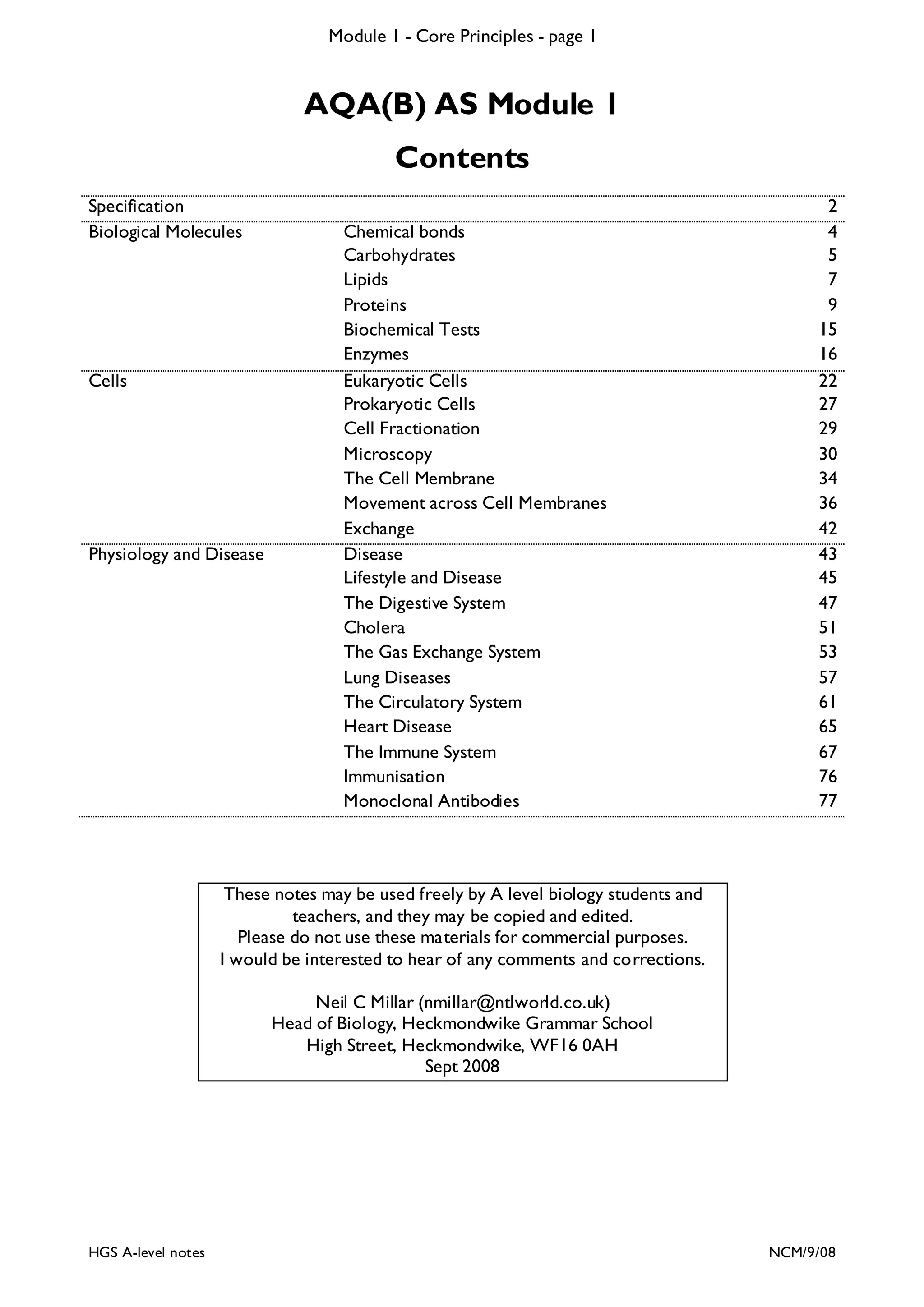
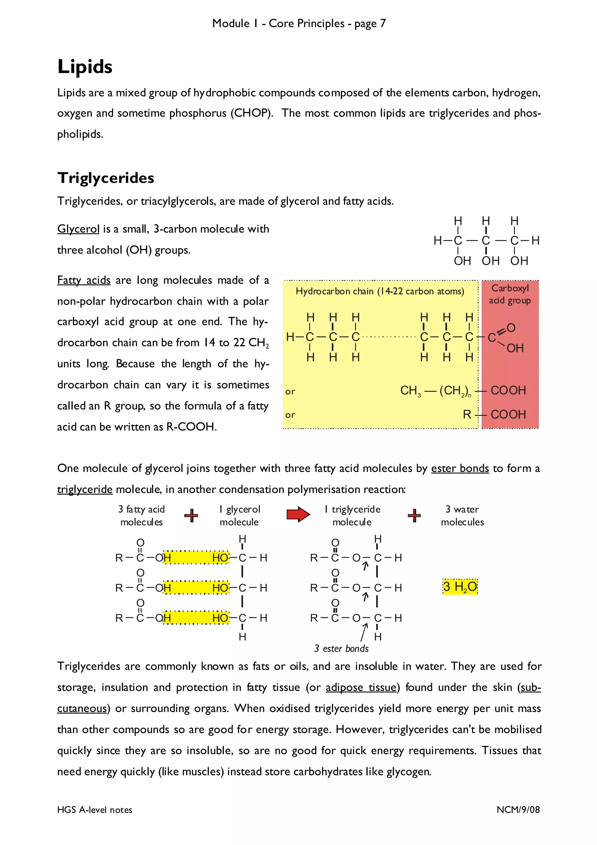
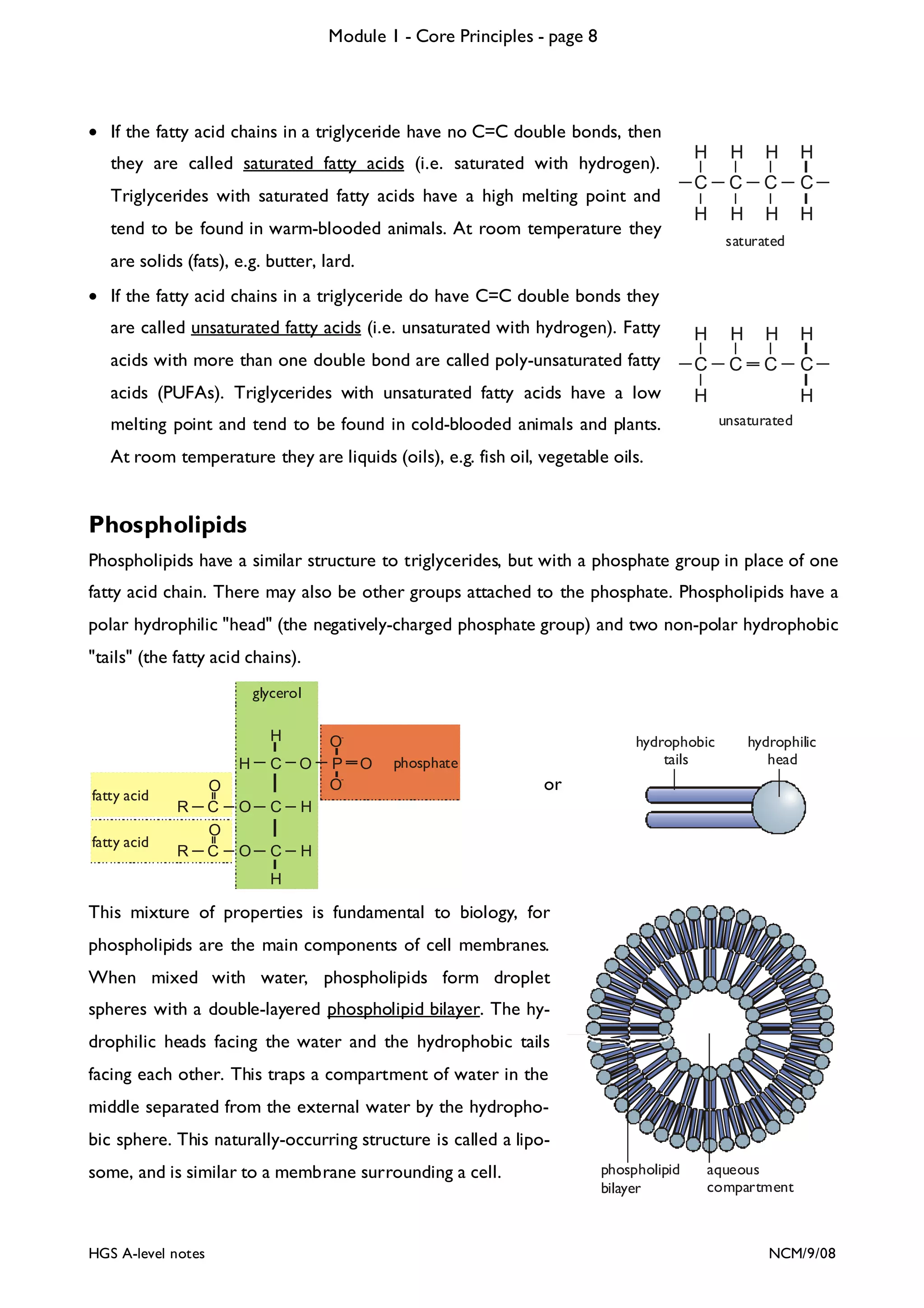
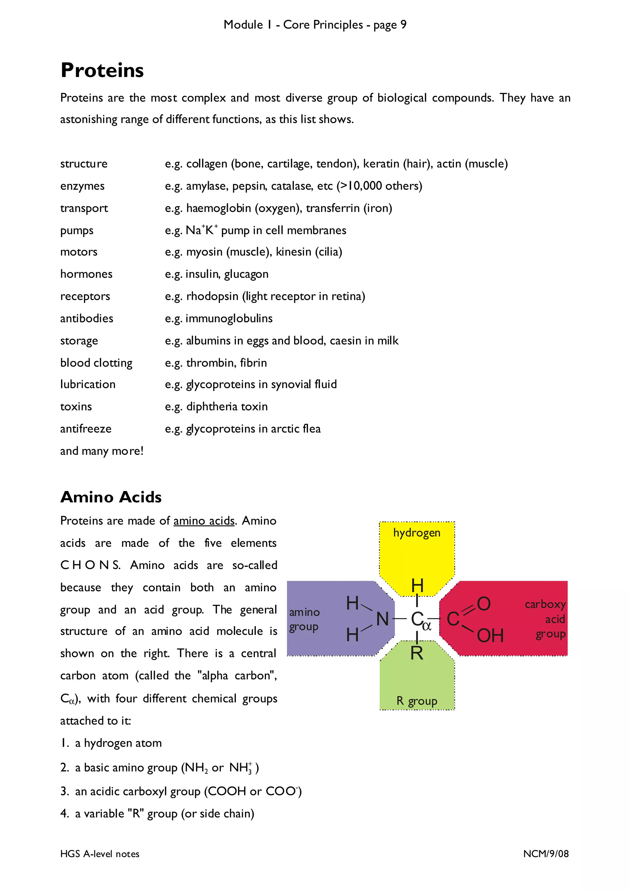
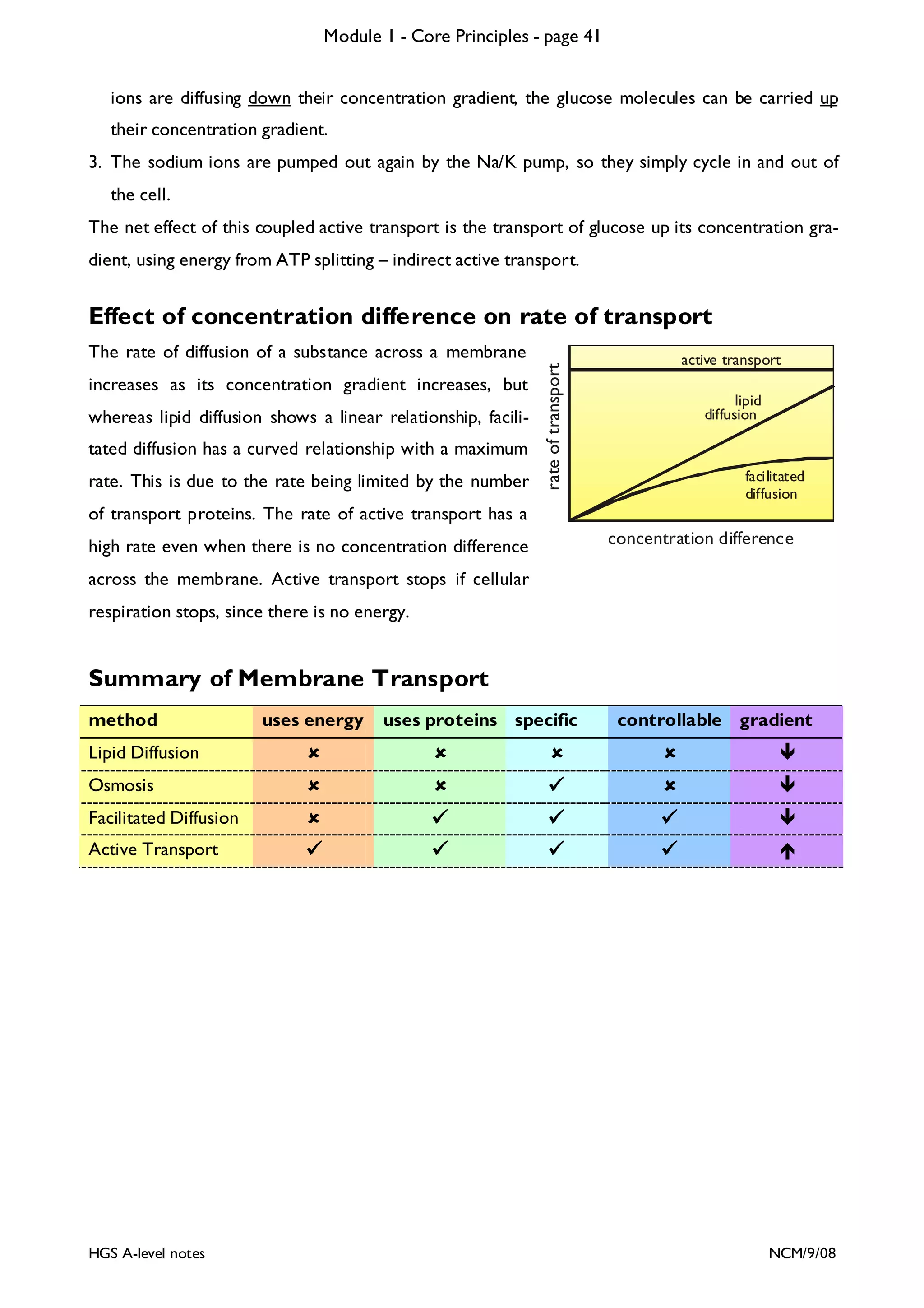
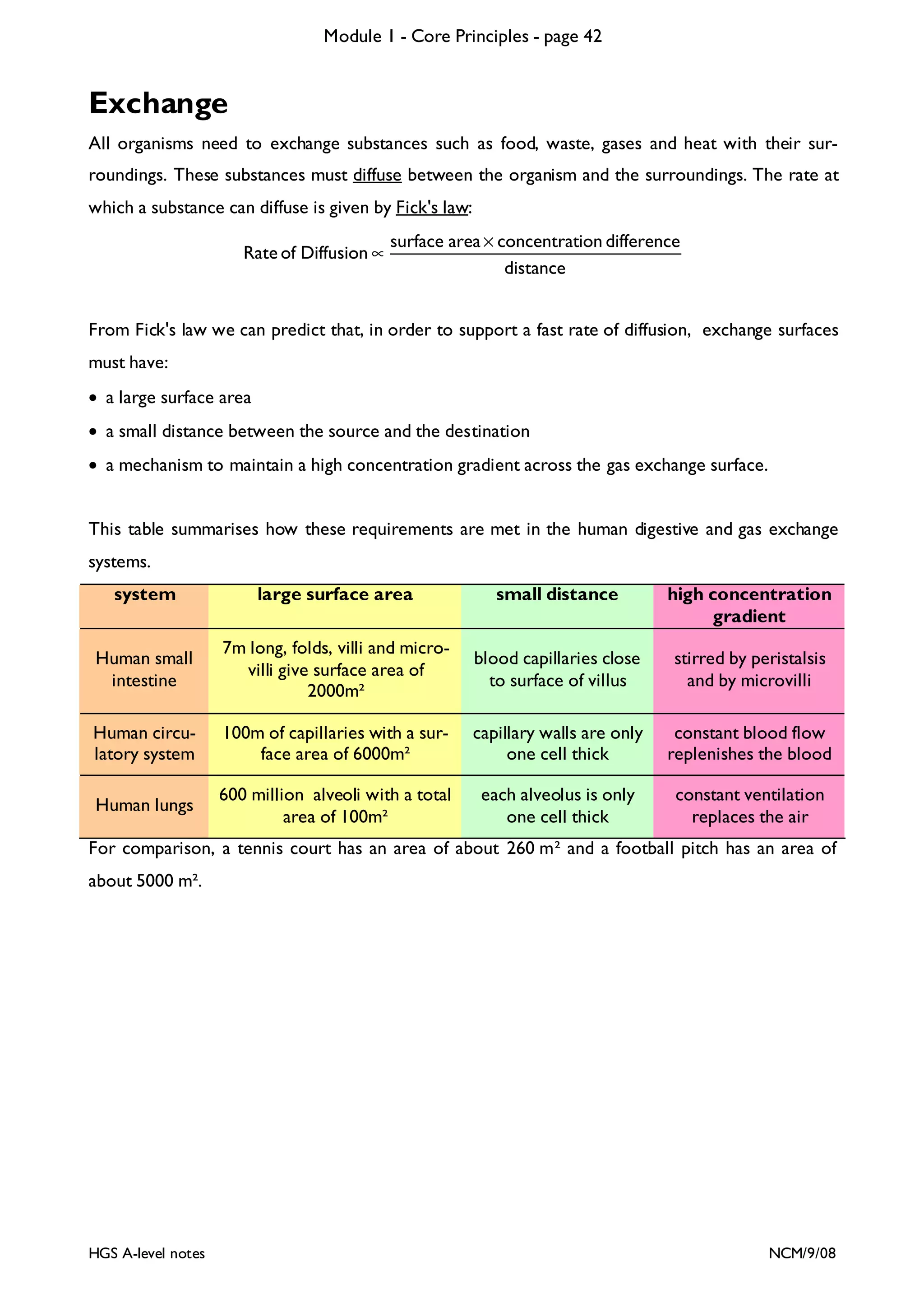
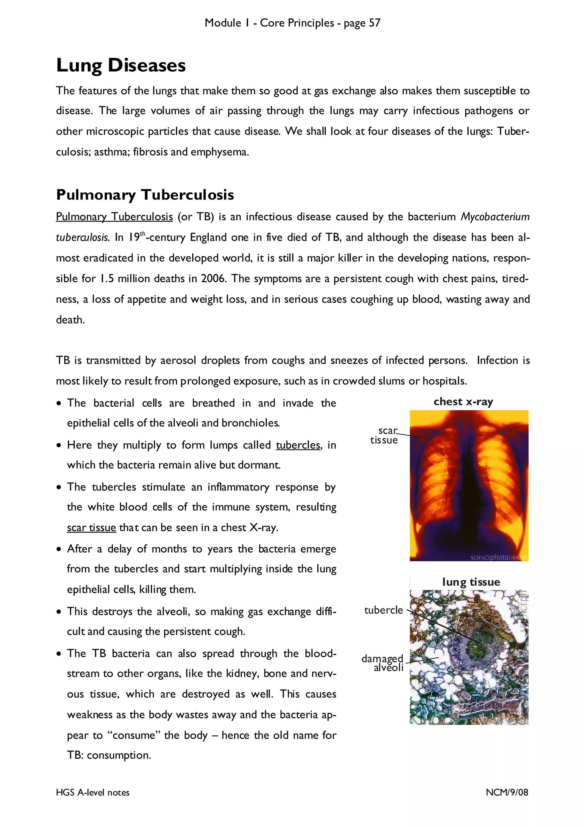
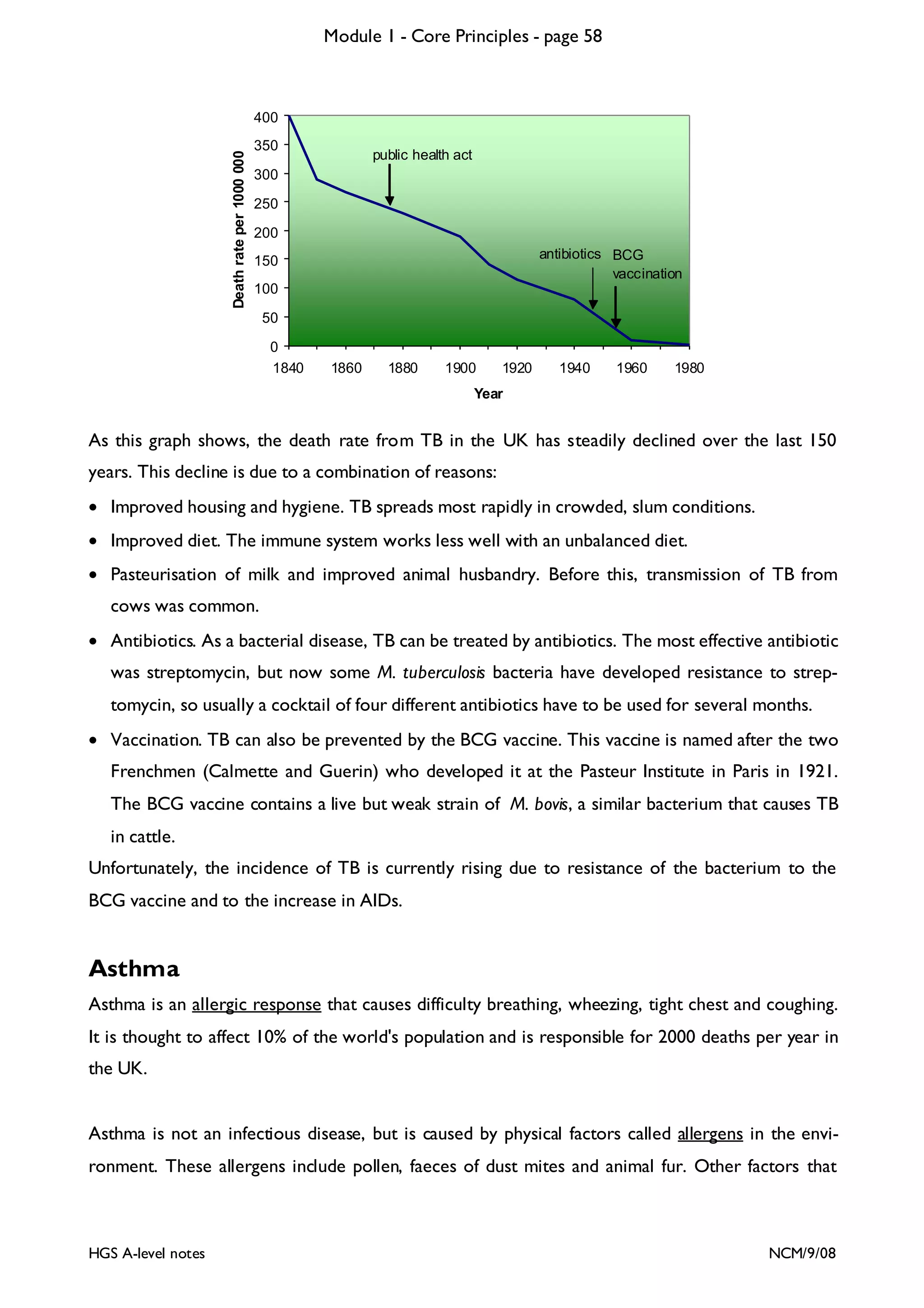
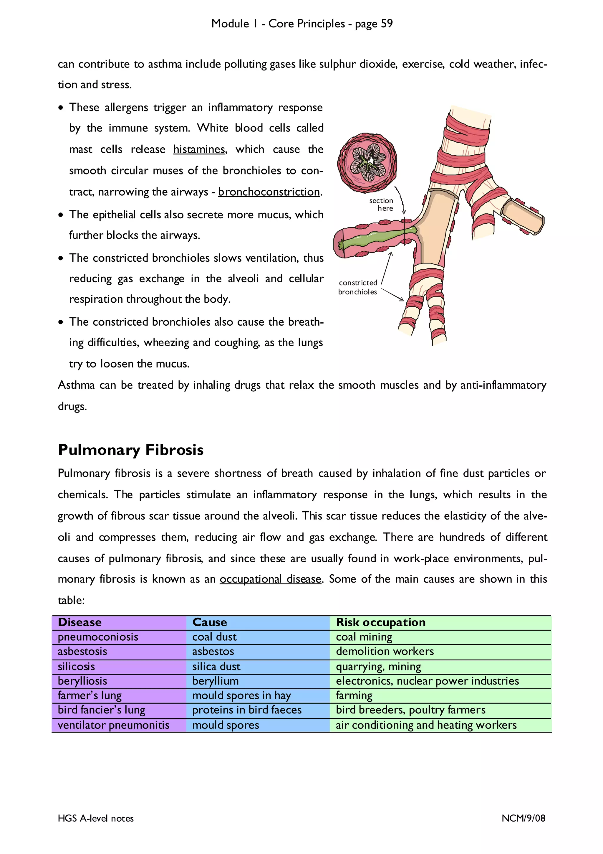
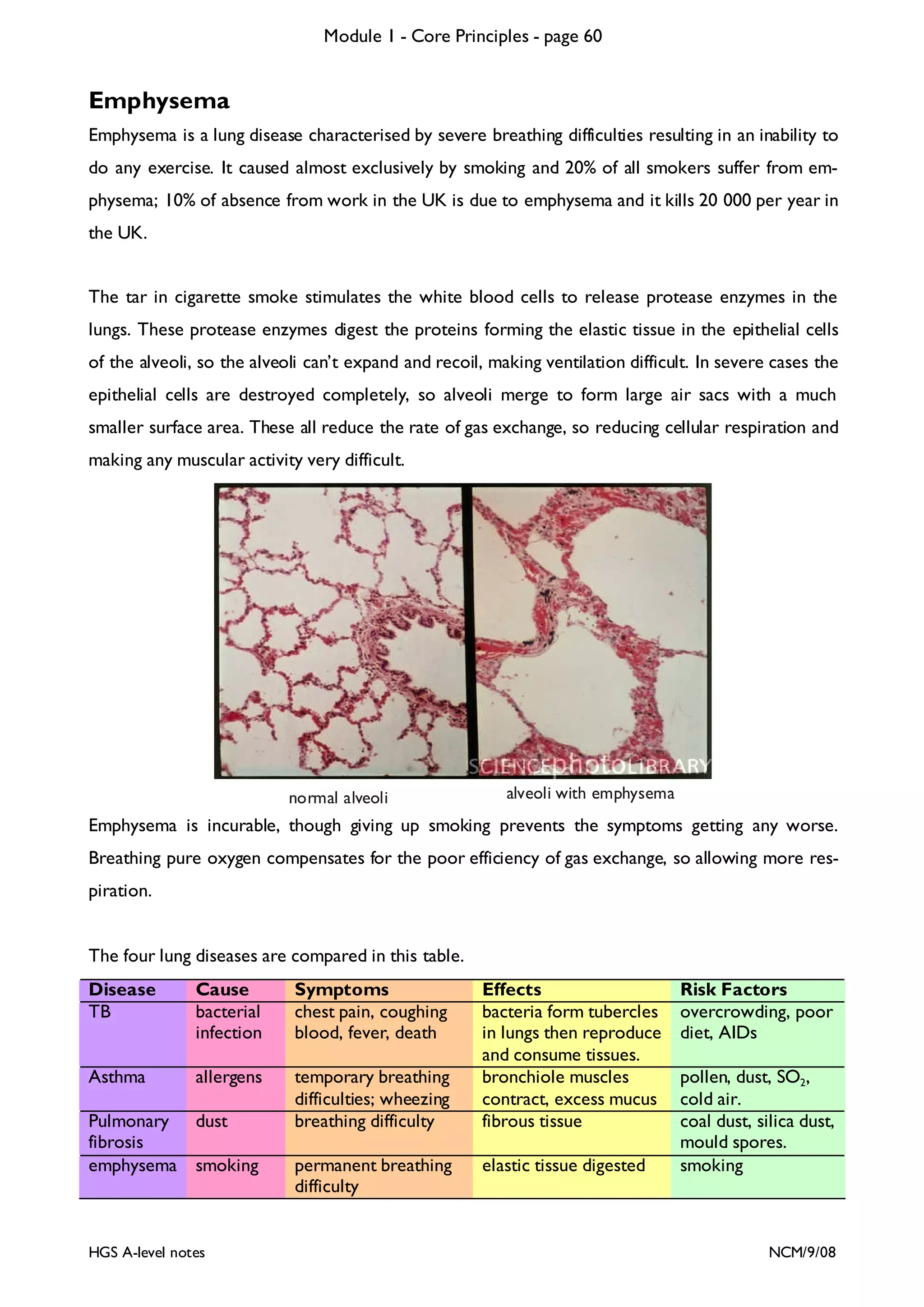
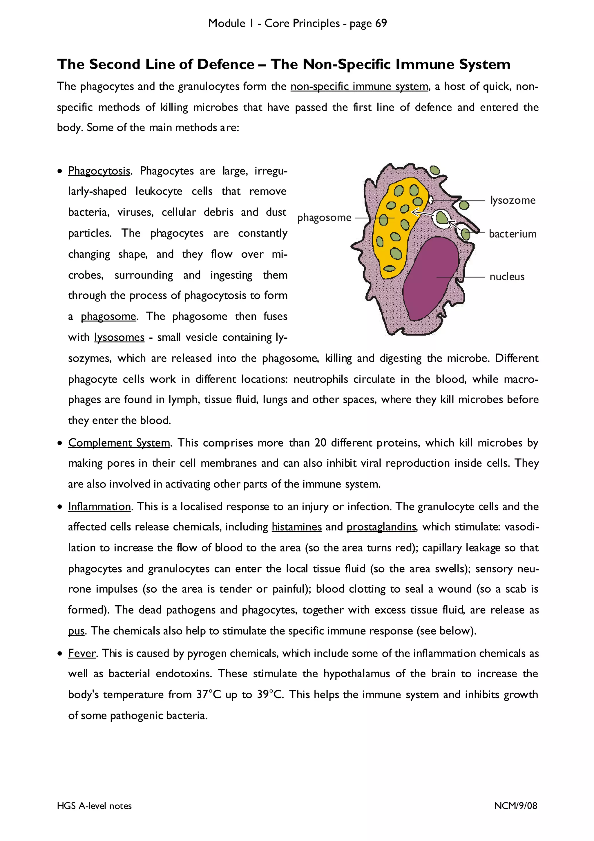
This document provides an overview of Module 1 - Core Principles for an AQA(B) AS biology course. It includes the following key points in 3 sentences:
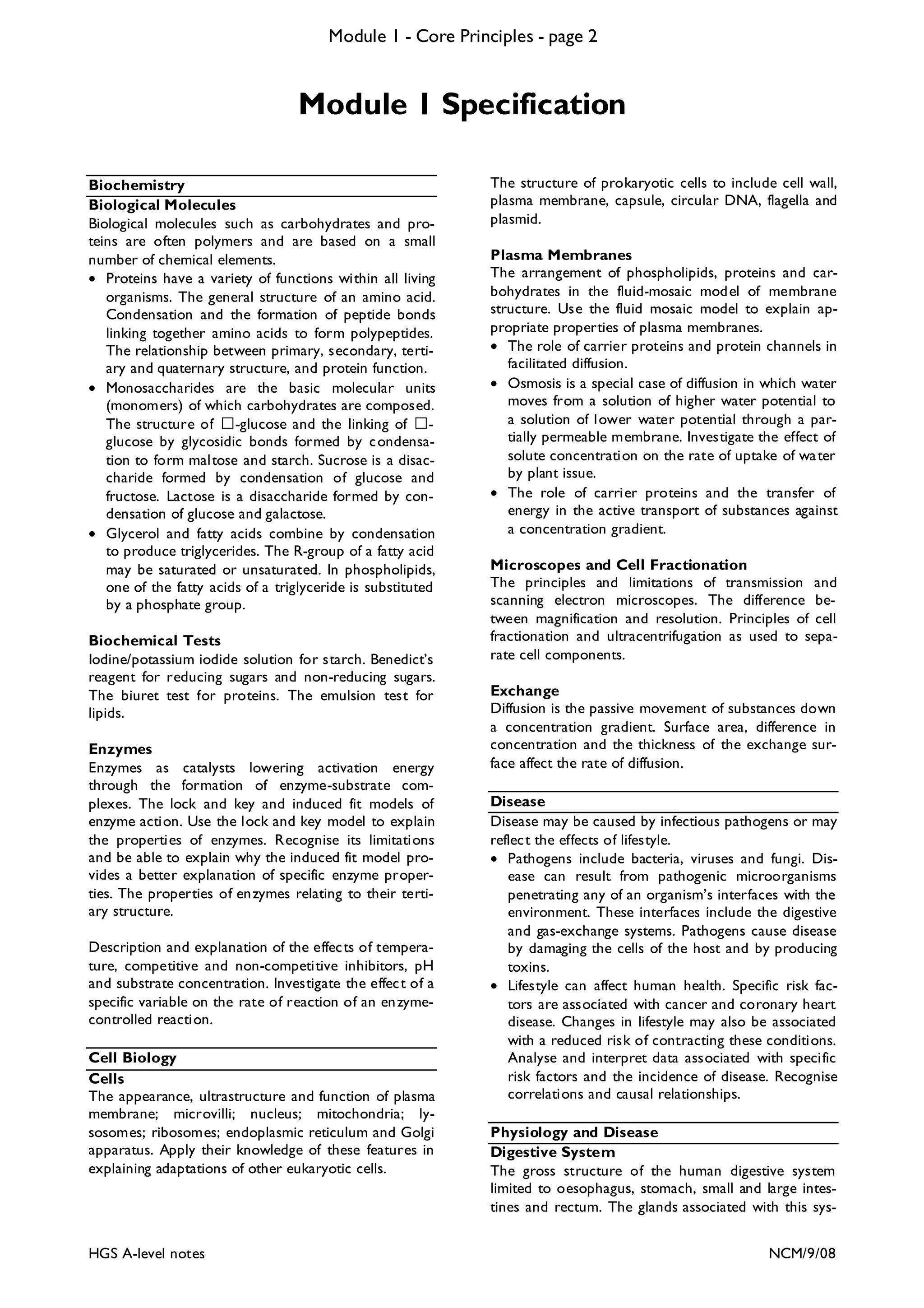
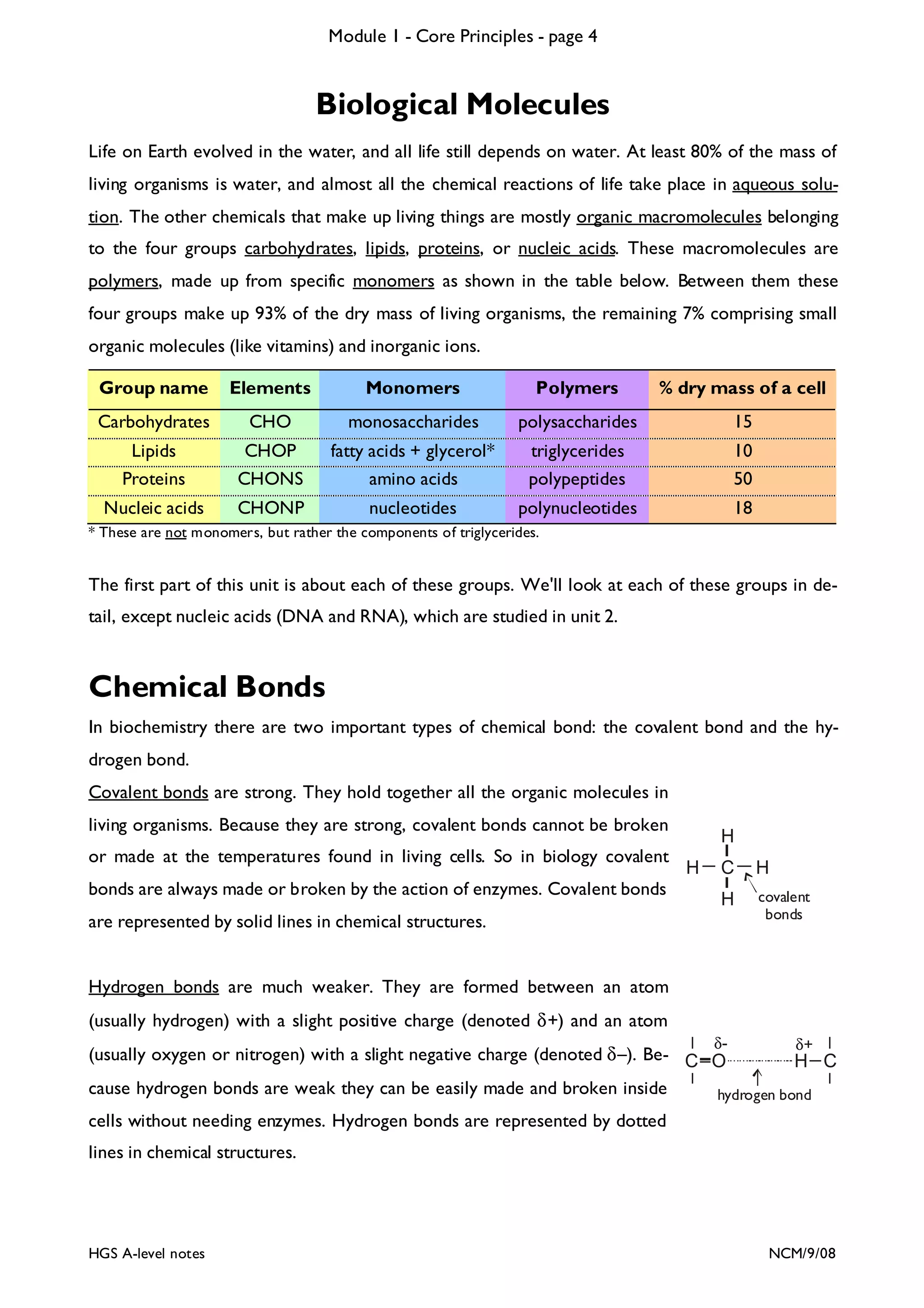
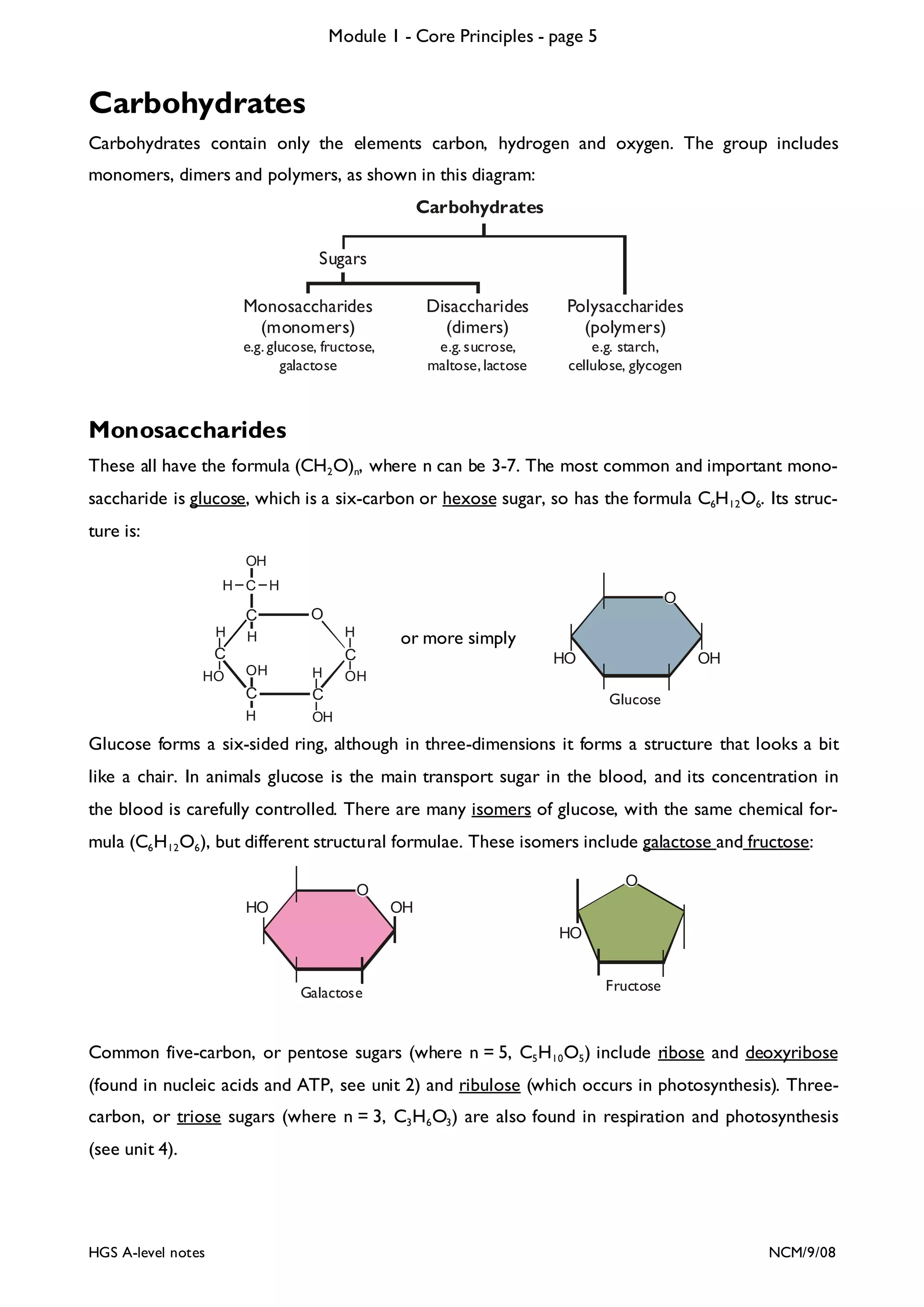
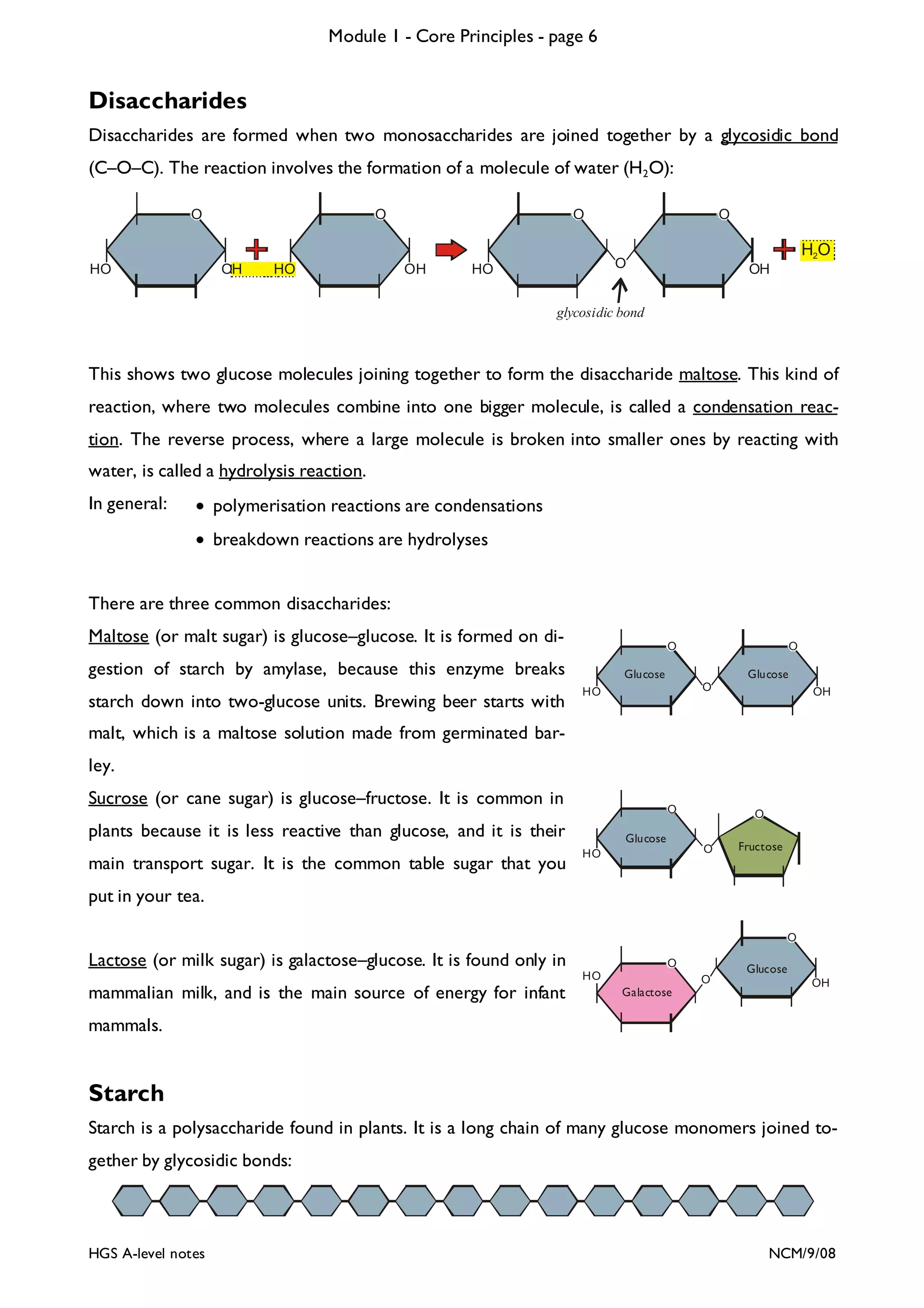
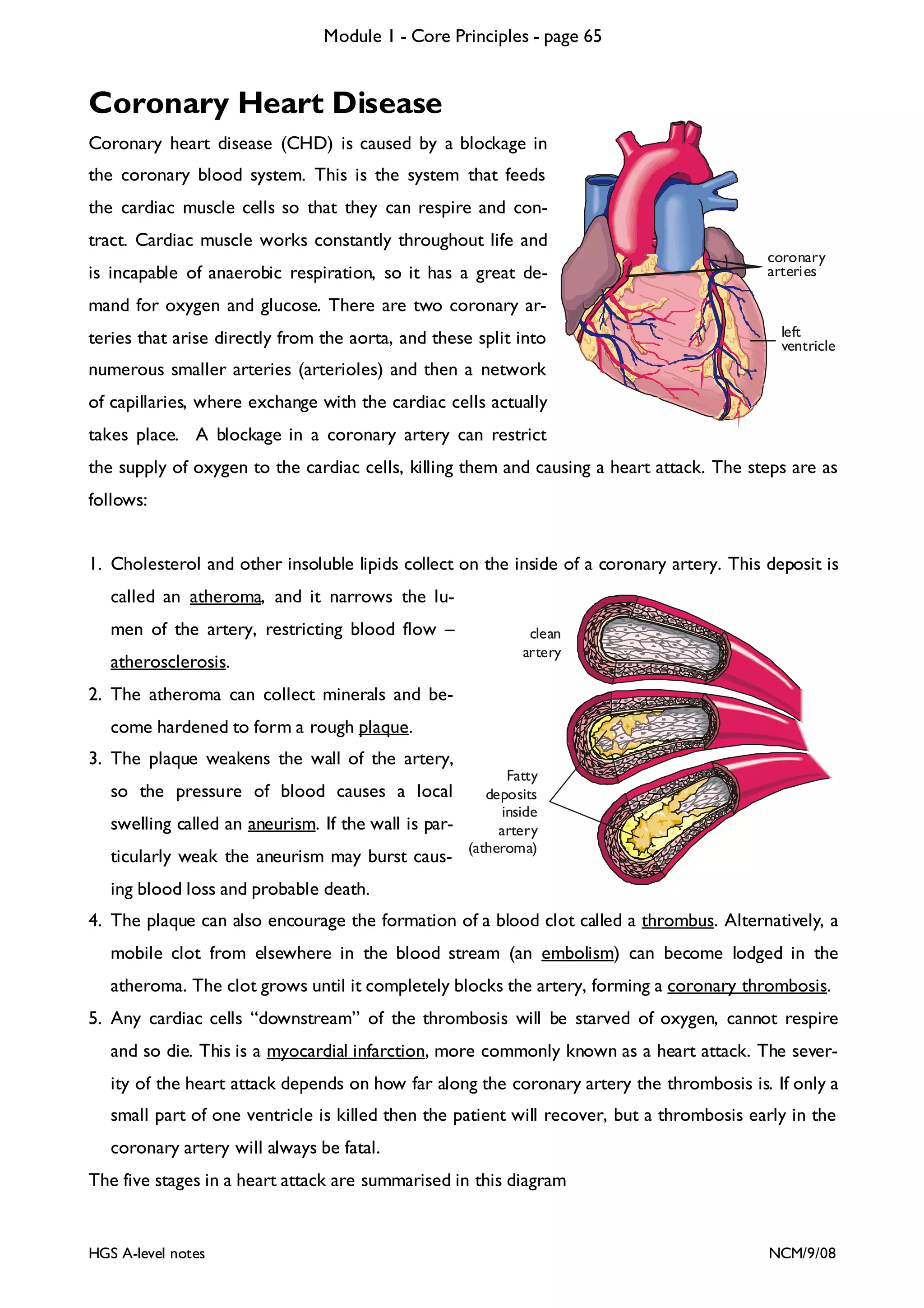
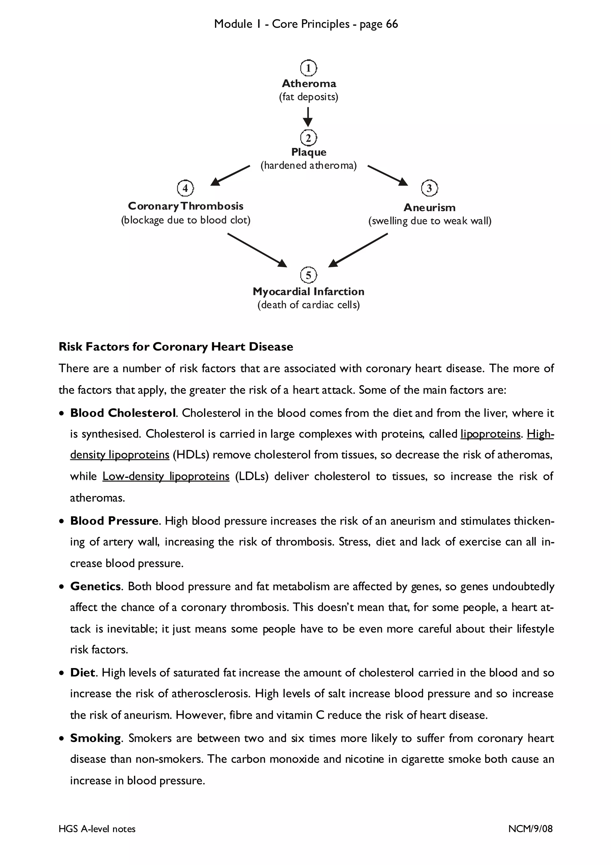
The document outlines the contents and topics to be covered in Module 1, including biological molecules, cells, physiology and disease. It provides a high-level summary of the main macromolecules that make up living things - carbohydrates, lipids, proteins and nucleic acids. The first pages provide more detailed information on the structures and properties of carbohydrates, lipids and the chemical bonds involved in forming these macromolecules.

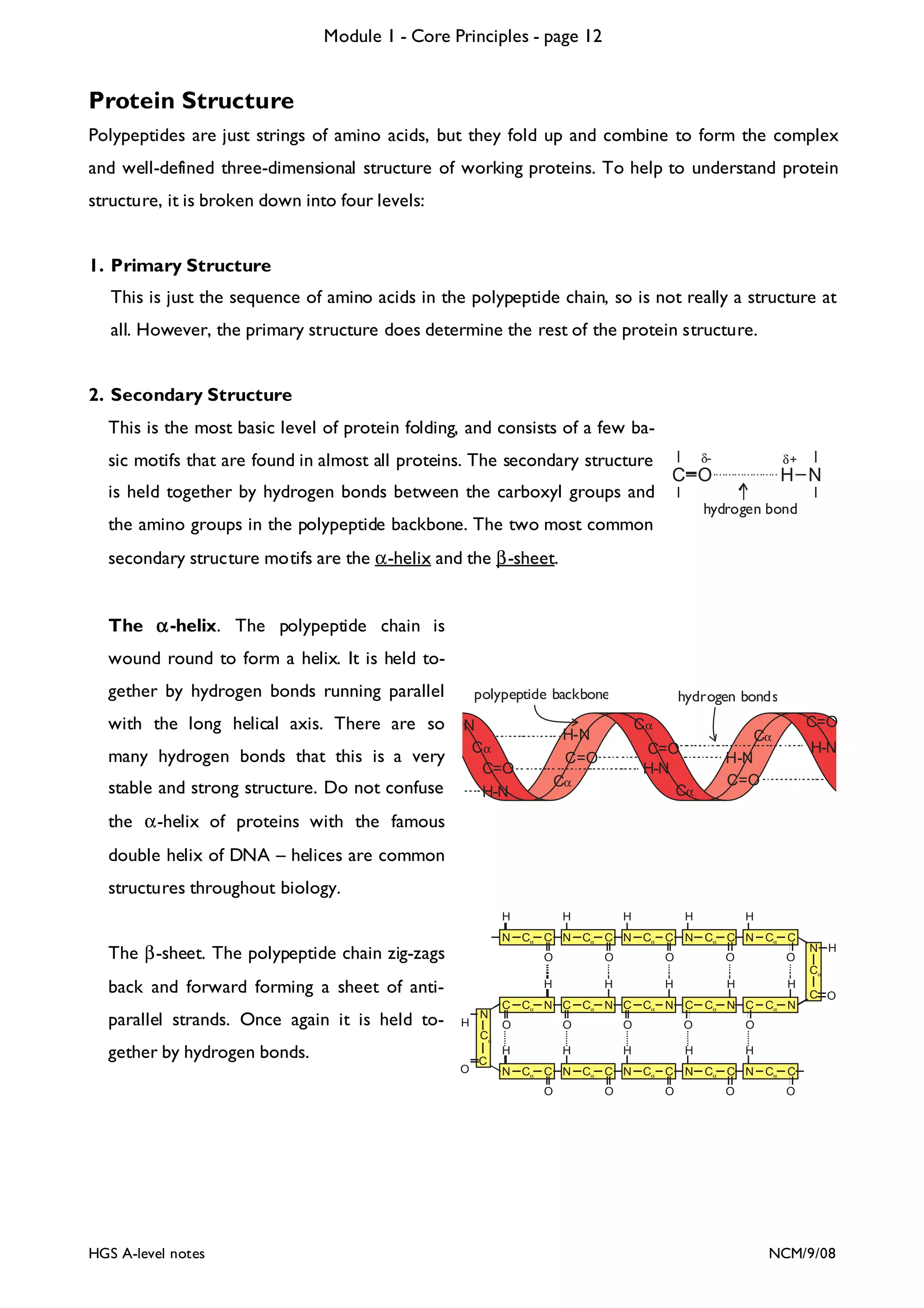
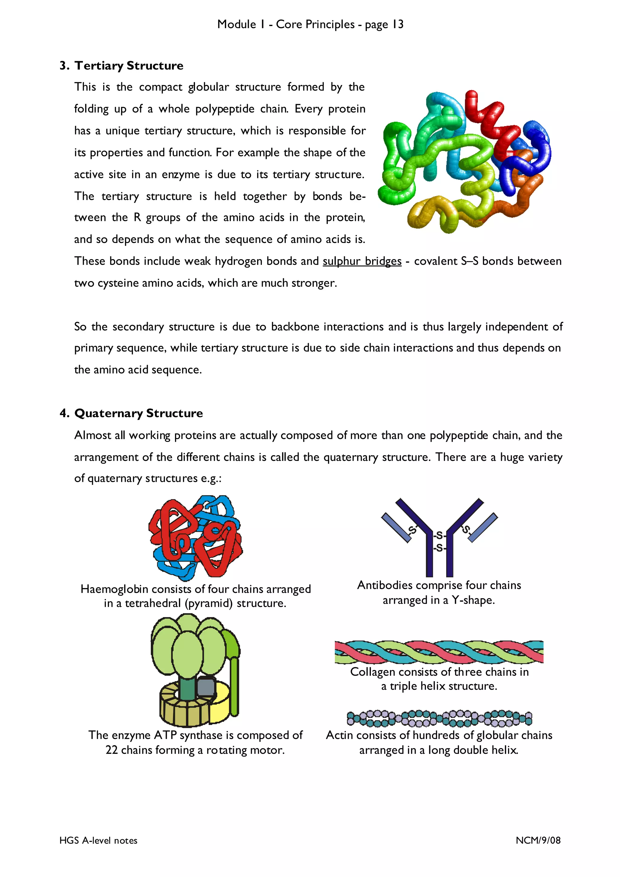
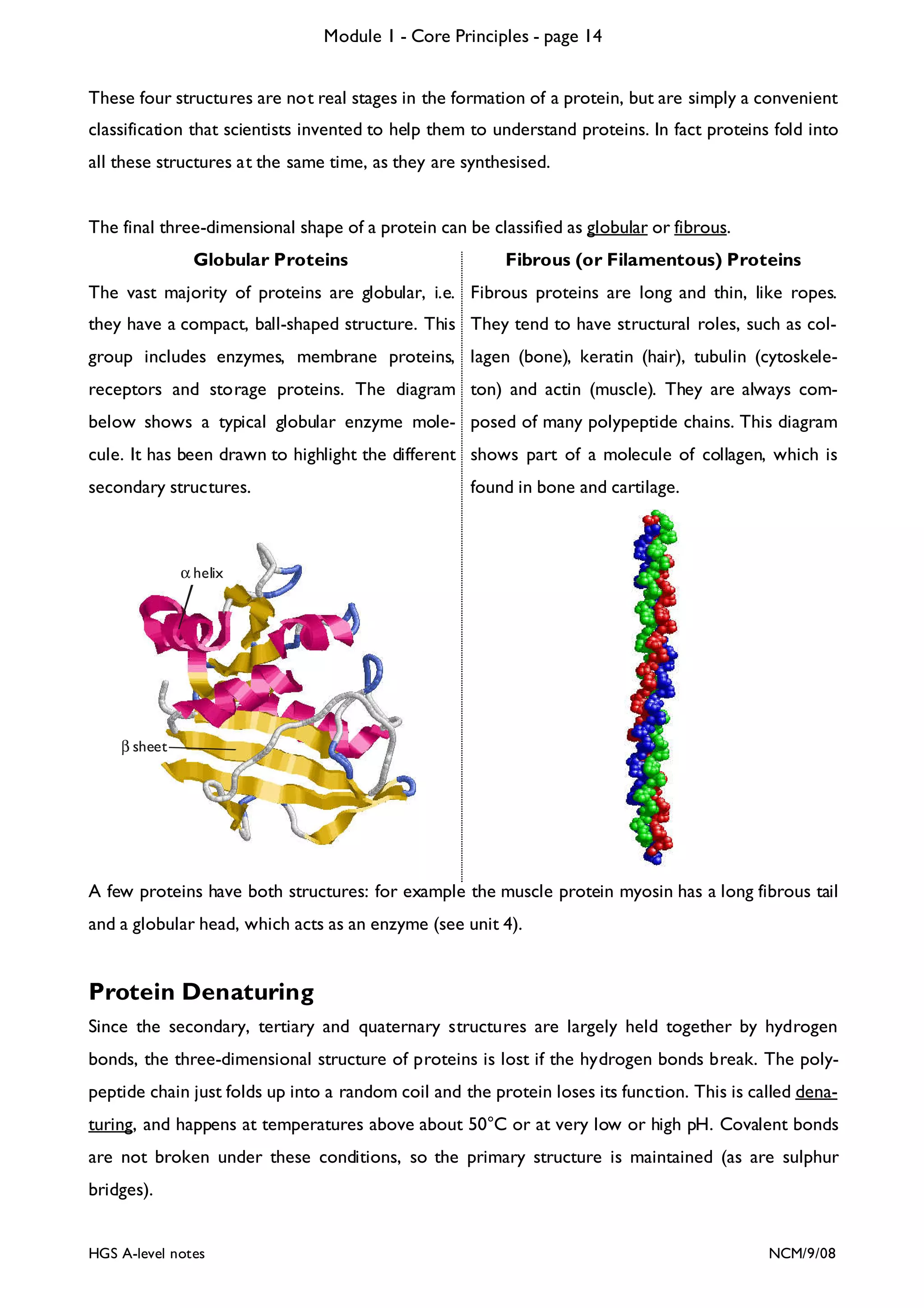

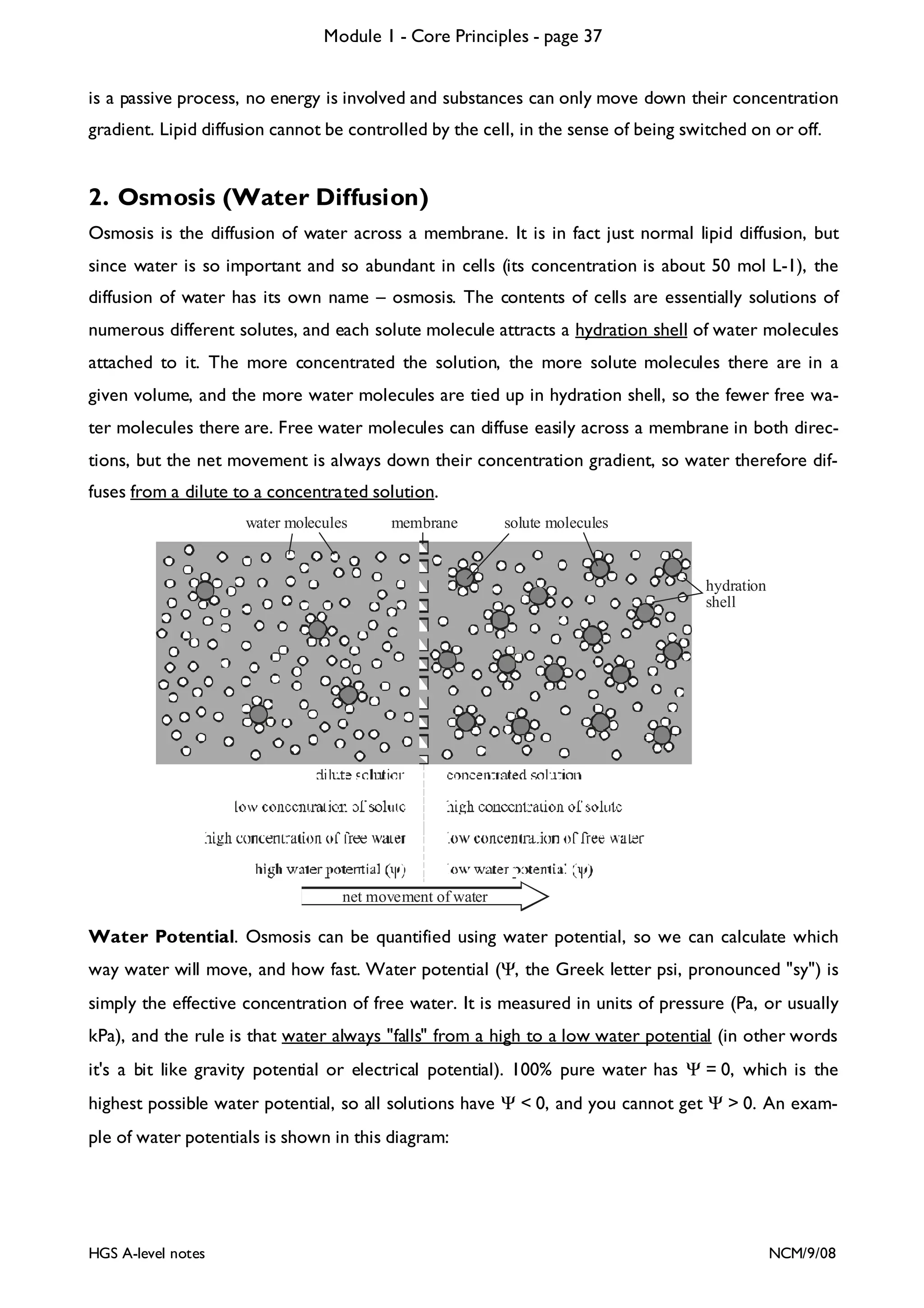
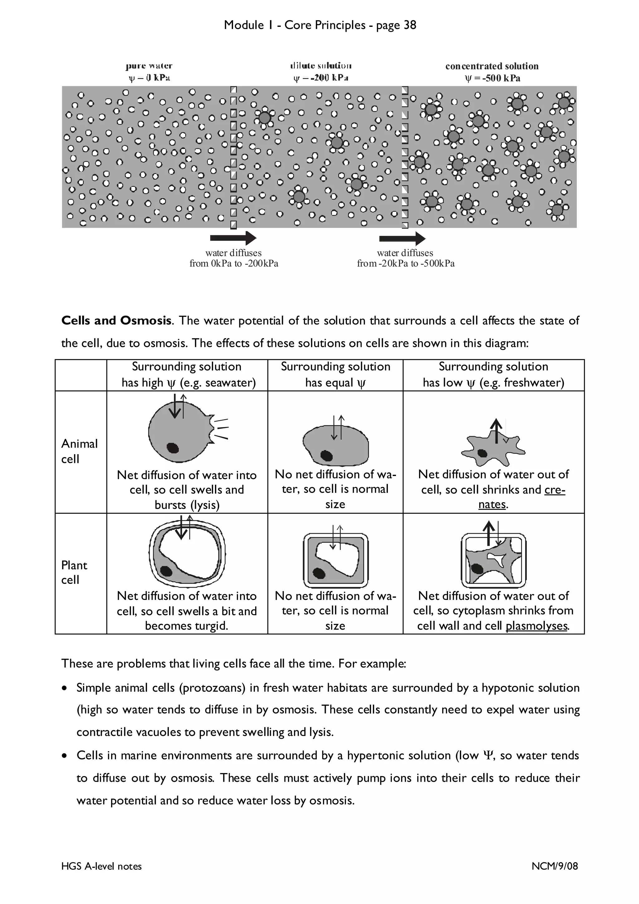
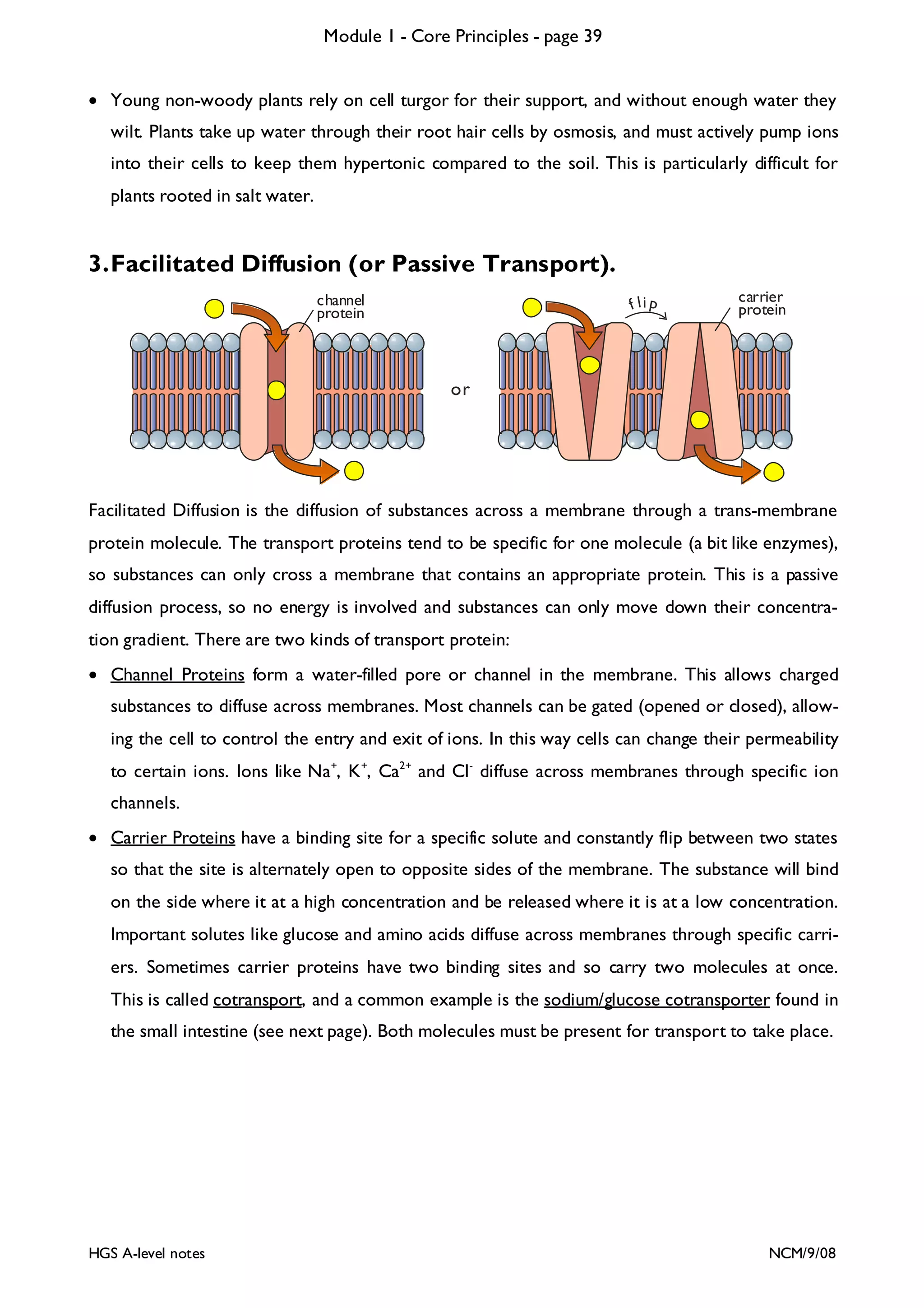
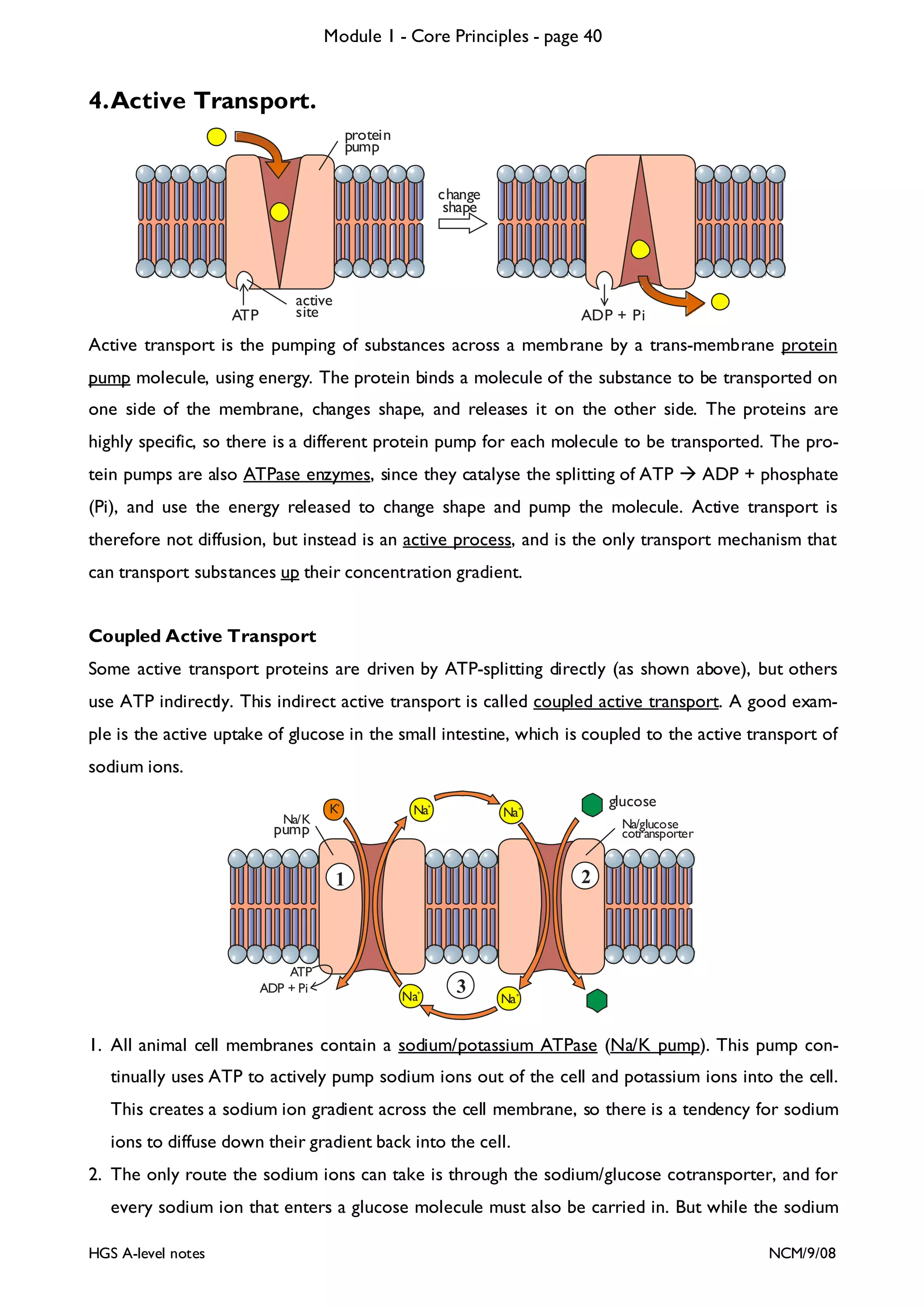
![Module 1 - Core Principles - page 77
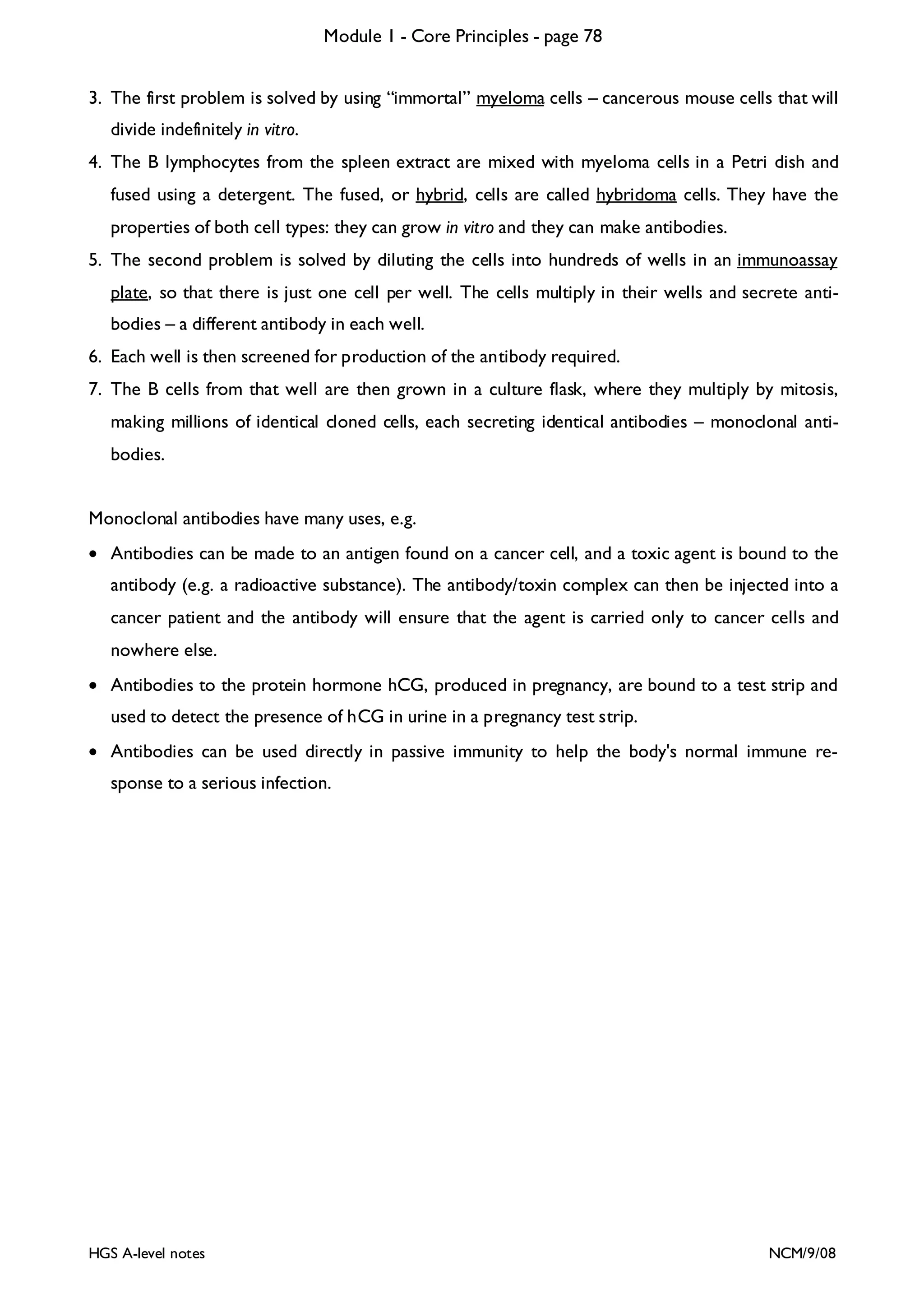
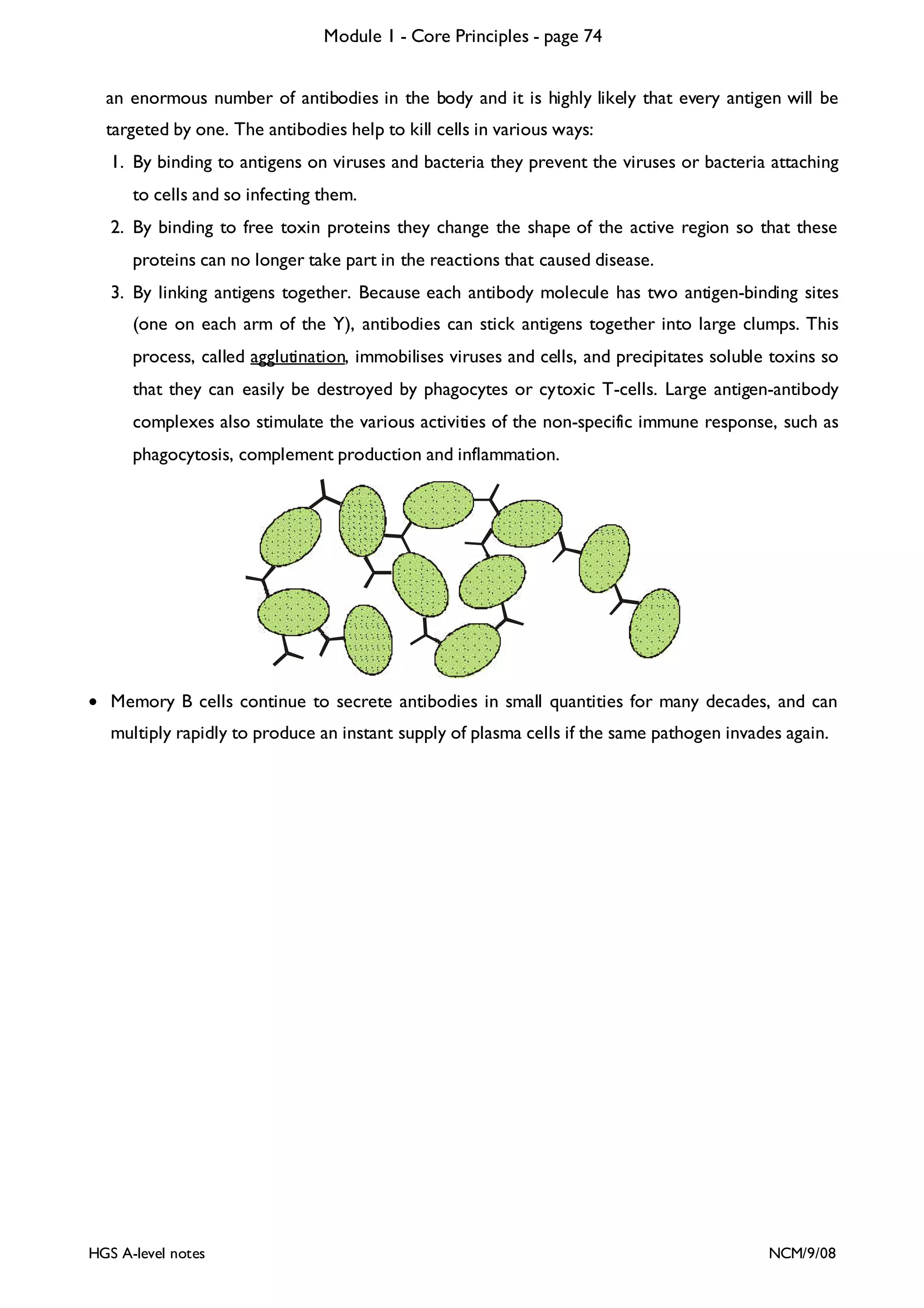
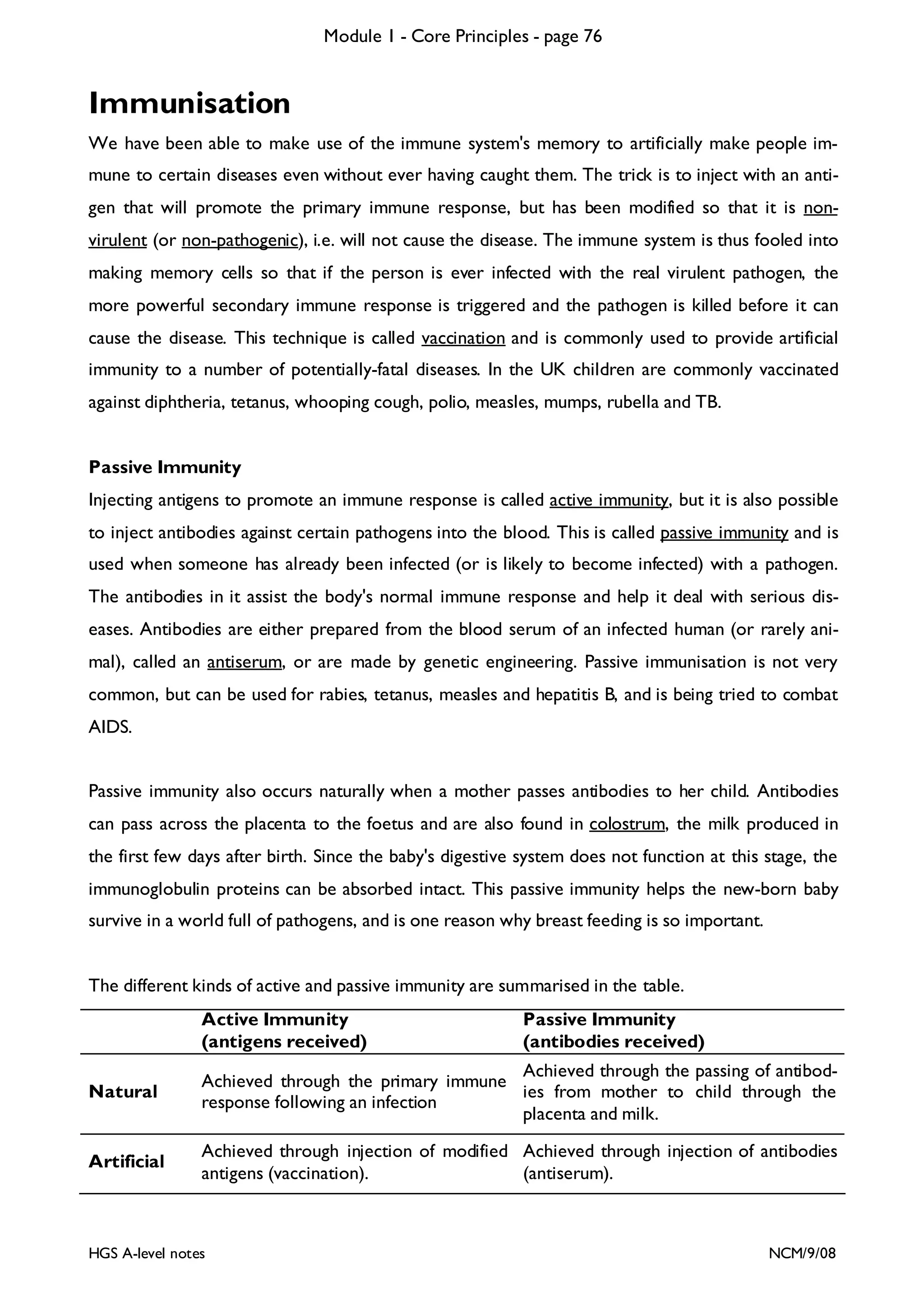
Monoclonal Antibodies
Scientists quickly realised that the remarkable specific binding property of antibody proteins in vivo
would make them very useful tools in medicine and research in vitro. [In vivo means “in life”, i.e. in a
living organism; and in vitro means “in glass”, i.e. in a test tube.] For example antigens could be used
as a “magic bullet” to target drugs at one specific cell type in the body, or antibodies could be used
to detect the presence of specific proteins in very low concentrations. So in 1975 Kohler and Milstein found a way to make pure monoclonal antibody proteins in the lab. This is their technique:
1
Inject mouse with antigens
spleen
2
3
obtain
B-lymphocytes
from spleen
myeloma cells
Fuse cells
4
hybridoma
cell
Put one cell
in each well of 5
immunoassay plate
test wells for
correct antibody 6
production
Clone this cell
in culture flask 7
and collect
monoclonal antibodies
1. Inject a mouse with the antigen proteins that you want antibodies for. These antigens could be
from a human cancer cell or a particular protein. The mouse will show a primary immune response and make a clone army of B lymphocytes with antibodies specific for that antigen.
2. After a few days, kill the mouse and extract B lymphocyte cells from its spleen. Although these
B cells will make antibodies, there are two problems: B lymphocyte cells won’t grow in vitro;
and the spleen extract contains a mixture of thousands of different B cells, each making their
own specific antibodies.
HGS A-level notes
NCM/9/08](https://image.slidesharecdn.com/unit1notes-131211075431-phpapp02/75/Unit-1-notes-77-2048.jpg)