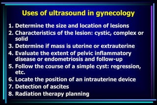
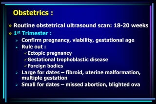
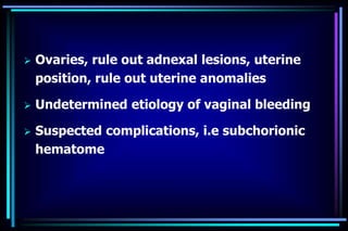
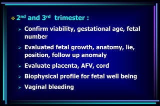
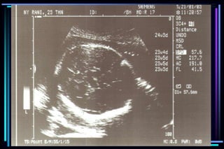
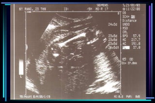
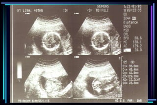
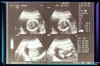
ULTRASOUND IN
OBSTETRICS & GYNECOLOGY
By:Muna Senan
Department of Obstetric & Gynecology
School Medicine Elmergib University /
 Ultrasound examination :
 Reveals vital information
 Fetal anatomy
 Fetal environment
 Growth
 Well being
 No biological hazard
 The first obstetrical application of USG
imaging : Ian Donald (1958)
 Sound waves of frequencies higher than the
hearing limit of the human ear are called
ultrasonic waves
 The limit is by convention 20 KHz
 Medical practice frequencies between 1-10
MHz are commonly used
Ultrasound
Technology :
 Sound waves reflecting back from the
fetus or image structure  displayed on
the ultrasound screen
 Alternating current is applied to a
transducer made of piezoelectric
material  intermittent high frequency
sound waves exceeding 20,000 cps are
generated
The transducer emits a pulse of sound waves that
passes through the layer of soft tissue

Interface between structures of
different tissue densities

Some of the energy is reflected back
to the transducer

A small electrical voltage

Display on a screen
 Bone is dense (echogenic)  white on the screen
 Fluid (anechoic)  black
 Soft tissues  varying shade of gray
1.Operator control 6. Visual information
2.Transducer drive 5. Electric information
3.Ultrasound pulses 4. Acoustic information
The diagnostic ultrasound visualization method
Operator
Instrument
Transducer
Tissue interaction
 Transabdominal Scanning :
 Performed with 3.5 – 7 MHz
 Safety : no confirmed biological effects
in mammalian tissue have been
demonstrated in the frequency range of
medical ultrasound (AIUM, 1991)
Choice of equipment :
 Transabdominal scanning
 Transvaginal scanning
 Doppler and color flow imaging
 3D
2D
Biophysical mechanism :
 Are not fully understood
 The effects of ultrasound on various biological
entities may be due to one or a combination of
any of the following :
1. Heating effects
2. Cavitational effects
3. Mechanical effects
Are associated only with high
average intensities and not
found in diagnostic equipment
Preparation of the patient
 Ultrasound beam cannot penetrate gas
 The interface created by gas  the sound beam
totally reflected
 Water  excellent transmission medium
 The distension of bladder  displaces intestinal
loop  acoustic window to the pelvic structure
 Patient should be scanned with a full bladder
 The full bladder technique offer :
1. It displaces the fetal head upwards
2. It allow visualization of the cervix
Coupling agents
 A gel or an oil  eliminates the air
interface between the transducer and the
patient’s skin
Scanning technique
 The patient is on your right
 The machine is on your left
 Work in dim lighting to help reflection on
the screen
Indications for ultrasound examination
 Gynecology : - Uterus, Ovaries, Adnexa
- Abnormal bleeding, pelvic pain &
discomfort
- IUCD location and complications
- Pelvic mass reveals mass or
enlargement of pelvic organ
- Evaluation of masses palpable
- Ovulation induction therapy
- Guidance for abscess drainage
Uses of ultrasound in gynecology
1. Determine the size and location of lesions
2. Characteristics of the lesion: cystic, complex or
solid
3. Determine if mass is uterine or extrauterine
4. Evaluate the extent of pelvic inflammatory
disease or endometriosis and follow-up
5. Follow the course of a simple cyst: regression,
etc.
6. Locate the position of an intrauterine device
7. Detection of ascites
8. Radiation therapy planning
Obstetrics :
 Routine obstetrical ultrasound scan: 18-20 weeks
 1st Trimester :
 Confirm pregnancy, viability, gestational age
 Rule out :
 Ectopic pregnancy
 Gestational trophoblastic disease
 Foreign bodies
 Large for dates – fibroid, uterine malformation,
multiple gestation
 Small for dates – missed abortion, blighted ova
 Ovaries, rule out adnexal lesions, uterine
position, rule out uterine anomalies
 Undetermined etiology of vaginal bleeding
 Suspected complications, i.e subchorionic
hematome
 2nd and 3rd trimester :
 Confirm viability, gestational age, fetal
number
 Evaluated fetal growth, anatomy, lie,
position, follow up anomaly
 Evaluate placenta, AFV, cord
 Biophysical profile for fetal well being
 Vaginal bleeding
Components of basic ultrasound examination
according to Trimester pregnancy
1st trimester
 Gestational sac location
 Embryo identification
 Crown rump length (CRL)
 Fetal heart rate motion
 Fetal number
 Uterus & adnexal
evaluation
2nd and 3rd trimester
 Fetal number
 Presentation
 Fetal heart motion
 Placental location
 Amniotic fluid volume
 Gestational age
 Survey of fetal anatomy
 Evaluation for maternal
pelvic mass
Ultrasonic Landmark Occurence
(weeks of gestation)
 Gestational sac 5 – 6 weeks
 Fetal embryonic pale 6 – 7 weeks
 Detection of fetal life 7 – 8 weeks
 Placenta 8 – 10 weeks
 Loss of gestational sac 11 – 13 weeks
 Fetal head 11 – 14 weeks
(Biparietal diameter)
Evaluation of ultrasound to determine
gestational age
 Crown Rump Length (CRL)
 Biparietal diameter (BPD)
 Femur Length (FL)
 Head Circumference (HC)
 Abdominal Circumference (AC)
Accuracy of ultrasound measurement for
different gestational groups
Ultrasound parameter Accuracy
 Gestational sac diameter + 7 days
 Crown Rump Length + 3-5 days
 BPD second trimester + 1 to 1.5 weeks
 BPD third trimester + 2 to 4 weeks
 FL second trimester + 1 to 1.5 weeks
 FL third trimester + 3 to 3.5 weeks
 Multiple parameter (2nd trimester) + 1.5 weeks
 Multiple parameter (3rd trimester) + 2.5 weeks
Summary
1. Physics of ultrasound
The ultrasound used in clinical practice is limited
to frequencies in the range of 1-10 cycles per
second (1-10 MHz)
In Obstetrics : 3.5 MHz and 5 MHz
2.Ultrasound
Is produced when transmitted pulses of sound
from the transducer cross body structures and
reflect energy back to the transducer from the
interfaces of organs
3. Ultrasound in Obstetrics
 Confirmation of an intrauterine pregnancies
 Exclusion of an ectopic pregnancies
 Assessment of an intrauterine pregnancies
 Determination of gestational age :
 Are uncertain of the date of conception
 Resolving discrepancies between gestational
age and uterine size :
 Large for dates
 Small for dates
 Investigation of uterine bleeding
 Visualization for high risk procedures
 Amniocentesis
 Chorionic villous sampling
 Cordocentesis
 Evaluation of fetal well-being
 Distinguish congenital malformation
 Biophysical profile
 Doppler flow velocimetry
 IUGR  60-80% of the time in cases where the
umbilical artery waveforms are abnormal due to
uteroplacental insufficiency
ultra sound O&Gy guide.ppt

ultra sound O&Gy guide.ppt

  • 1.
    ULTRASOUND IN OBSTETRICS &GYNECOLOGY By:Muna Senan Department of Obstetric & Gynecology School Medicine Elmergib University /
  • 2.
     Ultrasound examination:  Reveals vital information  Fetal anatomy  Fetal environment  Growth  Well being  No biological hazard  The first obstetrical application of USG imaging : Ian Donald (1958)
  • 3.
     Sound wavesof frequencies higher than the hearing limit of the human ear are called ultrasonic waves  The limit is by convention 20 KHz  Medical practice frequencies between 1-10 MHz are commonly used Ultrasound
  • 4.
    Technology :  Soundwaves reflecting back from the fetus or image structure  displayed on the ultrasound screen  Alternating current is applied to a transducer made of piezoelectric material  intermittent high frequency sound waves exceeding 20,000 cps are generated
  • 5.
    The transducer emitsa pulse of sound waves that passes through the layer of soft tissue  Interface between structures of different tissue densities  Some of the energy is reflected back to the transducer  A small electrical voltage  Display on a screen  Bone is dense (echogenic)  white on the screen  Fluid (anechoic)  black  Soft tissues  varying shade of gray
  • 6.
    1.Operator control 6.Visual information 2.Transducer drive 5. Electric information 3.Ultrasound pulses 4. Acoustic information The diagnostic ultrasound visualization method Operator Instrument Transducer Tissue interaction
  • 11.
     Transabdominal Scanning:  Performed with 3.5 – 7 MHz  Safety : no confirmed biological effects in mammalian tissue have been demonstrated in the frequency range of medical ultrasound (AIUM, 1991)
  • 12.
    Choice of equipment:  Transabdominal scanning  Transvaginal scanning  Doppler and color flow imaging  3D 2D
  • 13.
    Biophysical mechanism : Are not fully understood  The effects of ultrasound on various biological entities may be due to one or a combination of any of the following : 1. Heating effects 2. Cavitational effects 3. Mechanical effects Are associated only with high average intensities and not found in diagnostic equipment
  • 14.
    Preparation of thepatient  Ultrasound beam cannot penetrate gas  The interface created by gas  the sound beam totally reflected  Water  excellent transmission medium  The distension of bladder  displaces intestinal loop  acoustic window to the pelvic structure  Patient should be scanned with a full bladder  The full bladder technique offer : 1. It displaces the fetal head upwards 2. It allow visualization of the cervix
  • 15.
    Coupling agents  Agel or an oil  eliminates the air interface between the transducer and the patient’s skin Scanning technique  The patient is on your right  The machine is on your left  Work in dim lighting to help reflection on the screen
  • 16.
    Indications for ultrasoundexamination  Gynecology : - Uterus, Ovaries, Adnexa - Abnormal bleeding, pelvic pain & discomfort - IUCD location and complications - Pelvic mass reveals mass or enlargement of pelvic organ - Evaluation of masses palpable - Ovulation induction therapy - Guidance for abscess drainage
  • 17.
    Uses of ultrasoundin gynecology 1. Determine the size and location of lesions 2. Characteristics of the lesion: cystic, complex or solid 3. Determine if mass is uterine or extrauterine 4. Evaluate the extent of pelvic inflammatory disease or endometriosis and follow-up 5. Follow the course of a simple cyst: regression, etc. 6. Locate the position of an intrauterine device 7. Detection of ascites 8. Radiation therapy planning
  • 18.
    Obstetrics :  Routineobstetrical ultrasound scan: 18-20 weeks  1st Trimester :  Confirm pregnancy, viability, gestational age  Rule out :  Ectopic pregnancy  Gestational trophoblastic disease  Foreign bodies  Large for dates – fibroid, uterine malformation, multiple gestation  Small for dates – missed abortion, blighted ova
  • 19.
     Ovaries, ruleout adnexal lesions, uterine position, rule out uterine anomalies  Undetermined etiology of vaginal bleeding  Suspected complications, i.e subchorionic hematome
  • 20.
     2nd and3rd trimester :  Confirm viability, gestational age, fetal number  Evaluated fetal growth, anatomy, lie, position, follow up anomaly  Evaluate placenta, AFV, cord  Biophysical profile for fetal well being  Vaginal bleeding
  • 21.
    Components of basicultrasound examination according to Trimester pregnancy 1st trimester  Gestational sac location  Embryo identification  Crown rump length (CRL)  Fetal heart rate motion  Fetal number  Uterus & adnexal evaluation 2nd and 3rd trimester  Fetal number  Presentation  Fetal heart motion  Placental location  Amniotic fluid volume  Gestational age  Survey of fetal anatomy  Evaluation for maternal pelvic mass
  • 22.
    Ultrasonic Landmark Occurence (weeksof gestation)  Gestational sac 5 – 6 weeks  Fetal embryonic pale 6 – 7 weeks  Detection of fetal life 7 – 8 weeks  Placenta 8 – 10 weeks  Loss of gestational sac 11 – 13 weeks  Fetal head 11 – 14 weeks (Biparietal diameter)
  • 23.
    Evaluation of ultrasoundto determine gestational age  Crown Rump Length (CRL)  Biparietal diameter (BPD)  Femur Length (FL)  Head Circumference (HC)  Abdominal Circumference (AC)
  • 24.
    Accuracy of ultrasoundmeasurement for different gestational groups Ultrasound parameter Accuracy  Gestational sac diameter + 7 days  Crown Rump Length + 3-5 days  BPD second trimester + 1 to 1.5 weeks  BPD third trimester + 2 to 4 weeks  FL second trimester + 1 to 1.5 weeks  FL third trimester + 3 to 3.5 weeks  Multiple parameter (2nd trimester) + 1.5 weeks  Multiple parameter (3rd trimester) + 2.5 weeks
  • 25.
    Summary 1. Physics ofultrasound The ultrasound used in clinical practice is limited to frequencies in the range of 1-10 cycles per second (1-10 MHz) In Obstetrics : 3.5 MHz and 5 MHz 2.Ultrasound Is produced when transmitted pulses of sound from the transducer cross body structures and reflect energy back to the transducer from the interfaces of organs
  • 26.
    3. Ultrasound inObstetrics  Confirmation of an intrauterine pregnancies  Exclusion of an ectopic pregnancies  Assessment of an intrauterine pregnancies  Determination of gestational age :  Are uncertain of the date of conception  Resolving discrepancies between gestational age and uterine size :  Large for dates  Small for dates
  • 27.
     Investigation ofuterine bleeding  Visualization for high risk procedures  Amniocentesis  Chorionic villous sampling  Cordocentesis  Evaluation of fetal well-being  Distinguish congenital malformation  Biophysical profile  Doppler flow velocimetry  IUGR  60-80% of the time in cases where the umbilical artery waveforms are abnormal due to uteroplacental insufficiency