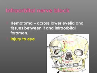
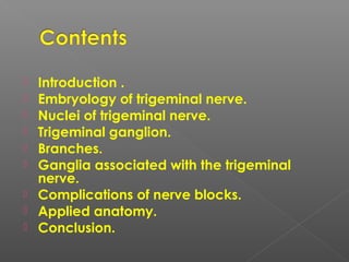
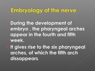
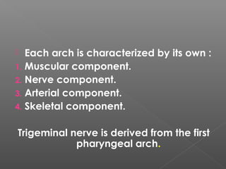
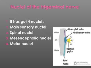
PRESENTED BY :
DR ISHANI SHARMA
PG FIRST YEAR
DEPT OF PAEDODONTICS AND
PREVENTIVE DENTISTRY.
MODERATER : DR SHIREEN
DR AMRITA
1. Malamed S. Handbook of local
anesthesia. St. Louis, Mo.:
Elsevier/Mosby; 2002.
2. Rajendran R, Sivapathasundharam B,
Shafer W. Shafer's textbook of oral
pathology. 4.
3. Ghom A. Textbook of oral medicine. St
Louis (USA): Jaypee Bros.; 2010.
 4. Chaurasia B. Bd chaurasia's human
anatomy regional and applied
dissection and clinical.; 2013.
 Introduction .
 Embryology of trigeminal nerve.
 Nuclei of trigeminal nerve.
 Trigeminal ganglion.
 Branches.
 Ganglia associated with the trigeminal
nerve.
 Complications of nerve blocks.
 Applied anatomy.
 Conclusion.
 Nerve-
a bundle of fibers that uses chemical
and electrical signals to transmit the sensory
and motor information from one body part to
another.
 Neuron –
these are specialized cells that
constitute the functional units of the nervous
system and has a special property of being
able to conduct impulses rapidly from one
part of body to another.
 During the development of
embryo , the pharyngeal arches
appear in the fourth and fifth
week.
 It gives rise to the six pharyngeal
arches, of which the fifth arch
dissappears.
 Each arch is characterized by its own :
1. Muscular component.
2. Nerve component.
3. Arterial component.
4. Skeletal component.
Trigeminal nerve is derived from the first
pharyngeal arch.
 It has got 4 nuclei :
1. Main sensory nuclei
2. Spinal nuclei
3. Mesencephalic nuclei
4. Motor nuclei
 The central processes large sensory
root attached to pons at
middle cerebellar peduncle.
 The peripheral processes three
divisions namely the ophthalmic ,
maxillary and mandibular nerve.
 The ganglion is supplied by twigs from:
1. Internal carotid.
2. Middle meningeal.
3. Accessory meningeal arteries.
4. By the meningeal branch of the
ascending pharyngeal artery.
 largest cranial nerve.
 composed of small motor and large
sensory root.
 motor root supplies -muscles of
mastication and other muscles in the
region.
 sensory root supply-the skin of the entire
face ,the mucous membrane of the
cranial viscera and oral cavity , except
for the pharynx and base of tongue.
 Injury to ophthalmic nerve:
There is a loss of corneal blink reflex.
 ‘shingles or zona ‘.
 acute viral infection.
 Caused by Varicella Zoster virus.
 extremely painful.
 characterized by inflammation of dorsal
root ganglion.
 vesicular eruptions of skin and mucous
membrane.
 Trauma
 Malignancy dorsal root ganglion.
 Radiation – local x-ray radiation can also
be pre disposing factor.
 Immunosuppressive therapy-lead to
reactivation of virus.
 Age and sex – Adults , no sex
prediliction.
 Prodromal symptoms- prodromal period
of 2 to 4 days , shooting pain ,
paresthesia , burning and tenderness
appears along the course of the affected
nerve.
 Appearance – unilateral vesicles on an
erythematous base appear in clusters.
 Corneal scarring
 Blindness
 Lesion of scalp
 Related to viral spread , neural damage ,
vasculitis and inflammatory immune
response.
 Known as herpes zoster ophthalmicus.
 Clinical diagnosis – lesions along the
nerve.
 Fluorescent antibody testing-smear is
obtained by scraping the lesion and
staining it with the fluorescent
conjugated monoclonal antibody.
 PCR – used to detect viral antigen.
 Biopsy –it will show multinucleated giant
cells.
 Recurrent herpes simplex infection –
vesiculoerosive lesion in crops and
clustures but not limited to a specific
dermatome.
 Herpangina – acute infection , palatal
vault is affected.
 Antiviral drug- ACYCLOVIR 800mg five
times a daily.
 Symptomatic treatment – antipyretic
medication with anti-pruritics
diphenhydramine can be administered
to decrease itching.
 Capsaicin – topical capsaicin 0.025%
four times a day for temporary relief.
1. Hematoma
2. Penetration of orbit – greater palatine
approach.
3. Penetration of nasal cavity.
1. Hematoma – a)syringe in pterygoid
plexus of veins.
b) intraoral hematoma within several
minutes.
2. Mandibular anesthesia – located lateral
to the PSA nerves.
 Hematoma and discoloration.
 Hematoma – across lower eyelid and
tissues between it and infraorbital
foramen.
 Injury to eye.
 Ischemia and necrosis of soft tissues.
 Hematoma – possible but rare due to the
density and the firm adherence of soft
tissues.
 Necrosis of soft tissues.
 Tender- interdental papilla between
maxillary incisors.
 Trigeminal neuralgia - 5th
cranial nerve.
 affects 2ND
and 3RD
division-exhibit a
trigger zone ,
 stimulation of which initiates paroxysm of
pain.
 The pain - brief facial tic or spasm.
 unilateral and lasts for few seconds to a
minute.
 Unknown
 Initially thought to be of dental origin.
 Older adults , seldom occurring
before the age of 35 years.
 Females are more commonly
affected (3:2) .
 Right side of the face is affected
more (1.7 to 1).
 searing , stabbing or lancinating
type of pain,
 Initiate- patient touches a “trigger
zone” on the face.
 ‘tic douloureux’ -spasmodic
contractions of facial muscles.
 Early pain – termed as ‘pre-
trigeminal neuralgia’ by MITCHELL
 sometimes described as dull ,
aching or burning or resembling a
sharp toothache.
 As attack occurs -patient may
clutch his face as if in terror of the
dreaded pain.
 The ‘trigger zones’- the vermillion
border of the lips , the alae of the
nose , the cheeks and around
eyes.
 In some cases it is not necessary that
skin actually be touched to initiate the
painful seizure; exposure to the strong
breeze or simply the act of eating or
smiling has been known to precipitate it.
1. Migraine
2. Sinusitis
3. Dental pain
4. Tumors of nasopharynx
5. Post herpetic neuralgia
 Abrupt onset of pain with trigger point.
 Pain is extreme in nature with less
duration of time.
 Pain is localized to known distribution of
trigeminal nerve.
 Spontaneous remission can occur.
 Diagnostic nerve block.
1. CARBAMAZEPINE has a special effect on
the paroxysmal pain.
 dose – 100mg twice daily
2. DILANTIN- diphenylhydantoin , an
anti-convulsant , 300-400mg per
day
3. BACLOFEN – antispastic drug.
4. COMBINATION THERAPY –
carbamezipine combined with dilantin.
5. CLORAZEPAM- anti-epileptic drug
belonging to benzodiazepine.
6. ANTI-INFLAMMATORY- indomethacin
and short course steroids.
7. TRICHLOROETHYLENE INHALATION.
 temporary relief
 area where a nerve exits.
 neurolysis occurs distal to the site of
injection.
 Nerve regeneration occurs in 6 to 24
months for most patients.
 If the injection in the ganglion Is
successful then the anesthesia will last for
6 months to 1 year.
 Generally -1-95% alcohol is used or
 2-procaine or monocaine 2% ,
 3-chloroform 5% ,
 4-absolute alcohol 70% ,
 5- ringer lactate solution 23% can also
be used.
Trigeminal nerve - max and opthalmic div

Trigeminal nerve - max and opthalmic div

  • 1.
    PRESENTED BY : DRISHANI SHARMA PG FIRST YEAR DEPT OF PAEDODONTICS AND PREVENTIVE DENTISTRY. MODERATER : DR SHIREEN DR AMRITA
  • 2.
    1. Malamed S.Handbook of local anesthesia. St. Louis, Mo.: Elsevier/Mosby; 2002. 2. Rajendran R, Sivapathasundharam B, Shafer W. Shafer's textbook of oral pathology. 4. 3. Ghom A. Textbook of oral medicine. St Louis (USA): Jaypee Bros.; 2010.
  • 3.
     4. ChaurasiaB. Bd chaurasia's human anatomy regional and applied dissection and clinical.; 2013.
  • 4.
     Introduction . Embryology of trigeminal nerve.  Nuclei of trigeminal nerve.  Trigeminal ganglion.  Branches.  Ganglia associated with the trigeminal nerve.  Complications of nerve blocks.  Applied anatomy.  Conclusion.
  • 5.
     Nerve- a bundleof fibers that uses chemical and electrical signals to transmit the sensory and motor information from one body part to another.  Neuron – these are specialized cells that constitute the functional units of the nervous system and has a special property of being able to conduct impulses rapidly from one part of body to another.
  • 6.
     During thedevelopment of embryo , the pharyngeal arches appear in the fourth and fifth week.  It gives rise to the six pharyngeal arches, of which the fifth arch dissappears.
  • 7.
     Each archis characterized by its own : 1. Muscular component. 2. Nerve component. 3. Arterial component. 4. Skeletal component. Trigeminal nerve is derived from the first pharyngeal arch.
  • 8.
     It hasgot 4 nuclei : 1. Main sensory nuclei 2. Spinal nuclei 3. Mesencephalic nuclei 4. Motor nuclei
  • 10.
     The centralprocesses large sensory root attached to pons at middle cerebellar peduncle.  The peripheral processes three divisions namely the ophthalmic , maxillary and mandibular nerve.
  • 11.
     The ganglionis supplied by twigs from: 1. Internal carotid. 2. Middle meningeal. 3. Accessory meningeal arteries. 4. By the meningeal branch of the ascending pharyngeal artery.
  • 13.
     largest cranialnerve.  composed of small motor and large sensory root.  motor root supplies -muscles of mastication and other muscles in the region.  sensory root supply-the skin of the entire face ,the mucous membrane of the cranial viscera and oral cavity , except for the pharynx and base of tongue.
  • 17.
     Injury toophthalmic nerve: There is a loss of corneal blink reflex.
  • 18.
     ‘shingles orzona ‘.  acute viral infection.  Caused by Varicella Zoster virus.  extremely painful.  characterized by inflammation of dorsal root ganglion.  vesicular eruptions of skin and mucous membrane.
  • 19.
     Trauma  Malignancydorsal root ganglion.  Radiation – local x-ray radiation can also be pre disposing factor.  Immunosuppressive therapy-lead to reactivation of virus.
  • 20.
     Age andsex – Adults , no sex prediliction.  Prodromal symptoms- prodromal period of 2 to 4 days , shooting pain , paresthesia , burning and tenderness appears along the course of the affected nerve.  Appearance – unilateral vesicles on an erythematous base appear in clusters.
  • 21.
     Corneal scarring Blindness  Lesion of scalp  Related to viral spread , neural damage , vasculitis and inflammatory immune response.  Known as herpes zoster ophthalmicus.
  • 22.
     Clinical diagnosis– lesions along the nerve.  Fluorescent antibody testing-smear is obtained by scraping the lesion and staining it with the fluorescent conjugated monoclonal antibody.  PCR – used to detect viral antigen.  Biopsy –it will show multinucleated giant cells.
  • 23.
     Recurrent herpessimplex infection – vesiculoerosive lesion in crops and clustures but not limited to a specific dermatome.  Herpangina – acute infection , palatal vault is affected.
  • 24.
     Antiviral drug-ACYCLOVIR 800mg five times a daily.  Symptomatic treatment – antipyretic medication with anti-pruritics diphenhydramine can be administered to decrease itching.  Capsaicin – topical capsaicin 0.025% four times a day for temporary relief.
  • 30.
    1. Hematoma 2. Penetrationof orbit – greater palatine approach. 3. Penetration of nasal cavity.
  • 31.
    1. Hematoma –a)syringe in pterygoid plexus of veins. b) intraoral hematoma within several minutes. 2. Mandibular anesthesia – located lateral to the PSA nerves.
  • 32.
     Hematoma anddiscoloration.
  • 33.
     Hematoma –across lower eyelid and tissues between it and infraorbital foramen.  Injury to eye.
  • 34.
     Ischemia andnecrosis of soft tissues.  Hematoma – possible but rare due to the density and the firm adherence of soft tissues.
  • 35.
     Necrosis ofsoft tissues.  Tender- interdental papilla between maxillary incisors.
  • 37.
     Trigeminal neuralgia- 5th cranial nerve.  affects 2ND and 3RD division-exhibit a trigger zone ,  stimulation of which initiates paroxysm of pain.
  • 38.
     The pain- brief facial tic or spasm.  unilateral and lasts for few seconds to a minute.
  • 39.
     Unknown  Initiallythought to be of dental origin.
  • 40.
     Older adults, seldom occurring before the age of 35 years.  Females are more commonly affected (3:2) .  Right side of the face is affected more (1.7 to 1).
  • 41.
     searing ,stabbing or lancinating type of pain,  Initiate- patient touches a “trigger zone” on the face.  ‘tic douloureux’ -spasmodic contractions of facial muscles.
  • 42.
     Early pain– termed as ‘pre- trigeminal neuralgia’ by MITCHELL  sometimes described as dull , aching or burning or resembling a sharp toothache.
  • 43.
     As attackoccurs -patient may clutch his face as if in terror of the dreaded pain.  The ‘trigger zones’- the vermillion border of the lips , the alae of the nose , the cheeks and around eyes.
  • 44.
     In somecases it is not necessary that skin actually be touched to initiate the painful seizure; exposure to the strong breeze or simply the act of eating or smiling has been known to precipitate it.
  • 45.
    1. Migraine 2. Sinusitis 3.Dental pain 4. Tumors of nasopharynx 5. Post herpetic neuralgia
  • 46.
     Abrupt onsetof pain with trigger point.  Pain is extreme in nature with less duration of time.  Pain is localized to known distribution of trigeminal nerve.  Spontaneous remission can occur.  Diagnostic nerve block.
  • 50.
    1. CARBAMAZEPINE hasa special effect on the paroxysmal pain.  dose – 100mg twice daily
  • 51.
    2. DILANTIN- diphenylhydantoin, an anti-convulsant , 300-400mg per day
  • 52.
    3. BACLOFEN –antispastic drug. 4. COMBINATION THERAPY – carbamezipine combined with dilantin. 5. CLORAZEPAM- anti-epileptic drug belonging to benzodiazepine. 6. ANTI-INFLAMMATORY- indomethacin and short course steroids. 7. TRICHLOROETHYLENE INHALATION.
  • 54.
  • 55.
     area wherea nerve exits.  neurolysis occurs distal to the site of injection.  Nerve regeneration occurs in 6 to 24 months for most patients.
  • 56.
     If theinjection in the ganglion Is successful then the anesthesia will last for 6 months to 1 year.  Generally -1-95% alcohol is used or  2-procaine or monocaine 2% ,  3-chloroform 5% ,  4-absolute alcohol 70% ,  5- ringer lactate solution 23% can also be used.