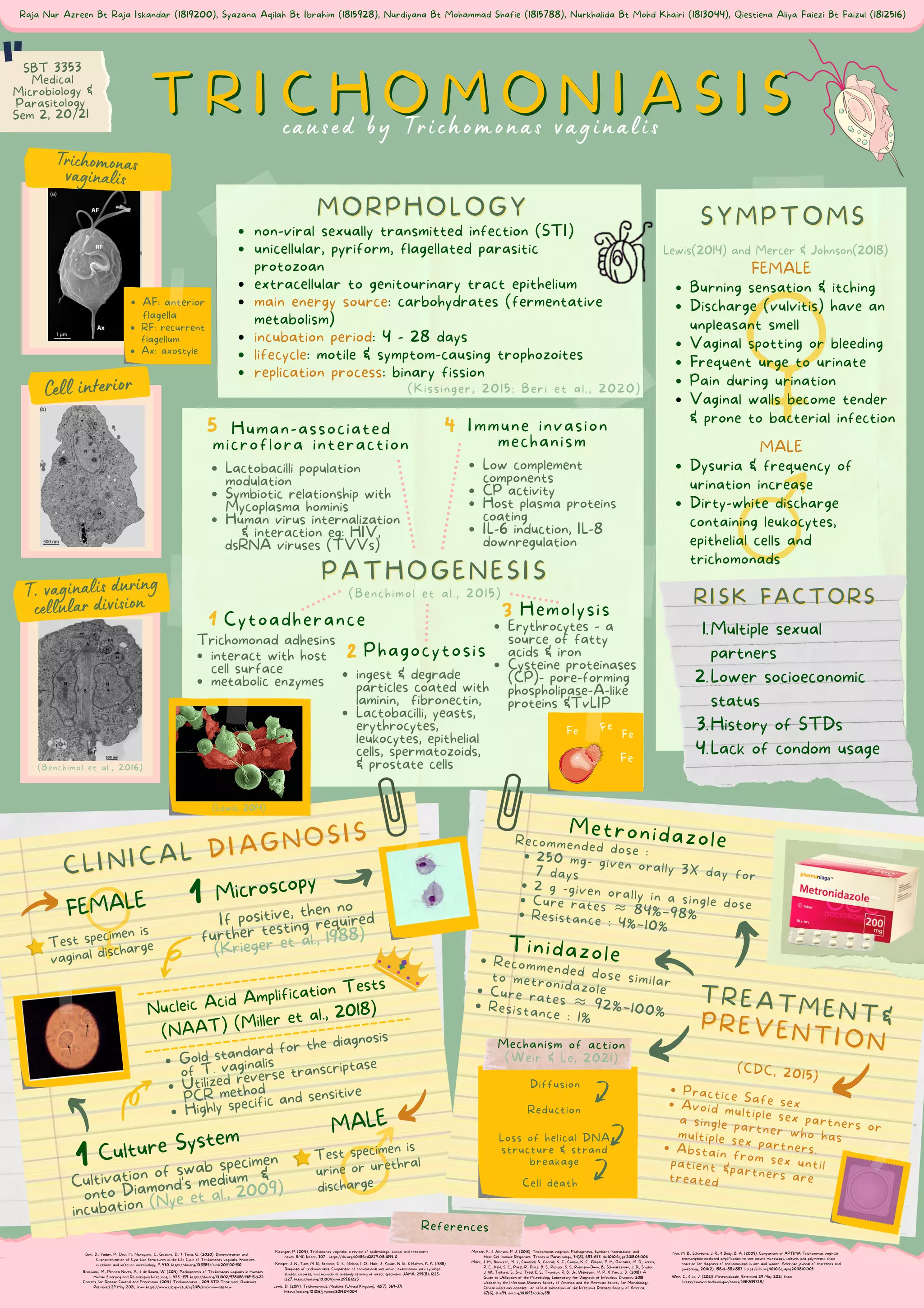
Diffusion
Recommended dose similar
to metronidazole
Cure rates ≈ 92%–100%
Resistance : 1%
(Benchimol et al., 2015)
Cytoadherance
Hemolysis
Phagocytosis
Immune invasion
mechanism
Cell death
Loss of helical DNA
structure & strand
breakage
AF: anterior
flagella
RF: recurrent
flagellum
Ax: axostyle
Nucleic Acid Amplification Tests
(NAAT) (Miller et al., 2018)
Reduction
non-viral sexually transmitted infection (STI)
unicellular, pyriform, flagellated parasitic
protozoan
extracellular to genitourinary tract epithelium
main energy source: carbohydrates (fermentative
metabolism)
incubation period: 4 - 28 days
lifecycle: motile & symptom-causing trophozoites
replication process: binary fission
Burning sensation & itching
Discharge (vulvitis) have an
unpleasant smell
Vaginal spotting or bleeding
Frequent urge to urinate
Pain during urination
Vaginal walls become tender
& prone to bacterial infection
Dysuria & frequency of
urination increase
Dirty-white discharge
containing leukocytes,
epithelial cells and
trichomonads
Lewis(2014) and Mercer & Johnson(2018)
FEMALE
MALE
250 mg- given orally 3X day for
7 days
2 g -given orally in a single dose
Cure rates ≈ 84%–98%
Resistance : 4%–10%
Recommended dose :
Multiple sexual
partners
Lower socioeconomic
status
History of STDs
Lack of condom usage
1.
2.
3.
4.
Practice Safe sex
Avoid multiple sex partners or
a single partner who has
multiple sex partners.
Abstain from sex until
(CDC, 2015)
patient &partners are
treated
Raja Nur Azreen Bt Raja Iskandar (1819200), Syazana Aqilah Bt Ibrahim (1815928), Nurdiyana Bt Mohammad Shafie (1815788), Nurkhalida Bt Mohd Khairi (1813044), Qiestiena Aliya Faiezi Bt Faizul (1812516)
Human-associated
microflora interaction
Lactobacilli population
modulation
Symbiotic relationship with
Mycoplasma hominis
Human virus internalization
& interaction eg: HIV,
dsRNA viruses (TVVs)
Low complement
components
CP activity
Host plasma proteins
coating
IL-6 induction, IL-8
downregulation
Gold standard for the diagnosis
of T. vaginalis
Utilized reverse transcriptase
PCR method
Highly specific and sensitive
interact with host
cell surface
metabolic enzymes
ingest & degrade
particles coated with
laminin, fibronectin,
Lactobacilli, yeasts,
erythrocytes,
leukocytes, epithelial
cells, spermatozoids,
& prostate cells
caused by Trichomonas vaginalis
Erythrocytes - a
source of fatty
acids & iron
Cysteine proteinases
(CP)- pore-forming
phospholipase-A-like
proteins &TvLIP
If positive, then no
further testing required
(Krieger et al., 1988)
Cultivation of swab specimen
onto Diamond's medium &
incubation (Nye et al., 2009)
Miller, J. M., Binnicker, M. J., Campbell, S., Carroll, K. C., Chapin, K. C., Gilligan, P. H., Gonzalez, M. D., Jerris,
R. C., Kehl, S. C., Patel, R., Pritt, B. S., Richter, S. S., Robinson-Dunn, B., Schwartzman, J. D., Snyder,
J. W., Telford, S., 3rd, Theel, E. S., Thomson, R. B., Jr, Weinstein, M. P., & Yao, J. D. (2018). A
Guide to Utilization of the Microbiology Laboratory for Diagnosis of Infectious Diseases: 2018
Update by the Infectious Diseases Society of America and the American Society for Microbiology.
Clinical infectious diseases : an official publication of the Infectious Diseases Society of America,
67(6), e1–e94. doi.org/10.1093/cid/ciy381
(Kissinger, 2015; Beri et al., 2020)
T. vaginalis during
cellular division
Trichomonas
vaginalis
Test specimen is
urine or urethral
discharge
1
Benchimol, M., Pereira‐Neves, A., & de Souza, W. (2015). Pathogenesis of Trichomonas vaginalis in Humans.
Human Emerging and Re‐emerging Infections, I, 423–439. https://doi.org/10.1002/9781118644843.ch22
Mechanism of action
(Weir & Le, 2021)
Diffusion
Microscopy
1 Culture System
Krieger, J. N., Tam, M. R., Stevens, C. E., Nielsen, I. O., Hale, J., Kiviat, N. B., & Holmes, K. K. (1988).
Diagnosis of trichomoniasis. Comparison of conventional wet-mount examination with cytologic
studies, cultures, and monoclonal antibody staining of direct specimens. JAMA, 259(8), 1223–
1227. https://doi.org/10.1001/jama.259.8.1223
Nye, M. B., Schwebke, J. R., & Body, B. A. (2009). Comparison of APTIMA Trichomonas vaginalis
transcription-mediated amplification to wet mount microscopy, culture, and polymerase chain
reaction for diagnosis of trichomoniasis in men and women. American journal of obstetrics and
gynecology, 200(2), 188.e1–188.e1887. https://doi.org/10.1016/j.ajog.2008.10.005
(Lewis, 2014)
Fe
Fe
Fe
Fe
1
2
3
4
5
Weir, C., & Le, J. (2021). Metronidazole. Retrieved 29 May 2021, from
https://www.ncbi.nlm.nih.gov/books/NBK539728/
(Benchimol et al., 2016)
Cell interior
Beri, D., Yadav, P., Devi, H., Narayana, C., Gadara, D., & Tatu, U. (2020). Demonstration and
Characterization of Cyst-Like Structures in the Life Cycle of Trichomonas vaginalis. Frontiers
in cellular and infection microbiology, 9, 430. https://doi.org/10.3389/fcimb.2019.00430
Metronidazole
Tinidazole
Mercer, F., & Johnson, P. J. (2018). Trichomonas vaginalis: Pathogenesis, Symbiont Interactions, and
Host Cell Immune Responses. Trends in Parasitology, 34(8), 683–693. doi:10.1016/j.pt.2018.05.006
Kissinger, P. (2015). Trichomonas vaginalis: a review of epidemiologic, clinical and treatment
issues. BMC Infect, 307 . https://doi.org/10.1186/s12879-015-1055-0
Lewis, D. (2014). Trichomoniasis. Medicine (United Kingdom), 42(7), 369–371.
https://doi.org/10.1016/j.mpmed.2014.04.004
Centers for Disease Control and Prevention. (2015). Trichomoniasis - 2015 STD Treatment Guidelines.
Retrieved 29 May 2021, from https://www.cdc.gov/std/tg2015/trichomoniasis.htm
References
Trichomonad adhesins
Test specimen is
vaginal discharge
SBT 3353
Medical
Microbiology &
Parasitology
Sem 2, 20/21

TRICHOMONIASIS.pdf

  • 1.
    Diffusion Recommended dose similar tometronidazole Cure rates ≈ 92%–100% Resistance : 1% (Benchimol et al., 2015) Cytoadherance Hemolysis Phagocytosis Immune invasion mechanism Cell death Loss of helical DNA structure & strand breakage AF: anterior flagella RF: recurrent flagellum Ax: axostyle Nucleic Acid Amplification Tests (NAAT) (Miller et al., 2018) Reduction non-viral sexually transmitted infection (STI) unicellular, pyriform, flagellated parasitic protozoan extracellular to genitourinary tract epithelium main energy source: carbohydrates (fermentative metabolism) incubation period: 4 - 28 days lifecycle: motile & symptom-causing trophozoites replication process: binary fission Burning sensation & itching Discharge (vulvitis) have an unpleasant smell Vaginal spotting or bleeding Frequent urge to urinate Pain during urination Vaginal walls become tender & prone to bacterial infection Dysuria & frequency of urination increase Dirty-white discharge containing leukocytes, epithelial cells and trichomonads Lewis(2014) and Mercer & Johnson(2018) FEMALE MALE 250 mg- given orally 3X day for 7 days 2 g -given orally in a single dose Cure rates ≈ 84%–98% Resistance : 4%–10% Recommended dose : Multiple sexual partners Lower socioeconomic status History of STDs Lack of condom usage 1. 2. 3. 4. Practice Safe sex Avoid multiple sex partners or a single partner who has multiple sex partners. Abstain from sex until (CDC, 2015) patient &partners are treated Raja Nur Azreen Bt Raja Iskandar (1819200), Syazana Aqilah Bt Ibrahim (1815928), Nurdiyana Bt Mohammad Shafie (1815788), Nurkhalida Bt Mohd Khairi (1813044), Qiestiena Aliya Faiezi Bt Faizul (1812516) Human-associated microflora interaction Lactobacilli population modulation Symbiotic relationship with Mycoplasma hominis Human virus internalization & interaction eg: HIV, dsRNA viruses (TVVs) Low complement components CP activity Host plasma proteins coating IL-6 induction, IL-8 downregulation Gold standard for the diagnosis of T. vaginalis Utilized reverse transcriptase PCR method Highly specific and sensitive interact with host cell surface metabolic enzymes ingest & degrade particles coated with laminin, fibronectin, Lactobacilli, yeasts, erythrocytes, leukocytes, epithelial cells, spermatozoids, & prostate cells caused by Trichomonas vaginalis Erythrocytes - a source of fatty acids & iron Cysteine proteinases (CP)- pore-forming phospholipase-A-like proteins &TvLIP If positive, then no further testing required (Krieger et al., 1988) Cultivation of swab specimen onto Diamond's medium & incubation (Nye et al., 2009) Miller, J. M., Binnicker, M. J., Campbell, S., Carroll, K. C., Chapin, K. C., Gilligan, P. H., Gonzalez, M. D., Jerris, R. C., Kehl, S. C., Patel, R., Pritt, B. S., Richter, S. S., Robinson-Dunn, B., Schwartzman, J. D., Snyder, J. W., Telford, S., 3rd, Theel, E. S., Thomson, R. B., Jr, Weinstein, M. P., & Yao, J. D. (2018). A Guide to Utilization of the Microbiology Laboratory for Diagnosis of Infectious Diseases: 2018 Update by the Infectious Diseases Society of America and the American Society for Microbiology. Clinical infectious diseases : an official publication of the Infectious Diseases Society of America, 67(6), e1–e94. doi.org/10.1093/cid/ciy381 (Kissinger, 2015; Beri et al., 2020) T. vaginalis during cellular division Trichomonas vaginalis Test specimen is urine or urethral discharge 1 Benchimol, M., Pereira‐Neves, A., & de Souza, W. (2015). Pathogenesis of Trichomonas vaginalis in Humans. Human Emerging and Re‐emerging Infections, I, 423–439. https://doi.org/10.1002/9781118644843.ch22 Mechanism of action (Weir & Le, 2021) Diffusion Microscopy 1 Culture System Krieger, J. N., Tam, M. R., Stevens, C. E., Nielsen, I. O., Hale, J., Kiviat, N. B., & Holmes, K. K. (1988). Diagnosis of trichomoniasis. Comparison of conventional wet-mount examination with cytologic studies, cultures, and monoclonal antibody staining of direct specimens. JAMA, 259(8), 1223– 1227. https://doi.org/10.1001/jama.259.8.1223 Nye, M. B., Schwebke, J. R., & Body, B. A. (2009). Comparison of APTIMA Trichomonas vaginalis transcription-mediated amplification to wet mount microscopy, culture, and polymerase chain reaction for diagnosis of trichomoniasis in men and women. American journal of obstetrics and gynecology, 200(2), 188.e1–188.e1887. https://doi.org/10.1016/j.ajog.2008.10.005 (Lewis, 2014) Fe Fe Fe Fe 1 2 3 4 5 Weir, C., & Le, J. (2021). Metronidazole. Retrieved 29 May 2021, from https://www.ncbi.nlm.nih.gov/books/NBK539728/ (Benchimol et al., 2016) Cell interior Beri, D., Yadav, P., Devi, H., Narayana, C., Gadara, D., & Tatu, U. (2020). Demonstration and Characterization of Cyst-Like Structures in the Life Cycle of Trichomonas vaginalis. Frontiers in cellular and infection microbiology, 9, 430. https://doi.org/10.3389/fcimb.2019.00430 Metronidazole Tinidazole Mercer, F., & Johnson, P. J. (2018). Trichomonas vaginalis: Pathogenesis, Symbiont Interactions, and Host Cell Immune Responses. Trends in Parasitology, 34(8), 683–693. doi:10.1016/j.pt.2018.05.006 Kissinger, P. (2015). Trichomonas vaginalis: a review of epidemiologic, clinical and treatment issues. BMC Infect, 307 . https://doi.org/10.1186/s12879-015-1055-0 Lewis, D. (2014). Trichomoniasis. Medicine (United Kingdom), 42(7), 369–371. https://doi.org/10.1016/j.mpmed.2014.04.004 Centers for Disease Control and Prevention. (2015). Trichomoniasis - 2015 STD Treatment Guidelines. Retrieved 29 May 2021, from https://www.cdc.gov/std/tg2015/trichomoniasis.htm References Trichomonad adhesins Test specimen is vaginal discharge SBT 3353 Medical Microbiology & Parasitology Sem 2, 20/21