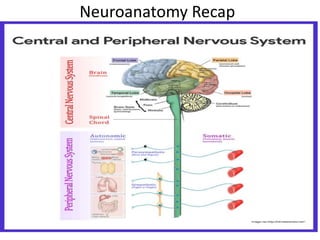
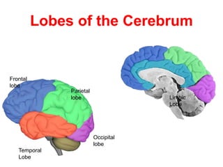
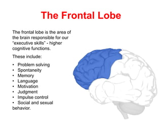
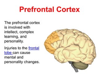
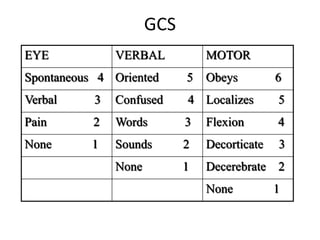
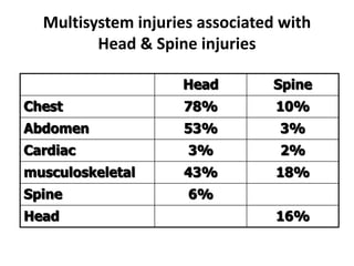
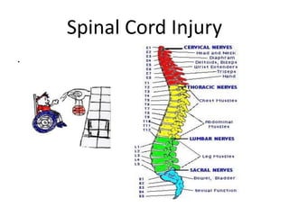
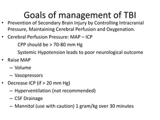
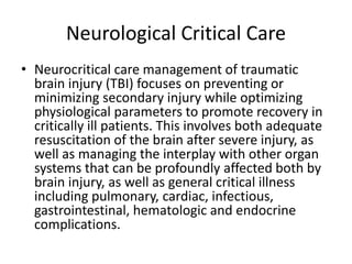
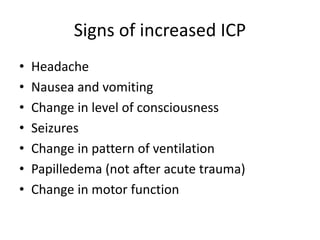
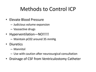
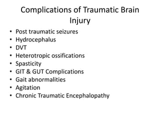
TRAUMATIC BRAIN INJURY & OTHER
ASSOCIATED NEUROTRAUMA
MIRITI .M.D
MASTERS OF CLINICAL MEDICINE
ACCIDENTS AND EMERGENCY
MCM/2017/73494
Definition
• According to the Common Data Elements
(CDE) Project, Traumatic Brain Injury TBI is an
alteration in brain function, or other evidence
of brain pathology, caused by an external
force, Examples of these forces include blows,
falls, sudden acceleration or deceleration of
the head, and blast waves.
Definition 2
• Traumatic Brain Injury (TBI) is a
nondegenerative, noncongenital insult to the
brain from an external mechanical force,
possibly leading to permanent or temporary
impairment of cognitive, physical and
psychosocial functions, with an associated
diminished or altered state of consciousness.
Monro-Kellie Doctrine
• Brain parenchyma—80%
• Cerebrospinal fluid—10%
• Blood—10%
• Monro-Kellie doctrine: Because the overall
volume of the cranial vault cannot change, an
increase in the volume of one component, or the
presence of pathologic components, necessitates
the displacement of other structures, an increase
in ICP, or both
Neuroanatomy Recap
Skull Anatomy
The skull is a rounded layer
of bone designed to protect
the brain from penetrating
injuries.
The base of the skull is rough, with
many bony protuberances.
These ridges can result in injury to
the temporal and frontal lobes of the
brain during rapid acceleration.
Bony ridges
Lobes of the Cerebrum
Frontal
lobe
Parietal
lobe
Occipital
lobe
Temporal
Lobe
Limbic
Lobe
The Frontal Lobe
The frontal lobe is the area of
the brain responsible for our
“executive skills” - higher
cognitive functions.
These include:
• Problem solving
• Spontaneity
• Memory
• Language
• Motivation
• Judgment
• Impulse control
• Social and sexual
behavior.
Prefrontal Cortex
The prefrontal cortex
is involved with
intellect, complex
learning, and
personality.
Injuries to the frontal
lobe can cause
mental and
personality changes.
Temporal Lobe
The temporal lobe plays a role
in emotions, and is also
responsible for smelling,
tasting, perception, memory,
understanding music,
aggressiveness, and sexual
behavior.
The temporal lobe also
contains the language area of
the brain.
Parietal Lobe
The parietal lobe plays a
role in our sensations of
touch, smell, and taste. It
also processes sensory
and spatial awareness, and
is a key component in eye-
hand co-ordination and
arm movement.
The parietal lobe also
contains a specialized area
called Wernicke’s area that
is responsible for matching
written words with the
sound of spoken speech.
Occipital Lobe
The occipital lobe
is at the rear of the
brain and controls
vision and
recognition.
The Limbic System
The limbic system is the area of the
brain that regulates emotion and
memory. It directly connects the
lower and higher brain functions.
The Neuron
Dendrites:
Collects
information from
other neurons
Cell Body
Axon:
Transmits information to
other neurons.
Epidemiology
• TBI epidemiology difficult due to inconsistency in the
definition and classification of Traumatic Brain Injury.
• Worldwide distribution
• TBI accounts for approx 40% of all deaths from acute
injuries in the United states, where annually 200000
victims of TBI need hospitalization, and 1.74 million
persons sustain mild TBI requiring an office visit or
temporary disability for at least 1 day.
• In the United States, the Centers for Disease Control
and Prevention (CDC) has reported that the total
combined rate for TBI-related emergency department
(ED) visits, hospitalizations, and deaths has reached
823.7 per 100,000 (available at
http://www.cdc.gov/traumaticbraininjury/index.html).
Traumatic Brain Injury Related Visits to
Emergency Departments
• Sex: Men more than Women
• Age group: Highest Risk (15-24 peak age) ,
Paediatric ages (0-15)
• External Cause: The external causes of TBI-
related death vary by age group. In 0- to 4-
year-olds, they are primarily associated with
assaults and falls, 15-34 MVT related
accidents, Above 65 Falls were among major
causes.
HIGH POPULATION RISK
• Young People
• Low income individuals
• Unmarried fellows
• Members of ethnic minority groups
• Residents of inner cities
• Men
• Individuals with a history of alcohol & substance
abuse
• Those who have suffered a previous TBI
Pathophysiology of TBI
• 1. Primary Injury: The type, direction, intensity, and duration
of forces all contribute to the characteristics and severity of
TBI. Forces that may contribute to TBI include angular,
rotational, shear, and translational forces.
• Even in the absence of an impact, significant acceleration or
deceleration of the head can cause TBI; however in most
cases a combination of impact and acceleration is probably to
blame.
• Forces involving the head striking or being struck by
something, termed contact or impact loading, are the cause
of most focal injuries, and movement of the brain within the
skull, termed noncontact or inertial loading, usually causes
diffuse injuries. The violent shaking of an infant that causes
shaken baby syndrome commonly manifests as diffuse injury.
Pathophysiology cont…….
• In impact loading, the force sends shock waves
through the skull and brain, resulting in tissue
damage. Shock waves caused by penetrating
injuries can also destroy tissue along the path of a
projectile, compounding the damage caused by the
missile itself.
• Damage may occur directly under the site of
impact, or it may occur on the side opposite the
impact (coup and contracoup injury,
respectively).When a moving object impacts the
stationary head, coup injuries are typical, while
contracoup injuries are usually produced when the
moving head strikes a stationary object
Pathophysiology………
• One type of focal injury, cerebral laceration, occurs
when the tissue is cut or torn e.g. Orbitofrontal cortex
due to Bony Protrusion of the skull.
• Cerebral contusion- Blood mixed with brain tissue
• Intracranial hemorrhage- Blood is not mixed with tissue
• Hematomas, also focal lesions, are collections of blood
in or around the brain that can result from
hemorrhage. Intra cerebral hemorrhage, with bleeding
in the brain tissue itself, is an intra-axial lesion.
• Extra-axial lesions include epidural hematoma,
subdural hematoma, subarachnoid hemorrhage, and
intra -ventricular hemorrhage.
Secondary Brain Injury
• Secondary types of Traumatic Brain Injury are attributable
to further cellular damage from the effects of primary
injuries.
• May develop over a period of hours or days following the
initial traumatic assault and is mediated through:
-Excitatory amino acids
-Endogenous opoid peptides
-Increased intracranial pressure
-Cerebral edema
-Hydrocephalus
-Brain Herniations
-Chronic traumatic encephalopathy
GCS
EYE VERBAL MOTOR
Spontaneous 4 Oriented 5 Obeys 6
Verbal 3 Confused 4 Localizes 5
Pain 2 Words 3 Flexion 4
None 1 Sounds 2 Decorticate 3
None 1 Decerebrate 2
None 1
Head Injury in combination with other
neuronal trauma
• Brain injury is chief among early concerns to those
providing care to patients with head trauma.
Providers should also be cognizant of the possibility
of injuries to associated structures and be aware of
strategies to recognize, evaluate, and treat
concomitant injuries. Multidisciplinary teams
composed of emergency department and trauma
providers, neurosurgery, otolaryngology,
ophthalmology, and facial trauma surgeons are
often needed to evaluate and treat these associated
injuries.
Multisystem injuries associated with
Head & Spine injuries
Head Spine
Chest 78% 10%
Abdomen 53% 3%
Cardiac 3% 2%
musculoskeletal 43% 18%
Spine 6%
Head 16%
Spinal Cord Injury
.
Spinal Cord Injury
• Mechanism of injury
The spine is usually injured
mainly in one of three
ways:
(a) a fall onto the back the
head and neck;
(b) a blow on the
forehead, Which forces
the neck into
hyperextension.
(c) - a fall onto legs or
buttocks
Scale of Motor Strength in SCI
• The American Spinal Injury Association:
– 0 - No contraction or movement
– 1 - Minimal movement
– 2 - Active movement, but not against gravity
– 3 - Active movement against gravity
– 4 - Active movement against resistance
– 5 - Active movement against full resistance
• Assessment of sensory function helps to identify the
different pathways for light touch, proprioception,
vibration, and pain. Use a pinprick to evaluate pain
sensation.
Types of Spinal Cord Paralysis
• Depending on the location and the extent of the
injury different forms of paralysis can occur.
• Monoplegia- paralysis of one limb
• Diplegia- paralysis of both upper or lower limbs
• Paraplegia- paralysis of both lower limbs
• Hemiplegia- paralysis of upper limb, torso and lower
leg on one side of the body
• Quadraplegia- paralysis of all four limbs
General Management Guidelines
• Strict spine precautions (immobilization)
• Emergency resuscitation (ABC..)
• Comprehensive approach
• Neurological and Radiological assessment.
• Always expect multiple trauma (neuroexam, chest,
abdomin, muskuloskeletal…)
• Differentiate hemorrhagic from neurogenic shock
The protocol of Initial
evaluation.(ATLS)
• Primary survey (cABCDE).Identify the injury
• Resuscitation; treat(ventilation, I.V. fluids & blood)
• Secondary survey ( a thorough head to toe exam)
• Definitive treatment or transfer to trauma center
(imaging, lab studies & surgeries)
• This is the essence of Advanced Trauma Life Support (ATLS).
Early identification & effective treatment for injury mainly in
early deaths it also decreases the No of late deaths
(Preventable deaths)
Primary Survey
• Patients are assessed and treatment priorities
established based on their injuries, vital signs,
and injury mechanisms
• cABCDE of trauma care
– C c-spine protection
– A Airway
– B Breathing and ventilation
– C Circulation with hemorrhage control
– D Disability/Neurologic status
– E Exposure/Environmental control
Secondary Survey
• AMPLE history
– Allergies, medications, PMH, last meal, events
• Physical exam from head to toe
• Frequent reassessment of vitals
• Diagnostic studies at this time
simultaneously
– X-rays, lab work, CT SCAN
Neurologic exam during secondary
survey
• (GCS) Mental Status
• Cranial Nerve Exam (pupils!!)
• Motor Exam of Upper and Lower Extremities
• Sensory Exam
• Reflexes (Babinski Sign?)
• Gait and Station/Ataxia (rarely done in the
acute situation
Goals of management of TBI
• Prevention of Secondary Brain Injury by Controlling Intracranial
Pressure, Maintaining Cerebral Perfusion and Oxygenation.
• Cerebral Perfusion Pressure: MAP – ICP
CPP should be > 70-80 mm Hg
Systemic Hypotension leads to poor neurological outcome
• Raise MAP
– Volume
– Vasopressors
• Decrease ICP (if > 20 mm Hg)
– Hyperventilation (not recommended)
– CSF Drainage
– Mannitol (use with caution) 1 gram/kg over 30 minutes
Neurological Critical Care
• Neurocritical care management of traumatic
brain injury (TBI) focuses on preventing or
minimizing secondary injury while optimizing
physiological parameters to promote recovery in
critically ill patients. This involves both adequate
resuscitation of the brain after severe injury, as
well as managing the interplay with other organ
systems that can be profoundly affected both by
brain injury, as well as general critical illness
including pulmonary, cardiac, infectious,
gastrointestinal, hematologic and endocrine
complications.
Signs of increased ICP
• Headache
• Nausea and vomiting
• Change in level of consciousness
• Seizures
• Change in pattern of ventilation
• Papilledema (not after acute trauma)
• Change in motor function
Methods to Control ICP
• Elevate Blood Pressure
– Judicious volume expansion
– Vasoactive drugs
• Hyperventilation—NO!!!!!
– Maintain pC02 around 35 mmHg
• Diuretics
– Mannitol
– Use with caution after neurosurgical consultation
• Drainage of CSF from Ventriculostomy Catheter
Complications of Traumatic Brain
Injury
• Post traumatic seizures
• Hydrocephalus
• DVT
• Heterotropic ossifications
• Spasticity
• GIT & GUT Complications
• Gait abnormalities
• Agitation
• Chronic Traumatic Encephalopathy

Traumatic brain injury 2018

  • 1.
    TRAUMATIC BRAIN INJURY& OTHER ASSOCIATED NEUROTRAUMA MIRITI .M.D MASTERS OF CLINICAL MEDICINE ACCIDENTS AND EMERGENCY MCM/2017/73494
  • 2.
    Definition • According tothe Common Data Elements (CDE) Project, Traumatic Brain Injury TBI is an alteration in brain function, or other evidence of brain pathology, caused by an external force, Examples of these forces include blows, falls, sudden acceleration or deceleration of the head, and blast waves.
  • 3.
    Definition 2 • TraumaticBrain Injury (TBI) is a nondegenerative, noncongenital insult to the brain from an external mechanical force, possibly leading to permanent or temporary impairment of cognitive, physical and psychosocial functions, with an associated diminished or altered state of consciousness.
  • 4.
    Monro-Kellie Doctrine • Brainparenchyma—80% • Cerebrospinal fluid—10% • Blood—10% • Monro-Kellie doctrine: Because the overall volume of the cranial vault cannot change, an increase in the volume of one component, or the presence of pathologic components, necessitates the displacement of other structures, an increase in ICP, or both
  • 5.
  • 6.
    Skull Anatomy The skullis a rounded layer of bone designed to protect the brain from penetrating injuries. The base of the skull is rough, with many bony protuberances. These ridges can result in injury to the temporal and frontal lobes of the brain during rapid acceleration. Bony ridges
  • 7.
    Lobes of theCerebrum Frontal lobe Parietal lobe Occipital lobe Temporal Lobe Limbic Lobe
  • 8.
    The Frontal Lobe Thefrontal lobe is the area of the brain responsible for our “executive skills” - higher cognitive functions. These include: • Problem solving • Spontaneity • Memory • Language • Motivation • Judgment • Impulse control • Social and sexual behavior.
  • 9.
    Prefrontal Cortex The prefrontalcortex is involved with intellect, complex learning, and personality. Injuries to the frontal lobe can cause mental and personality changes.
  • 10.
    Temporal Lobe The temporallobe plays a role in emotions, and is also responsible for smelling, tasting, perception, memory, understanding music, aggressiveness, and sexual behavior. The temporal lobe also contains the language area of the brain.
  • 11.
    Parietal Lobe The parietallobe plays a role in our sensations of touch, smell, and taste. It also processes sensory and spatial awareness, and is a key component in eye- hand co-ordination and arm movement. The parietal lobe also contains a specialized area called Wernicke’s area that is responsible for matching written words with the sound of spoken speech.
  • 12.
    Occipital Lobe The occipitallobe is at the rear of the brain and controls vision and recognition.
  • 13.
    The Limbic System Thelimbic system is the area of the brain that regulates emotion and memory. It directly connects the lower and higher brain functions.
  • 14.
    The Neuron Dendrites: Collects information from otherneurons Cell Body Axon: Transmits information to other neurons.
  • 15.
    Epidemiology • TBI epidemiologydifficult due to inconsistency in the definition and classification of Traumatic Brain Injury. • Worldwide distribution • TBI accounts for approx 40% of all deaths from acute injuries in the United states, where annually 200000 victims of TBI need hospitalization, and 1.74 million persons sustain mild TBI requiring an office visit or temporary disability for at least 1 day. • In the United States, the Centers for Disease Control and Prevention (CDC) has reported that the total combined rate for TBI-related emergency department (ED) visits, hospitalizations, and deaths has reached 823.7 per 100,000 (available at http://www.cdc.gov/traumaticbraininjury/index.html).
  • 16.
    Traumatic Brain InjuryRelated Visits to Emergency Departments • Sex: Men more than Women • Age group: Highest Risk (15-24 peak age) , Paediatric ages (0-15) • External Cause: The external causes of TBI- related death vary by age group. In 0- to 4- year-olds, they are primarily associated with assaults and falls, 15-34 MVT related accidents, Above 65 Falls were among major causes.
  • 17.
    HIGH POPULATION RISK •Young People • Low income individuals • Unmarried fellows • Members of ethnic minority groups • Residents of inner cities • Men • Individuals with a history of alcohol & substance abuse • Those who have suffered a previous TBI
  • 18.
    Pathophysiology of TBI •1. Primary Injury: The type, direction, intensity, and duration of forces all contribute to the characteristics and severity of TBI. Forces that may contribute to TBI include angular, rotational, shear, and translational forces. • Even in the absence of an impact, significant acceleration or deceleration of the head can cause TBI; however in most cases a combination of impact and acceleration is probably to blame. • Forces involving the head striking or being struck by something, termed contact or impact loading, are the cause of most focal injuries, and movement of the brain within the skull, termed noncontact or inertial loading, usually causes diffuse injuries. The violent shaking of an infant that causes shaken baby syndrome commonly manifests as diffuse injury.
  • 19.
    Pathophysiology cont……. • Inimpact loading, the force sends shock waves through the skull and brain, resulting in tissue damage. Shock waves caused by penetrating injuries can also destroy tissue along the path of a projectile, compounding the damage caused by the missile itself. • Damage may occur directly under the site of impact, or it may occur on the side opposite the impact (coup and contracoup injury, respectively).When a moving object impacts the stationary head, coup injuries are typical, while contracoup injuries are usually produced when the moving head strikes a stationary object
  • 20.
    Pathophysiology……… • One typeof focal injury, cerebral laceration, occurs when the tissue is cut or torn e.g. Orbitofrontal cortex due to Bony Protrusion of the skull. • Cerebral contusion- Blood mixed with brain tissue • Intracranial hemorrhage- Blood is not mixed with tissue • Hematomas, also focal lesions, are collections of blood in or around the brain that can result from hemorrhage. Intra cerebral hemorrhage, with bleeding in the brain tissue itself, is an intra-axial lesion. • Extra-axial lesions include epidural hematoma, subdural hematoma, subarachnoid hemorrhage, and intra -ventricular hemorrhage.
  • 21.
    Secondary Brain Injury •Secondary types of Traumatic Brain Injury are attributable to further cellular damage from the effects of primary injuries. • May develop over a period of hours or days following the initial traumatic assault and is mediated through: -Excitatory amino acids -Endogenous opoid peptides -Increased intracranial pressure -Cerebral edema -Hydrocephalus -Brain Herniations -Chronic traumatic encephalopathy
  • 22.
    GCS EYE VERBAL MOTOR Spontaneous4 Oriented 5 Obeys 6 Verbal 3 Confused 4 Localizes 5 Pain 2 Words 3 Flexion 4 None 1 Sounds 2 Decorticate 3 None 1 Decerebrate 2 None 1
  • 23.
    Head Injury incombination with other neuronal trauma • Brain injury is chief among early concerns to those providing care to patients with head trauma. Providers should also be cognizant of the possibility of injuries to associated structures and be aware of strategies to recognize, evaluate, and treat concomitant injuries. Multidisciplinary teams composed of emergency department and trauma providers, neurosurgery, otolaryngology, ophthalmology, and facial trauma surgeons are often needed to evaluate and treat these associated injuries.
  • 24.
    Multisystem injuries associatedwith Head & Spine injuries Head Spine Chest 78% 10% Abdomen 53% 3% Cardiac 3% 2% musculoskeletal 43% 18% Spine 6% Head 16%
  • 25.
  • 26.
    Spinal Cord Injury •Mechanism of injury The spine is usually injured mainly in one of three ways: (a) a fall onto the back the head and neck; (b) a blow on the forehead, Which forces the neck into hyperextension. (c) - a fall onto legs or buttocks
  • 27.
    Scale of MotorStrength in SCI • The American Spinal Injury Association: – 0 - No contraction or movement – 1 - Minimal movement – 2 - Active movement, but not against gravity – 3 - Active movement against gravity – 4 - Active movement against resistance – 5 - Active movement against full resistance • Assessment of sensory function helps to identify the different pathways for light touch, proprioception, vibration, and pain. Use a pinprick to evaluate pain sensation.
  • 28.
    Types of SpinalCord Paralysis • Depending on the location and the extent of the injury different forms of paralysis can occur. • Monoplegia- paralysis of one limb • Diplegia- paralysis of both upper or lower limbs • Paraplegia- paralysis of both lower limbs • Hemiplegia- paralysis of upper limb, torso and lower leg on one side of the body • Quadraplegia- paralysis of all four limbs
  • 29.
    General Management Guidelines •Strict spine precautions (immobilization) • Emergency resuscitation (ABC..) • Comprehensive approach • Neurological and Radiological assessment. • Always expect multiple trauma (neuroexam, chest, abdomin, muskuloskeletal…) • Differentiate hemorrhagic from neurogenic shock
  • 30.
    The protocol ofInitial evaluation.(ATLS) • Primary survey (cABCDE).Identify the injury • Resuscitation; treat(ventilation, I.V. fluids & blood) • Secondary survey ( a thorough head to toe exam) • Definitive treatment or transfer to trauma center (imaging, lab studies & surgeries) • This is the essence of Advanced Trauma Life Support (ATLS). Early identification & effective treatment for injury mainly in early deaths it also decreases the No of late deaths (Preventable deaths)
  • 31.
    Primary Survey • Patientsare assessed and treatment priorities established based on their injuries, vital signs, and injury mechanisms • cABCDE of trauma care – C c-spine protection – A Airway – B Breathing and ventilation – C Circulation with hemorrhage control – D Disability/Neurologic status – E Exposure/Environmental control
  • 32.
    Secondary Survey • AMPLEhistory – Allergies, medications, PMH, last meal, events • Physical exam from head to toe • Frequent reassessment of vitals • Diagnostic studies at this time simultaneously – X-rays, lab work, CT SCAN
  • 33.
    Neurologic exam duringsecondary survey • (GCS) Mental Status • Cranial Nerve Exam (pupils!!) • Motor Exam of Upper and Lower Extremities • Sensory Exam • Reflexes (Babinski Sign?) • Gait and Station/Ataxia (rarely done in the acute situation
  • 34.
    Goals of managementof TBI • Prevention of Secondary Brain Injury by Controlling Intracranial Pressure, Maintaining Cerebral Perfusion and Oxygenation. • Cerebral Perfusion Pressure: MAP – ICP CPP should be > 70-80 mm Hg Systemic Hypotension leads to poor neurological outcome • Raise MAP – Volume – Vasopressors • Decrease ICP (if > 20 mm Hg) – Hyperventilation (not recommended) – CSF Drainage – Mannitol (use with caution) 1 gram/kg over 30 minutes
  • 35.
    Neurological Critical Care •Neurocritical care management of traumatic brain injury (TBI) focuses on preventing or minimizing secondary injury while optimizing physiological parameters to promote recovery in critically ill patients. This involves both adequate resuscitation of the brain after severe injury, as well as managing the interplay with other organ systems that can be profoundly affected both by brain injury, as well as general critical illness including pulmonary, cardiac, infectious, gastrointestinal, hematologic and endocrine complications.
  • 36.
    Signs of increasedICP • Headache • Nausea and vomiting • Change in level of consciousness • Seizures • Change in pattern of ventilation • Papilledema (not after acute trauma) • Change in motor function
  • 37.
    Methods to ControlICP • Elevate Blood Pressure – Judicious volume expansion – Vasoactive drugs • Hyperventilation—NO!!!!! – Maintain pC02 around 35 mmHg • Diuretics – Mannitol – Use with caution after neurosurgical consultation • Drainage of CSF from Ventriculostomy Catheter
  • 38.
    Complications of TraumaticBrain Injury • Post traumatic seizures • Hydrocephalus • DVT • Heterotropic ossifications • Spasticity • GIT & GUT Complications • Gait abnormalities • Agitation • Chronic Traumatic Encephalopathy