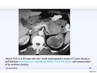
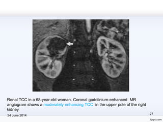
TRANSITIONAL CELL CARCINOMA
Upper urinary tract
Transitional Cell Carcinoma
• Originates from Transitional epithelium of urinary tract.
• Most common in urinary bladder, then in renal pelvis,
least in ureter(125:2.5:1)
• 5-10% of upper urinary tract neoplasms.
• Renal TCC most common --extrarenal part of the pelvis,
followed by the infundibulocaliceal region
• 2%–4% ---bilaterally.
24 June 2014 2
Clinical features:
• most common in 7th decade, rare in childhood
• males 3 times > female
• typically presents with hematuria
• 1/3 -- flank pain or acute renal colic
• discovered incidentally at radiologic examination
24 June 2014 3
Tumor spreads by
• mucosal extension
• local
• Hematogenous
• lymphatic invasion
• The most common sites for metastases are the liver,
bone, and lungs
24 June 2014 4
ETIOLOGY
• Increasing age
• Male gender
• Most important risk factor is smoking, 2-3 times
• Chemical carcinogens (aniline, benzidine, aromatic
amine, azo dyes),
24 June 2014 5
• Cyclo-phosphamide therapy
• Heavy caffeine consumption.
• Stasis of urine and structural abnormalities such as
horseshoe kidney.
24 June 2014 6
IMAGING MODALITIES
INTRAVENOUS UROGRAPHY
• noninvasive method of choice.
• detailed anatomy of the pelvicalyceal system and
ureters.
24 June 2014 7
• a filling defect within the contrast-enhanced collecting
system, single or multiple & smooth, irregular or stippled
• Stipple sign---tracking of contrast material into the
interstices of a papillary lesion
• Tumor-filled, distended calyces --“oncocalyces.”
• If these fail to opacify with contrast-- “phantom calyces.”
24 June 2014 8
Retrograde Pyelography
• in inadequately excreting kidneys,
• in cases of contrast allergy.
• facilitates ureterorendoscopy with biopsy or brushing &
cytology of urine
• an intraluminal filling defect,-- smooth, irregular, or
stippled.
24 June 2014 9
• An “apple core” appearance-- eccentric or encircling
ureteric lesions
• localized ureteric dilatation around and distal to the filling
defect may give rise to the “goblet” sign.
24 June 2014 10
Ultrasonography
• a central soft-tissue mass in the echogenic renal sinus,
with or without hydronephrosis.
• TCC is usually slightly hyperechoic relative to
surrounding renal parenchyma; occasionally, areas of
mixed echogenicity.
• typically TCC is infiltrative and does not distort the renal
contour.
24 June 2014 11
• US has a limited role in the evaluation of ureteric TCC
• If visualized, these tumors are typically intraluminal soft-
tissue masses with proximal distention of the ureter
• US also allows limited assessment of periureteric
tissues.
24 June 2014 12
Computed Tomography
• CT is well established in the preoperative staging and
assessment of upper tract TCC.
CT urography
• single breath-hold coverage of the entire urinary tract,
• has improved resolution
• has the ability to capture multiple phases of contrast
material excretion
24 June 2014 13
• hyperdense (5–30 HU) to urine and renal parenchyma
but hypodense than other pelvic filling defects such as
clot or calculus.
• typically seen as a sessile filling defect or
• pelvicaliceal irregularity, focal or diffuse mural thickening,
oncocalyx, and focally obstructed calyces.
24 June 2014 14
• Advanced TCC extends into the renal parenchyma in an
infiltrating pattern --- distorts normal architecture
• However, reniform shape is typically preserved (unlike
in renal cell carcinoma)
• enhances poorly after IV contrast
24 June 2014 15
• Hydronephrosis and hydroureter
• Ureteric TCC-- Ureteric wall thickening (eccentric or
circumferential), luminal narrowing, or an infiltrating
mass.
• A thickened enhancing ureteric wall with periureteric fat
stranding -- suggestive of extramural spread
24 June 2014 16
24 June 2014 17
TCC of the renal pelvis in a 60-year-old man with painless hematuria. Fifteen-
minute IVU image shows a large irregular filling defect (arrow) involving the right
renal pelvis and extending into the lower pole calyceal system
24 June 2014 18
TCC of the renal pelvis in a 65-year-old man. Fifteen-minute IVU image
shows a large stippled filling defect involving the collecting system of the right
kidney.
24 June 2014 19
TCC of the upper pole collecting system in a 55-year-old woman. Fifteen-
minute IVU image shows amputation of the upper pole calyx secondary to
TCC.
24 June 2014 20
Ureteric TCC in a 68-year-old woman. RP image shows a long irregular
stricture of the left distal ureter with proximal hydroureter and “shouldering” .
24 June 2014 21
Renal TCC in a 59-year-old woman. Sagittal US scan shows a well defined
hyerechoic mass in the upper pole. Tumor tissue is more echogenic than the
surrounding renal cortex but less echogenic than renal sinus fat.
24 June 2014 22
Renal TCC in a 65-year-old woman. Sagittal US scan shows a large mass of
mixed echogenicity (arrows) involving the upper pole and overlying renal
parenchyma.
24 June 2014 23
TCC of the renal pelvis in a 43-year-old man with flank pain and
hematuria. Axial nonenhanced CT scan shows a mass in the right renal
pelvis. The mass is slightly hyperdense relative to the urine and renal
parenchyma.
24 June 2014 24
Post contrast image shows characteristic early enhancement of the
mass, which is less than that of the surrounding renal parenchyma.
24 June 2014 25
Renal TCC in a 53-year-old man. Axial nephrographic phase CT scan shows a
well defined heterogenous hypodense lesion in the left kidney with preservation
of its reniform contour
24 June 2014 26
Bilateral ureteric TCC in a 57-year-old woman. Coronal T2-weighted MR image
show low-signal-intensity tumors in the distal right and distal left ureters.
24 June 2014 27
Renal TCC in a 68-year-old woman. Coronal gadolinium-enhanced MR
angiogram shows a moderately enhancing TCC in the upper pole of the right
kidney
Thank You
24 June 2014 28

Transitional cell carcinoma

  • 1.
  • 2.
    Transitional Cell Carcinoma •Originates from Transitional epithelium of urinary tract. • Most common in urinary bladder, then in renal pelvis, least in ureter(125:2.5:1) • 5-10% of upper urinary tract neoplasms. • Renal TCC most common --extrarenal part of the pelvis, followed by the infundibulocaliceal region • 2%–4% ---bilaterally. 24 June 2014 2
  • 3.
    Clinical features: • mostcommon in 7th decade, rare in childhood • males 3 times > female • typically presents with hematuria • 1/3 -- flank pain or acute renal colic • discovered incidentally at radiologic examination 24 June 2014 3
  • 4.
    Tumor spreads by •mucosal extension • local • Hematogenous • lymphatic invasion • The most common sites for metastases are the liver, bone, and lungs 24 June 2014 4
  • 5.
    ETIOLOGY • Increasing age •Male gender • Most important risk factor is smoking, 2-3 times • Chemical carcinogens (aniline, benzidine, aromatic amine, azo dyes), 24 June 2014 5
  • 6.
    • Cyclo-phosphamide therapy •Heavy caffeine consumption. • Stasis of urine and structural abnormalities such as horseshoe kidney. 24 June 2014 6
  • 7.
    IMAGING MODALITIES INTRAVENOUS UROGRAPHY •noninvasive method of choice. • detailed anatomy of the pelvicalyceal system and ureters. 24 June 2014 7
  • 8.
    • a fillingdefect within the contrast-enhanced collecting system, single or multiple & smooth, irregular or stippled • Stipple sign---tracking of contrast material into the interstices of a papillary lesion • Tumor-filled, distended calyces --“oncocalyces.” • If these fail to opacify with contrast-- “phantom calyces.” 24 June 2014 8
  • 9.
    Retrograde Pyelography • ininadequately excreting kidneys, • in cases of contrast allergy. • facilitates ureterorendoscopy with biopsy or brushing & cytology of urine • an intraluminal filling defect,-- smooth, irregular, or stippled. 24 June 2014 9
  • 10.
    • An “applecore” appearance-- eccentric or encircling ureteric lesions • localized ureteric dilatation around and distal to the filling defect may give rise to the “goblet” sign. 24 June 2014 10
  • 11.
    Ultrasonography • a centralsoft-tissue mass in the echogenic renal sinus, with or without hydronephrosis. • TCC is usually slightly hyperechoic relative to surrounding renal parenchyma; occasionally, areas of mixed echogenicity. • typically TCC is infiltrative and does not distort the renal contour. 24 June 2014 11
  • 12.
    • US hasa limited role in the evaluation of ureteric TCC • If visualized, these tumors are typically intraluminal soft- tissue masses with proximal distention of the ureter • US also allows limited assessment of periureteric tissues. 24 June 2014 12
  • 13.
    Computed Tomography • CTis well established in the preoperative staging and assessment of upper tract TCC. CT urography • single breath-hold coverage of the entire urinary tract, • has improved resolution • has the ability to capture multiple phases of contrast material excretion 24 June 2014 13
  • 14.
    • hyperdense (5–30HU) to urine and renal parenchyma but hypodense than other pelvic filling defects such as clot or calculus. • typically seen as a sessile filling defect or • pelvicaliceal irregularity, focal or diffuse mural thickening, oncocalyx, and focally obstructed calyces. 24 June 2014 14
  • 15.
    • Advanced TCCextends into the renal parenchyma in an infiltrating pattern --- distorts normal architecture • However, reniform shape is typically preserved (unlike in renal cell carcinoma) • enhances poorly after IV contrast 24 June 2014 15
  • 16.
    • Hydronephrosis andhydroureter • Ureteric TCC-- Ureteric wall thickening (eccentric or circumferential), luminal narrowing, or an infiltrating mass. • A thickened enhancing ureteric wall with periureteric fat stranding -- suggestive of extramural spread 24 June 2014 16
  • 17.
    24 June 201417 TCC of the renal pelvis in a 60-year-old man with painless hematuria. Fifteen- minute IVU image shows a large irregular filling defect (arrow) involving the right renal pelvis and extending into the lower pole calyceal system
  • 18.
    24 June 201418 TCC of the renal pelvis in a 65-year-old man. Fifteen-minute IVU image shows a large stippled filling defect involving the collecting system of the right kidney.
  • 19.
    24 June 201419 TCC of the upper pole collecting system in a 55-year-old woman. Fifteen- minute IVU image shows amputation of the upper pole calyx secondary to TCC.
  • 20.
    24 June 201420 Ureteric TCC in a 68-year-old woman. RP image shows a long irregular stricture of the left distal ureter with proximal hydroureter and “shouldering” .
  • 21.
    24 June 201421 Renal TCC in a 59-year-old woman. Sagittal US scan shows a well defined hyerechoic mass in the upper pole. Tumor tissue is more echogenic than the surrounding renal cortex but less echogenic than renal sinus fat.
  • 22.
    24 June 201422 Renal TCC in a 65-year-old woman. Sagittal US scan shows a large mass of mixed echogenicity (arrows) involving the upper pole and overlying renal parenchyma.
  • 23.
    24 June 201423 TCC of the renal pelvis in a 43-year-old man with flank pain and hematuria. Axial nonenhanced CT scan shows a mass in the right renal pelvis. The mass is slightly hyperdense relative to the urine and renal parenchyma.
  • 24.
    24 June 201424 Post contrast image shows characteristic early enhancement of the mass, which is less than that of the surrounding renal parenchyma.
  • 25.
    24 June 201425 Renal TCC in a 53-year-old man. Axial nephrographic phase CT scan shows a well defined heterogenous hypodense lesion in the left kidney with preservation of its reniform contour
  • 26.
    24 June 201426 Bilateral ureteric TCC in a 57-year-old woman. Coronal T2-weighted MR image show low-signal-intensity tumors in the distal right and distal left ureters.
  • 27.
    24 June 201427 Renal TCC in a 68-year-old woman. Coronal gadolinium-enhanced MR angiogram shows a moderately enhancing TCC in the upper pole of the right kidney
  • 28.